 Movie
Movie Controller

[English] 日本語
 Yorodumi
- EMDB-44797: C-terminus truncated (last two residues) mutant of Human light ch... -
+ Open data

- Basic information
| Entry |  | |||||||||
|---|---|---|---|---|---|---|---|---|---|---|
| Title | C-terminus truncated (last two residues) mutant of Human light chain ferritin reacted with iron (3 Fe2+ to ferritin monomer ratio). Reconstruction of particles with no nanoparticle. | |||||||||
 Map data Map data | C-terminus truncated (last two residues) mutant of Human light chain ferritin reacted with Ferrous salt(3 Fe2 per ferritin subunit) .Map based on 2D classes which show no iron nanoparticle. | |||||||||
 Sample Sample |
| |||||||||
 Keywords Keywords | Iron oxide binding protein / METAL BINDING PROTEIN | |||||||||
| Function / homology |  Function and homology information Function and homology informationferritin complex / Scavenging by Class A Receptors / Golgi Associated Vesicle Biogenesis / autolysosome / ferric iron binding / autophagosome / iron ion transport / ferrous iron binding / Iron uptake and transport / azurophil granule lumen ...ferritin complex / Scavenging by Class A Receptors / Golgi Associated Vesicle Biogenesis / autolysosome / ferric iron binding / autophagosome / iron ion transport / ferrous iron binding / Iron uptake and transport / azurophil granule lumen / intracellular iron ion homeostasis / iron ion binding / Neutrophil degranulation / extracellular exosome / extracellular region / membrane / identical protein binding / cytoplasm / cytosol Similarity search - Function | |||||||||
| Biological species |  Homo sapiens (human) Homo sapiens (human) | |||||||||
| Method | single particle reconstruction / cryo EM / Resolution: 2.36 Å | |||||||||
 Authors Authors | Sen S / Nannenga BL / Williams D | |||||||||
| Funding support |  United States, 1 items United States, 1 items
| |||||||||
 Citation Citation | Journal: J Am Chem Soc / Year: 2025 Title: Observation of the Protein-Inorganic Interface of Ferritin by Cryo-Electron Microscopy. Authors: Sagnik Sen / Amar Thaker / Alison Haymaker / Dewight Williams / Po-Lin Chiu / Brent L Nannenga / Abstract: Visualizing the structure of the protein-inorganic interface is critically important for a more complete understanding of biomineralization. Unfortunately, there are limited approaches for the direct ...Visualizing the structure of the protein-inorganic interface is critically important for a more complete understanding of biomineralization. Unfortunately, there are limited approaches for the direct and detailed study of biomolecules that interact with inorganic materials. Here, we use single-particle cryo-electron microscopy (cryo-EM) to study the protein-nanoparticle (NP) interactions of human light chain ferritin and visualize the high-resolution details of the protein-inorganic interface. In this work, we determined the 2.85 Å structure of human light chain ferritin bound to its native iron oxide NP substrate. The resulting cryo-EM maps confirmed and enhanced previously proposed interactions of the protein with the material along the B-helix and revealed new interaction at the C-terminus of light chain ferritin. This work sheds new light on the mechanisms of ferritin biomineralization and further demonstrates the application of cryo-EM for the study of protein-inorganic systems. | |||||||||
| History |
|
- Structure visualization
| Supplemental images |
|---|

- Downloads & links
-EMDB archive
| Map data | emd_44797.map.gz | 12.6 MB | EMDB map data format | |
|---|---|---|---|---|
| Header (meta data) | emd-44797-v30.xmlemd-44797.xml | 14.7 KB 14.7 KB | Display Display | EMDB header |
| FSC (resolution estimation) | emd_44797_fsc.xml | 6.1 KB | Display | FSC data file |
| Images |  emd_44797.png emd_44797.png | 69.9 KB | ||
| Filedesc metadata | emd-44797.cif.gz | 5.4 KB | ||
| Others | emd_44797_half_map_1.map.gzemd_44797_half_map_2.map.gz | 23.4 MB 23.4 MB | ||
| Archive directory |  http://ftp.pdbj.org/pub/emdb/structures/EMD-44797ftp://ftp.pdbj.org/pub/emdb/structures/EMD-44797 http://ftp.pdbj.org/pub/emdb/structures/EMD-44797ftp://ftp.pdbj.org/pub/emdb/structures/EMD-44797 | HTTPS FTP |
-Related structure data
| Related structure data | 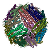 9bq5MC  9bpiC 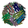 9bpjC  9bpkC C: citing same article ( M: atomic model generated by this map |
|---|---|
| Similar structure data |


-Links
| EMDB pages | EMDB (EBI/PDBe) / EMDataResource |
|---|---|
| Related items in Molecule of the Month |


-Map
| File | Download / File: emd_44797.map.gz / Format: CCP4 / Size: 25.3 MB / Type: IMAGE STORED AS FLOATING POINT NUMBER (4 BYTES) | ||||||||||||||||||||||||||||||||||||
|---|---|---|---|---|---|---|---|---|---|---|---|---|---|---|---|---|---|---|---|---|---|---|---|---|---|---|---|---|---|---|---|---|---|---|---|---|---|
| Annotation | C-terminus truncated (last two residues) mutant of Human light chain ferritin reacted with Ferrous salt(3 Fe2 per ferritin subunit) .Map based on 2D classes which show no iron nanoparticle. | ||||||||||||||||||||||||||||||||||||
| Projections & slices | Image control
Images are generated by Spider. | ||||||||||||||||||||||||||||||||||||
| Voxel size | X=Y=Z: 1.03 Å | ||||||||||||||||||||||||||||||||||||
| Density |
| ||||||||||||||||||||||||||||||||||||
| Symmetry | Space group: 1 | ||||||||||||||||||||||||||||||||||||
| Details | EMDB XML:
|
 Z (Sec.)
 Y (Row.)
 X (Col.)


-Supplemental data
-Half map: Half map B.
| File | emd_44797_half_map_1.map | ||||||||||||
|---|---|---|---|---|---|---|---|---|---|---|---|---|---|
| Annotation | Half map B. | ||||||||||||
| Projections & Slices |
| ||||||||||||
| Density Histograms |


-Half map: Half map A.
| File | emd_44797_half_map_2.map | ||||||||||||
|---|---|---|---|---|---|---|---|---|---|---|---|---|---|
| Annotation | Half map A. | ||||||||||||
| Projections & Slices |
| ||||||||||||
| Density Histograms |


- Sample components
-Entire : Human Light Chain Ferritin
| Entire | Name: Human Light Chain Ferritin |
|---|---|
| Components |
|
-Supramolecule #1: Human Light Chain Ferritin
| Supramolecule | Name: Human Light Chain Ferritin / type: complex / ID: 1 / Parent: 0 / Macromolecule list: all |
|---|---|
| Source (natural) | Organism: Homo sapiens (human) |
| Molecular weight | Theoretical: 474 KDa |
-Macromolecule #1: Ferritin light chain
| Macromolecule | Name: Ferritin light chain / type: protein_or_peptide / ID: 1 / Number of copies: 24 / Enantiomer: LEVO |
|---|---|
| Source (natural) | Organism: Homo sapiens (human) |
| Molecular weight | Theoretical: 19.66425 KDa |
| Recombinant expression | Organism:  |
| Sequence | String: SSQIRQNYST DVEAAVNSLV NLYLQASYTY LSLGFYFDRD DVALEGVSHF FRELAEEKRE GYERLLKMQN QRGGRALFQD IKKPAEDEW GKTPDAMKAA MALEKKLNQA LLDLHALGSA RTDPHLCDFL ETHFLDEEVK LIKKMGDHLT NLHRLGGPEA G LGEYLFER LTLK UniProtKB: Ferritin light chain |
-Experimental details
-Structure determination
| Method | cryo EM |
|---|---|
 Processing Processing | single particle reconstruction |
| Aggregation state | particle |
-Sample preparation
| Buffer | pH: 7.5 |
|---|---|
| Vitrification | Cryogen name: ETHANE |
- Electron microscopy
| Microscope | TFS KRIOS |
|---|---|
| Image recording | Film or detector model: GATAN K2 SUMMIT (4k x 4k) / Average electron dose: 9.58 e/Å2 |
| Electron beam | Acceleration voltage: 300 kV / Electron source:  FIELD EMISSION GUN FIELD EMISSION GUN |
| Electron optics | Illumination mode: FLOOD BEAM / Imaging mode: BRIGHT FIELD / Nominal defocus max: 2.8000000000000003 µm / Nominal defocus min: 0.1 µm |
| Experimental equipment |  Model: Titan Krios / Image courtesy: FEI Company |

