+ Open data
- Basic information
| Entry |  | |||||||||
|---|---|---|---|---|---|---|---|---|---|---|
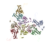
| Title | Pseudomonas phage DEV 5-fold vertex (major coat protein) | |||||||||
 Map data Map data | ||||||||||
 Sample Sample |
| |||||||||
 Keywords Keywords | virion coat / complex / STRUCTURAL PROTEIN / gp77 | |||||||||
| Function / homology | Phage capsid protein / Major coat protein Function and homology information Function and homology information | |||||||||
| Biological species |  Pseudomonas phage vB_PaeP_DEV (virus) Pseudomonas phage vB_PaeP_DEV (virus) | |||||||||
| Method | single particle reconstruction / cryo EM / Resolution: 3.3 Å | |||||||||
 Authors Authors | Iglesias SM / Hou CFD / Li F / Cingolani G | |||||||||
| Funding support |  United States, 2 items United States, 2 items
| |||||||||
 Citation Citation |  Journal: Nat Commun / Year: 2024 Journal: Nat Commun / Year: 2024Title: Integrative structural analysis of Pseudomonas phage DEV reveals a genome ejection motor. Authors: Ravi K Lokareddy / Chun-Feng David Hou / Francesca Forti / Stephano M Iglesias / Fenglin Li / Mikhail Pavlenok / David S Horner / Michael Niederweis / Federica Briani / Gino Cingolani /   Abstract: DEV is an obligatory lytic Pseudomonas phage of the N4-like genus, recently reclassified as Schitoviridae. The DEV genome encodes 91 ORFs, including a 3398 amino acid virion-associated RNA polymerase ...DEV is an obligatory lytic Pseudomonas phage of the N4-like genus, recently reclassified as Schitoviridae. The DEV genome encodes 91 ORFs, including a 3398 amino acid virion-associated RNA polymerase (vRNAP). Here, we describe the complete architecture of DEV, determined using a combination of cryo-electron microscopy localized reconstruction, biochemical methods, and genetic knockouts. We built de novo structures of all capsid factors and tail components involved in host attachment. We demonstrate that DEV long tail fibers are essential for infection of Pseudomonas aeruginosa but dispensable for infecting mutants with a truncated lipopolysaccharide devoid of the O-antigen. We determine that DEV vRNAP is part of a three-gene operon conserved in 191 Schitoviridae genomes. We propose these three proteins are ejected into the host to form a genome ejection motor spanning the cell envelope. We posit that the design principles of the DEV ejection apparatus are conserved in all Schitoviridae. | |||||||||
| History |
|
- Structure visualization
| Supplemental images |
|---|
- Downloads & links
-EMDB archive
| Map data |  emd_44518.map.gz emd_44518.map.gz | 472.9 MB |  EMDB map data format EMDB map data format | |
|---|---|---|---|---|
| Header (meta data) |  emd-44518-v30.xml emd-44518-v30.xml emd-44518.xml emd-44518.xml | 14.1 KB 14.1 KB | Display Display |  EMDB header EMDB header |
| FSC (resolution estimation) |  emd_44518_fsc.xml emd_44518_fsc.xml | 18.1 KB | Display |  FSC data file FSC data file |
| Images |  emd_44518.png emd_44518.png | 104.1 KB | ||
| Masks |  emd_44518_msk_1.map emd_44518_msk_1.map | 512 MB |  Mask map Mask map | |
| Filedesc metadata |  emd-44518.cif.gz emd-44518.cif.gz | 5.5 KB | ||
| Others |  emd_44518_half_map_1.map.gz emd_44518_half_map_1.map.gz emd_44518_half_map_2.map.gz emd_44518_half_map_2.map.gz | 412.9 MB 411.4 MB | ||
| Archive directory |  http://ftp.pdbj.org/pub/emdb/structures/EMD-44518 http://ftp.pdbj.org/pub/emdb/structures/EMD-44518 ftp://ftp.pdbj.org/pub/emdb/structures/EMD-44518 ftp://ftp.pdbj.org/pub/emdb/structures/EMD-44518 | HTTPS FTP |
-Validation report
| Summary document |  emd_44518_validation.pdf.gz emd_44518_validation.pdf.gz | 1.3 MB | Display |  EMDB validaton report EMDB validaton report |
|---|---|---|---|---|
| Full document |  emd_44518_full_validation.pdf.gz emd_44518_full_validation.pdf.gz | 1.3 MB | Display | |
| Data in XML |  emd_44518_validation.xml.gz emd_44518_validation.xml.gz | 26.3 KB | Display | |
| Data in CIF |  emd_44518_validation.cif.gz emd_44518_validation.cif.gz | 35.4 KB | Display | |
| Arichive directory |  https://ftp.pdbj.org/pub/emdb/validation_reports/EMD-44518 https://ftp.pdbj.org/pub/emdb/validation_reports/EMD-44518 ftp://ftp.pdbj.org/pub/emdb/validation_reports/EMD-44518 ftp://ftp.pdbj.org/pub/emdb/validation_reports/EMD-44518 | HTTPS FTP |
-Related structure data
| Related structure data |  9bgnMC  8vxqC  9bgmC  9bgoC  9codC M: atomic model generated by this map C: citing same article ( |
|---|---|
| Similar structure data | Similarity search - Function & homology  F&H Search F&H Search |
- Links
Links
| EMDB pages |  EMDB (EBI/PDBe) / EMDB (EBI/PDBe) /  EMDataResource EMDataResource |
|---|
- Map
Map
| File |  Download / File: emd_44518.map.gz / Format: CCP4 / Size: 512 MB / Type: IMAGE STORED AS FLOATING POINT NUMBER (4 BYTES) Download / File: emd_44518.map.gz / Format: CCP4 / Size: 512 MB / Type: IMAGE STORED AS FLOATING POINT NUMBER (4 BYTES) | ||||||||||||||||||||||||||||||||||||
|---|---|---|---|---|---|---|---|---|---|---|---|---|---|---|---|---|---|---|---|---|---|---|---|---|---|---|---|---|---|---|---|---|---|---|---|---|---|
| Projections & slices | Image control
Images are generated by Spider. | ||||||||||||||||||||||||||||||||||||
| Voxel size | X=Y=Z: 1.12 Å | ||||||||||||||||||||||||||||||||||||
| Density |
| ||||||||||||||||||||||||||||||||||||
| Symmetry | Space group: 1 | ||||||||||||||||||||||||||||||||||||
| Details | EMDB XML:
|
-Supplemental data
-Mask #1
| File |  emd_44518_msk_1.map emd_44518_msk_1.map | ||||||||||||
|---|---|---|---|---|---|---|---|---|---|---|---|---|---|
| Projections & Slices |
| ||||||||||||
| Density Histograms |
-Half map: #1
| File | emd_44518_half_map_1.map | ||||||||||||
|---|---|---|---|---|---|---|---|---|---|---|---|---|---|
| Projections & Slices |
| ||||||||||||
| Density Histograms |
-Half map: #2
| File | emd_44518_half_map_2.map | ||||||||||||
|---|---|---|---|---|---|---|---|---|---|---|---|---|---|
| Projections & Slices |
| ||||||||||||
| Density Histograms |
- Sample components
-Entire : Major coat protein of Pseudomonas phage DEV
| Entire | Name: Major coat protein of Pseudomonas phage DEV |
|---|---|
| Components |
|
-Supramolecule #1: Major coat protein of Pseudomonas phage DEV
| Supramolecule | Name: Major coat protein of Pseudomonas phage DEV / type: complex / ID: 1 / Parent: 0 / Macromolecule list: all |
|---|---|
| Source (natural) | Organism:  Pseudomonas phage vB_PaeP_DEV (virus) Pseudomonas phage vB_PaeP_DEV (virus) |
-Macromolecule #1: gp77 major coat protein
| Macromolecule | Name: gp77 major coat protein / type: protein_or_peptide / ID: 1 / Number of copies: 9 / Enantiomer: LEVO |
|---|---|
| Source (natural) | Organism:  Pseudomonas phage vB_PaeP_DEV (virus) Pseudomonas phage vB_PaeP_DEV (virus) |
| Molecular weight | Theoretical: 44.114113 KDa |
| Recombinant expression | Organism:  Pseudomonas (RNA similarity group I) Pseudomonas (RNA similarity group I) |
| Sequence | String: MAGPVDNIKP MKYNDPANGV ESSIGPQIHT RYWYKRALID AAKEAYFGQL ADTFSMPKHY GKEIVRLHYI PLLDDRNVND QGIDASGAT IANGNLYGSS RDVGNITAKM PTLTEIGGRV NRVGFKRVEI KGKLEKYGFF REYTQEQLDF DSDPAMEGHV T TEMVKGAN ...String: MAGPVDNIKP MKYNDPANGV ESSIGPQIHT RYWYKRALID AAKEAYFGQL ADTFSMPKHY GKEIVRLHYI PLLDDRNVND QGIDASGAT IANGNLYGSS RDVGNITAKM PTLTEIGGRV NRVGFKRVEI KGKLEKYGFF REYTQEQLDF DSDPAMEGHV T TEMVKGAN EITEDLLQID LLNSAGTVRY PGAATSDAEV DASTEVTYDS LMRLRLDLDN ARAPTKIKMI TGTRMIDTRT VG NARALYV GSDLVPTIEA MKDNHGNPAF IPIEKYAAGG ATMHGEVGQL GRFRVIVNPQ MMHWAGVGKA VDPNDQVPMH ESG GKYSVF PMLCVASEAF TTVGFATDGK NVKFKIITKR PGEATADRSD PYGEMGFMSI KWYYGFMVFR PEWIALLKTV ARL UniProtKB: Major coat protein |
-Experimental details
-Structure determination
| Method | cryo EM |
|---|---|
 Processing Processing | single particle reconstruction |
| Aggregation state | particle |
- Sample preparation
| Buffer | pH: 7.5 |
|---|---|
| Vitrification | Cryogen name: ETHANE |
- Electron microscopy
| Microscope | FEI TITAN KRIOS |
|---|---|
| Image recording | Film or detector model: GATAN K3 (6k x 4k) / Average electron dose: 50.0 e/Å2 |
| Electron beam | Acceleration voltage: 300 kV / Electron source:  FIELD EMISSION GUN FIELD EMISSION GUN |
| Electron optics | Illumination mode: FLOOD BEAM / Imaging mode: BRIGHT FIELD / Nominal defocus max: 1.6 µm / Nominal defocus min: 0.8 µm |
| Experimental equipment |  Model: Titan Krios / Image courtesy: FEI Company |
 Movie
Movie Controller


 X (Sec.)
 Y (Row.)
 Z (Col.)


