 Movie
Movie Controller

[English] 日本語
 Yorodumi
- EMDB-44453: MicroED structure of bovine liver catalase with missing cone solv... -
+ Open data

- Basic information
| Entry | 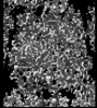 | ||||||||||||
|---|---|---|---|---|---|---|---|---|---|---|---|---|---|
| Title | MicroED structure of bovine liver catalase with missing cone solved by suspended drop | ||||||||||||
 Map data Map data | |||||||||||||
 Sample Sample |
| ||||||||||||
 Keywords Keywords | Heme-containing enzyme / OXIDOREDUCTASE | ||||||||||||
| Function / homology |  Function and homology information Function and homology informationcatalase complex / Detoxification of Reactive Oxygen Species / Peroxisomal protein import / cellular detoxification of hydrogen peroxide / catalase / catalase activity / Neutrophil degranulation / peroxisomal matrix / positive regulation of cell division / hydrogen peroxide catabolic process ...catalase complex / Detoxification of Reactive Oxygen Species / Peroxisomal protein import / cellular detoxification of hydrogen peroxide / catalase / catalase activity / Neutrophil degranulation / peroxisomal matrix / positive regulation of cell division / hydrogen peroxide catabolic process / response to hydrogen peroxide / peroxisome / heme binding / enzyme binding / mitochondrion / metal ion binding / cytoplasm Similarity search - Function | ||||||||||||
| Biological species |  | ||||||||||||
| Method | electron crystallography / cryo EM / Resolution: 4.0 Å | ||||||||||||
 Authors Authors | Gillman C / Bu G / Gonen T | ||||||||||||
| Funding support |  United States, 3 items United States, 3 items
| ||||||||||||
 Citation Citation | Journal: J Struct Biol X / Year: 2024 Title: Eliminating the missing cone challenge through innovative approaches. Authors: Cody Gillman / Guanhong Bu / Emma Danelius / Johan Hattne / Brent L Nannenga / Tamir Gonen / Abstract: Microcrystal electron diffraction (MicroED) has emerged as a powerful technique for unraveling molecular structures from microcrystals too small for X-ray diffraction. However, a significant hurdle ...Microcrystal electron diffraction (MicroED) has emerged as a powerful technique for unraveling molecular structures from microcrystals too small for X-ray diffraction. However, a significant hurdle arises with plate-like crystals that consistently orient themselves flat on the electron microscopy grid. If the normal of the plate correlates with the axes of the crystal lattice, the crystal orientations accessible for measurement are restricted because the crystal cannot be arbitrarily rotated. This limits the information that can be acquired, resulting in a missing cone of information. We recently introduced a novel crystallization strategy called suspended drop crystallization and proposed that crystals in a suspended drop could effectively address the challenge of preferred crystal orientation. Here we demonstrate the success of the suspended drop approach in eliminating the missing cone in two samples that crystallize as thin plates: bovine liver catalase and the SARS‑CoV‑2 main protease (Mpro). This innovative solution proves indispensable for crystals exhibiting systematic preferred orientations, unlocking new possibilities for structure determination by MicroED. | ||||||||||||
| History |
|
- Structure visualization
| Supplemental images |
|---|

- Downloads & links
-EMDB archive
| Map data | emd_44453.map.gz | 1.4 MB | EMDB map data format | |
|---|---|---|---|---|
| Header (meta data) | emd-44453-v30.xmlemd-44453.xml | 14.3 KB 14.3 KB | Display Display | EMDB header |
| Images |  emd_44453.png emd_44453.png | 234.4 KB | ||
| Filedesc metadata | emd-44453.cif.gz | 5.6 KB | ||
| Archive directory |  http://ftp.pdbj.org/pub/emdb/structures/EMD-44453ftp://ftp.pdbj.org/pub/emdb/structures/EMD-44453 http://ftp.pdbj.org/pub/emdb/structures/EMD-44453ftp://ftp.pdbj.org/pub/emdb/structures/EMD-44453 | HTTPS FTP |
-Validation report
| Summary document | emd_44453_validation.pdf.gz | 621.5 KB | Display | EMDB validaton report |
|---|---|---|---|---|
| Full document | emd_44453_full_validation.pdf.gz | 621.1 KB | Display | |
| Data in XML | emd_44453_validation.xml.gz | 4.4 KB | Display | |
| Data in CIF | emd_44453_validation.cif.gz | 4.9 KB | Display | |
| Arichive directory | https://ftp.pdbj.org/pub/emdb/validation_reports/EMD-44453ftp://ftp.pdbj.org/pub/emdb/validation_reports/EMD-44453 | HTTPS FTP |
-Related structure data
| Related structure data | 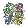 9bdjMC 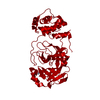 8vd7C M: atomic model generated by this map C: citing same article ( |
|---|---|
| Similar structure data |
-Links
| EMDB pages | EMDB (EBI/PDBe) / EMDataResource |
|---|---|
| Related items in Molecule of the Month |


-Map
| File | Download / File: emd_44453.map.gz / Format: CCP4 / Size: 2.1 MB / Type: IMAGE STORED AS FLOATING POINT NUMBER (4 BYTES) | ||||||||||||||||||||||||||||||||||||
|---|---|---|---|---|---|---|---|---|---|---|---|---|---|---|---|---|---|---|---|---|---|---|---|---|---|---|---|---|---|---|---|---|---|---|---|---|---|
| Projections & slices | Image control
Images are generated by Spider. generated in cubic-lattice coordinate | ||||||||||||||||||||||||||||||||||||
| Voxel size | X: 1.32 Å / Y: 1.313 Å / Z: 1.305 Å | ||||||||||||||||||||||||||||||||||||
| Density |
| ||||||||||||||||||||||||||||||||||||
| Symmetry | Space group: 19 | ||||||||||||||||||||||||||||||||||||
| Details | EMDB XML:
|
 Z (Sec.)
 X (Row.)
 Y (Col.)


-Supplemental data
- Sample components
-Entire : Bovine liver catalase
| Entire | Name: Bovine liver catalase |
|---|---|
| Components |
|
-Supramolecule #1: Bovine liver catalase
| Supramolecule | Name: Bovine liver catalase / type: complex / ID: 1 / Parent: 0 / Macromolecule list: #1 |
|---|---|
| Source (natural) | Organism: |
-Macromolecule #1: Catalase
| Macromolecule | Name: Catalase / type: protein_or_peptide / ID: 1 / Details: from bovine liver / Number of copies: 4 / Enantiomer: LEVO / EC number: catalase |
|---|---|
| Source (natural) | Organism: |
| Molecular weight | Theoretical: 59.99916 KDa |
| Sequence | String: MADNRDPASD QMKHWKEQRA AQKPDVLTTG GGNPVGDKLN SLTVGPRGPL LVQDVVFTDE MAHFDRERIP ERVVHAKGAG AFGYFEVTH DITRYSKAKV FEHIGKRTPI AVRFSTVAGE SGSADTVRDP RGFAVKFYTE DGNWDLVGNN TPIFFIRDAL L FPSFIHSQ ...String: MADNRDPASD QMKHWKEQRA AQKPDVLTTG GGNPVGDKLN SLTVGPRGPL LVQDVVFTDE MAHFDRERIP ERVVHAKGAG AFGYFEVTH DITRYSKAKV FEHIGKRTPI AVRFSTVAGE SGSADTVRDP RGFAVKFYTE DGNWDLVGNN TPIFFIRDAL L FPSFIHSQ KRNPQTHLKD PDMVWDFWSL RPESLHQVSF LFSDRGIPDG HRHMNGYGSH TFKLVNANGE AVYCKFHYKT DQ GIKNLSV EDAARLAHED PDYGLRDLFN AIATGNYPSW TLYIQVMTFS EAEIFPFNPF DLTKVWPHGD YPLIPVGKLV LNR NPVNYF AEVEQLAFDP SNMPPGIEPS PDKMLQGRLF AYPDTHRHRL GPNYLQIPVN CPYRARVANY QRDGPMCMMD NQGG APNYY PNSFSAPEHQ PSALEHRTHF SGDVQRFNSA NDDNVTQVRT FYLKVLNEEQ RKRLCENIAG HLKDAQLFIQ KKAVK NFSD VHPEYGSRIQ ALLDKYNEEK PKNAVHTYVQ HGSHLSAREK ANL UniProtKB: Catalase |
-Macromolecule #2: PROTOPORPHYRIN IX CONTAINING FE
| Macromolecule | Name: PROTOPORPHYRIN IX CONTAINING FE / type: ligand / ID: 2 / Number of copies: 4 / Formula: HEM |
|---|---|
| Molecular weight | Theoretical: 616.487 Da |
| Chemical component information |  ChemComp-HEM: |
-Macromolecule #3: NADPH DIHYDRO-NICOTINAMIDE-ADENINE-DINUCLEOTIDE PHOSPHATE
| Macromolecule | Name: NADPH DIHYDRO-NICOTINAMIDE-ADENINE-DINUCLEOTIDE PHOSPHATE type: ligand / ID: 3 / Number of copies: 4 / Formula: NDP |
|---|---|
| Molecular weight | Theoretical: 745.421 Da |
| Chemical component information |  ChemComp-NDP: |
-Experimental details
-Structure determination
| Method | cryo EM |
|---|---|
 Processing Processing | electron crystallography |
| Aggregation state | 3D array |
-Sample preparation
| Buffer | pH: 6.3 |
|---|---|
| Vitrification | Cryogen name: ETHANE |
- Electron microscopy
| Microscope | FEI TITAN KRIOS |
|---|---|
| Image recording | Film or detector model: FEI FALCON IV (4k x 4k) / Average electron dose: 0.0025 e/Å2 |
| Electron beam | Acceleration voltage: 300 kV / Electron source:  FIELD EMISSION GUN FIELD EMISSION GUN |
| Electron optics | Illumination mode: OTHER / Imaging mode: DIFFRACTION / Nominal defocus max: 0.0 µm / Nominal defocus min: 0.0 µm / Camera length: 2941 mm |
| Experimental equipment |  Model: Titan Krios / Image courtesy: FEI Company |
-Image processing
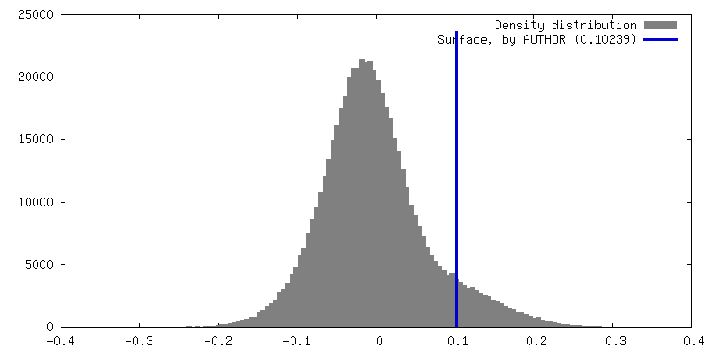
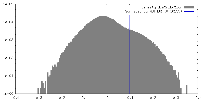
| Final reconstruction | Resolution.type: BY AUTHOR / Resolution: 4.0 Å / Resolution method: DIFFRACTION PATTERN/LAYERLINES | |||||||||||||||||||||||||||||||||||||||||||||||||
|---|---|---|---|---|---|---|---|---|---|---|---|---|---|---|---|---|---|---|---|---|---|---|---|---|---|---|---|---|---|---|---|---|---|---|---|---|---|---|---|---|---|---|---|---|---|---|---|---|---|---|
| Molecular replacement | Software - Name: MOLREP | |||||||||||||||||||||||||||||||||||||||||||||||||
| Crystallography statistics | Number intensities measured: 89263 / Number structure factors: 18157 / Fourier space coverage: 94.859 / R merge: 65.2 / Overall phase residual: 0 / Phase error rejection criteria: Rfree / High resolution: 4.0 Å Shell:
|
-Atomic model buiding 1
| Refinement | Protocol: OTHER |
|---|---|
| Output model | PDB-9bdj: |
