 Movie
Movie Controller

[English] 日本語
 Yorodumi
- EMDB-4392: In situ subtomogram average of a bacterial encapsulin expressed i... -
+ Open data

- Basic information
| Entry | Database: EMDB / ID: EMD-4392 | |||||||||
|---|---|---|---|---|---|---|---|---|---|---|
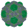
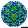
| Title | In situ subtomogram average of a bacterial encapsulin expressed in HEK293T cells (no iron treatment) | |||||||||
 Map data Map data | structure of EncABCD without iron treatment | |||||||||
 Sample Sample |
| |||||||||
| Biological species |  Myxococcus xanthus (bacteria) Myxococcus xanthus (bacteria) | |||||||||
| Method | subtomogram averaging / cryo EM / Resolution: 19.6 Å | |||||||||
 Authors Authors | Erdmann PS / Plitzko JM | |||||||||
 Citation Citation | Journal: Nat Commun / Year: 2018 Title: Bacterial encapsulins as orthogonal compartments for mammalian cell engineering. Authors: Felix Sigmund / Christoph Massner / Philipp Erdmann / Anja Stelzl / Hannes Rolbieski / Mitul Desai / Sarah Bricault / Tobias P Wörner / Joost Snijder / Arie Geerlof / Helmut Fuchs / Martin ...Authors: Felix Sigmund / Christoph Massner / Philipp Erdmann / Anja Stelzl / Hannes Rolbieski / Mitul Desai / Sarah Bricault / Tobias P Wörner / Joost Snijder / Arie Geerlof / Helmut Fuchs / Martin Hrabĕ de Angelis / Albert J R Heck / Alan Jasanoff / Vasilis Ntziachristos / Jürgen Plitzko / Gil G Westmeyer /    Abstract: We genetically controlled compartmentalization in eukaryotic cells by heterologous expression of bacterial encapsulin shell and cargo proteins to engineer enclosed enzymatic reactions and size- ...We genetically controlled compartmentalization in eukaryotic cells by heterologous expression of bacterial encapsulin shell and cargo proteins to engineer enclosed enzymatic reactions and size-constrained metal biomineralization. The shell protein (EncA) from Myxococcus xanthus auto-assembles into nanocompartments inside mammalian cells to which sets of native (EncB,C,D) and engineered cargo proteins self-target enabling localized bimolecular fluorescence and enzyme complementation. Encapsulation of the enzyme tyrosinase leads to the confinement of toxic melanin production for robust detection via multispectral optoacoustic tomography (MSOT). Co-expression of ferritin-like native cargo (EncB,C) results in efficient iron sequestration producing substantial contrast by magnetic resonance imaging (MRI) and allowing for magnetic cell sorting. The monodisperse, spherical, and iron-loading nanoshells are also excellent genetically encoded reporters for electron microscopy (EM). In general, eukaryotically expressed encapsulins enable cellular engineering of spatially confined multicomponent processes with versatile applications in multiscale molecular imaging, as well as intriguing implications for metabolic engineering and cellular therapy. | |||||||||
| History |
|
- Structure visualization
| Movie |
 Movie viewer Movie viewer |
|---|---|
| Structure viewer | EM map: SurfViewMolmilJmol/JSmol |
| Supplemental images |

- Downloads & links
-EMDB archive
| Map data | emd_4392.map.gz | 21.4 MB | EMDB map data format | |
|---|---|---|---|---|
| Header (meta data) | emd-4392-v30.xmlemd-4392.xml | 11.5 KB 11.5 KB | Display Display | EMDB header |
| Images | 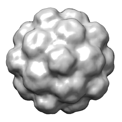 emd_4392.png emd_4392.png | 66.2 KB | ||
| Archive directory |  http://ftp.pdbj.org/pub/emdb/structures/EMD-4392ftp://ftp.pdbj.org/pub/emdb/structures/EMD-4392 http://ftp.pdbj.org/pub/emdb/structures/EMD-4392ftp://ftp.pdbj.org/pub/emdb/structures/EMD-4392 | HTTPS FTP |
-Related structure data


-Links
| EMDB pages | EMDB (EBI/PDBe) / EMDataResource |
|---|
-Map
| File | Download / File: emd_4392.map.gz / Format: CCP4 / Size: 23.8 MB / Type: IMAGE STORED AS FLOATING POINT NUMBER (4 BYTES) | ||||||||||||||||||||||||||||||||||||||||||||||||||||||||||||
|---|---|---|---|---|---|---|---|---|---|---|---|---|---|---|---|---|---|---|---|---|---|---|---|---|---|---|---|---|---|---|---|---|---|---|---|---|---|---|---|---|---|---|---|---|---|---|---|---|---|---|---|---|---|---|---|---|---|---|---|---|---|
| Annotation | structure of EncABCD without iron treatment | ||||||||||||||||||||||||||||||||||||||||||||||||||||||||||||
| Projections & slices | Image control
Images are generated by Spider. | ||||||||||||||||||||||||||||||||||||||||||||||||||||||||||||
| Voxel size | X=Y=Z: 3.42 Å | ||||||||||||||||||||||||||||||||||||||||||||||||||||||||||||
| Density |
| ||||||||||||||||||||||||||||||||||||||||||||||||||||||||||||
| Symmetry | Space group: 1 | ||||||||||||||||||||||||||||||||||||||||||||||||||||||||||||
| Details | EMDB XML:
CCP4 map header:
| ||||||||||||||||||||||||||||||||||||||||||||||||||||||||||||
 Z (Sec.)
 Y (Row.)
 X (Col.)


-Supplemental data
- Sample components
-Entire : Encapsulin from M. xanthus in its T = 3 configuration
| Entire | Name: Encapsulin from M. xanthus in its T = 3 configuration |
|---|---|
| Components |
|
-Supramolecule #1: Encapsulin from M. xanthus in its T = 3 configuration
| Supramolecule | Name: Encapsulin from M. xanthus in its T = 3 configuration / type: complex / ID: 1 / Parent: 0 |
|---|---|
| Source (natural) | Organism: Myxococcus xanthus (bacteria) |
| Recombinant expression | Organism:  Homo sapiens (human) / Recombinant cell: HEK293T Homo sapiens (human) / Recombinant cell: HEK293T |
| Molecular weight | Theoretical: 1.2 MDa |
-Experimental details
-Structure determination
| Method | cryo EM |
|---|---|
 Processing Processing | subtomogram averaging |
| Aggregation state | cell |
-Sample preparation
| Buffer | pH: 7 / Details: cells were plunge frozen in DMEM with 10% FBS |
|---|---|
| Grid | Model: Quantifoil R2/1 / Material: COPPER / Mesh: 200 / Pretreatment - Type: GLOW DISCHARGE / Pretreatment - Atmosphere: AIR |
| Vitrification | Cryogen name: ETHANE-PROPANE / Chamber humidity: 100 % / Chamber temperature: 310 K / Instrument: FEI VITROBOT MARK IV / Details: blot force: 10 blot time: 10. |
| Details | the sample was prepared by focused ion beam milling of vitrified HEK293T cells |
- Electron microscopy
| Microscope | FEI TITAN KRIOS |
|---|---|
| Image recording | Film or detector model: GATAN K2 SUMMIT (4k x 4k) / Detector mode: COUNTING / Average electron dose: 1.5 e/Å2 |
| Electron beam | Acceleration voltage: 300 kV / Electron source:  FIELD EMISSION GUN FIELD EMISSION GUN |
| Electron optics | C2 aperture diameter: 70.0 µm / Illumination mode: FLOOD BEAM / Imaging mode: BRIGHT FIELD / Cs: 2.7 mm / Nominal defocus min: -5.0 µm / Nominal magnification: 42000 |
| Sample stage | Specimen holder model: FEI TITAN KRIOS AUTOGRID HOLDER / Cooling holder cryogen: NITROGEN |
| Experimental equipment |  Model: Titan Krios / Image courtesy: FEI Company |
