 ムービー
ムービー コントローラー


+ データを開く

- 基本情報
基本情報
| 登録情報 | データベース: EMDB / ID: EMD-4376 | |||||||||
|---|---|---|---|---|---|---|---|---|---|---|
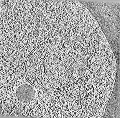
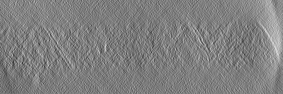
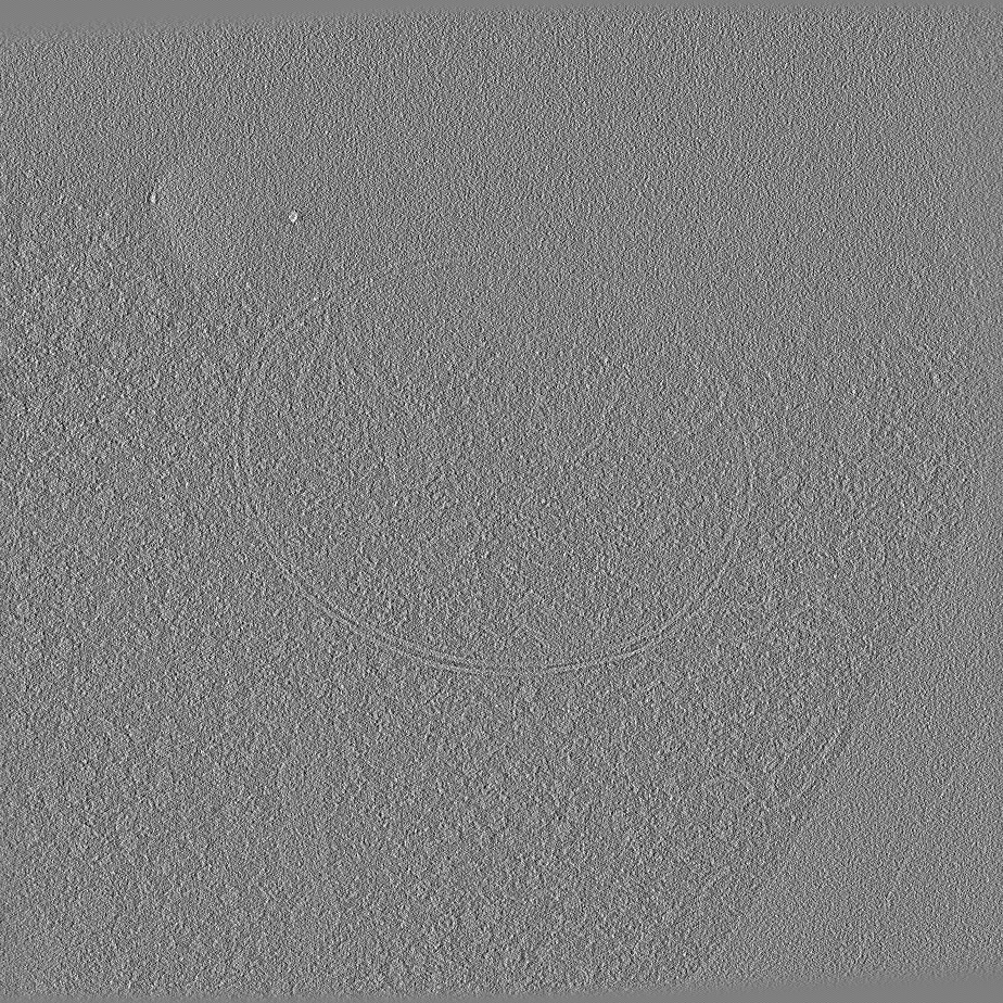
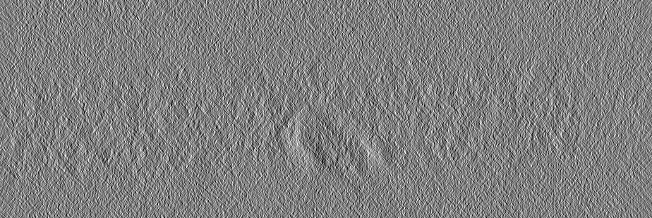
| タイトル | Tomogram obtained for control yeast cells | |||||||||
 マップデータ マップデータ | Tomogram obtained for control yeast cells | |||||||||
 試料 試料 |
| |||||||||
| 生物種 |  | |||||||||
| 手法 | 電子線トモグラフィー法 / クライオ電子顕微鏡法 | |||||||||
 データ登録者 データ登録者 | Pfeffer S / Engel BD / Schaffer M | |||||||||
 引用 引用 | ジャーナル: Cell / 年: 2018 タイトル: mTORC1 Controls Phase Separation and the Biophysical Properties of the Cytoplasm by Tuning Crowding. 著者: M Delarue / G P Brittingham / S Pfeffer / I V Surovtsev / S Pinglay / K J Kennedy / M Schaffer / J I Gutierrez / D Sang / G Poterewicz / J K Chung / J M Plitzko / J T Groves / C Jacobs-Wagner ...著者: M Delarue / G P Brittingham / S Pfeffer / I V Surovtsev / S Pinglay / K J Kennedy / M Schaffer / J I Gutierrez / D Sang / G Poterewicz / J K Chung / J M Plitzko / J T Groves / C Jacobs-Wagner / B D Engel / L J Holt /   要旨: Macromolecular crowding has a profound impact on reaction rates and the physical properties of the cell interior, but the mechanisms that regulate crowding are poorly understood. We developed ...Macromolecular crowding has a profound impact on reaction rates and the physical properties of the cell interior, but the mechanisms that regulate crowding are poorly understood. We developed genetically encoded multimeric nanoparticles (GEMs) to dissect these mechanisms. GEMs are homomultimeric scaffolds fused to a fluorescent protein that self-assemble into bright, stable particles of defined size and shape. By combining tracking of GEMs with genetic and pharmacological approaches, we discovered that the mTORC1 pathway can modulate the effective diffusion coefficient of particles ≥20 nm in diameter more than 2-fold by tuning ribosome concentration, without any discernable effect on the motion of molecules ≤5 nm. This change in ribosome concentration affected phase separation both in vitro and in vivo. Together, these results establish a role for mTORC1 in controlling both the mesoscale biophysical properties of the cytoplasm and biomolecular condensation. | |||||||||
| 履歴 |
|
- 構造の表示
構造の表示
| ムービー |
 ムービービューア ムービービューア |
|---|---|
| 添付画像 |
- ダウンロードとリンク
-EMDBアーカイブ
| マップデータ | emd_4376.map.gz | 934 MB | EMDBマップデータ形式 | |
|---|---|---|---|---|
| ヘッダ (付随情報) | emd-4376-v30.xmlemd-4376.xml | 10.8 KB 10.8 KB | 表示 表示 | EMDBヘッダ |
| 画像 |  emd_4376.png emd_4376.png | 189.9 KB | ||
| アーカイブディレクトリ |  http://ftp.pdbj.org/pub/emdb/structures/EMD-4376ftp://ftp.pdbj.org/pub/emdb/structures/EMD-4376 http://ftp.pdbj.org/pub/emdb/structures/EMD-4376ftp://ftp.pdbj.org/pub/emdb/structures/EMD-4376 | HTTPS FTP |
-検証レポート
| 文書・要旨 | emd_4376_validation.pdf.gz | 178.5 KB | 表示 | EMDB検証レポート |
|---|---|---|---|---|
| 文書・詳細版 | emd_4376_full_validation.pdf.gz | 177.7 KB | 表示 | |
| XML形式データ | emd_4376_validation.xml.gz | 4.2 KB | 表示 | |
| アーカイブディレクトリ | https://ftp.pdbj.org/pub/emdb/validation_reports/EMD-4376ftp://ftp.pdbj.org/pub/emdb/validation_reports/EMD-4376 | HTTPS FTP |
-関連構造データ

-リンク
| EMDBのページ | EMDB (EBI/PDBe) / EMDataResource |
|---|
-マップ
| ファイル | ダウンロード / ファイル: emd_4376.map.gz / 形式: CCP4 / 大きさ: 1006.4 MB / タイプ: IMAGE STORED AS FLOATING POINT NUMBER (4 BYTES) | ||||||||||||||||||||||||||||||||||||||||||||||||||||||||||||
|---|---|---|---|---|---|---|---|---|---|---|---|---|---|---|---|---|---|---|---|---|---|---|---|---|---|---|---|---|---|---|---|---|---|---|---|---|---|---|---|---|---|---|---|---|---|---|---|---|---|---|---|---|---|---|---|---|---|---|---|---|---|
| 注釈 | Tomogram obtained for control yeast cells | ||||||||||||||||||||||||||||||||||||||||||||||||||||||||||||
| 投影像・断面図 | 画像のコントロール
画像は Spider により作成 これらの図は立方格子座標系で作成されたものです | ||||||||||||||||||||||||||||||||||||||||||||||||||||||||||||
| ボクセルのサイズ | X=Y=Z: 13.68 Å | ||||||||||||||||||||||||||||||||||||||||||||||||||||||||||||
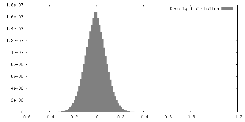
| 密度 |
| ||||||||||||||||||||||||||||||||||||||||||||||||||||||||||||
| 対称性 | 空間群: 1 | ||||||||||||||||||||||||||||||||||||||||||||||||||||||||||||
| 詳細 | EMDB XML:
CCP4マップ ヘッダ情報:
| ||||||||||||||||||||||||||||||||||||||||||||||||||||||||||||
 Z (Sec.)
 Y (Row.)
 X (Col.)


-添付データ
- 試料の構成要素
-全体 : Control yeast cell
| 全体 | 名称: Control yeast cell |
|---|---|
| 要素 |
|
-超分子 #1: Control yeast cell
| 超分子 | 名称: Control yeast cell / タイプ: cell / ID: 1 / 親要素: 0 |
|---|---|
| 由来(天然) | 生物種: |
-実験情報
-構造解析
| 手法 | クライオ電子顕微鏡法 |
|---|---|
 解析 解析 | 電子線トモグラフィー法 |
| 試料の集合状態 | cell |
-試料調製
| 緩衝液 | pH: 7 |
|---|---|
| グリッド | モデル: Quantifoil R2/1 / 材質: COPPER / 支持フィルム - 材質: CARBON / 支持フィルム - トポロジー: HOLEY |
| 凍結 | 凍結剤: ETHANE-PROPANE / 装置: FEI VITROBOT MARK IV / 詳細: 10 seconds blot time, blot force 10. |
| 詳細 | Control yeast cell thinned by focused ion beam milling |
| 切片作成 | 集束イオンビーム - 装置: OTHER / 集束イオンビーム - イオン: OTHER / 集束イオンビーム - 電圧: 30 kV / 集束イオンビーム - 電流: 0.3 nA / 集束イオンビーム - Dose rate: 3.3 / 集束イオンビーム - 時間: 3500 sec. / 集束イオンビーム - 温度: 92 K / 集束イオンビーム - Initial thickness: 10000 / 集束イオンビーム - 最終 厚さ: 150 集束イオンビーム - 詳細: 3.3E14. The value given for _emd_sectioning_focused_ion_beam.instrument is FEI Quanta FIB. This is not in a list of allowed values set(['DB235', 'OTHER']) so OTHER ...集束イオンビーム - 詳細: 3.3E14. The value given for _emd_sectioning_focused_ion_beam.instrument is FEI Quanta FIB. This is not in a list of allowed values set(['DB235', 'OTHER']) so OTHER is written into the XML file. |
- 電子顕微鏡法
| 顕微鏡 | FEI TITAN KRIOS |
|---|---|
| 撮影 | フィルム・検出器のモデル: GATAN K2 SUMMIT (4k x 4k) 検出モード: COUNTING / 平均電子線量: 1.5 e/Å2 |
| 電子線 | 加速電圧: 300 kV / 電子線源:  FIELD EMISSION GUN FIELD EMISSION GUN |
| 電子光学系 | 照射モード: FLOOD BEAM / 撮影モード: BRIGHT FIELD / 最大 デフォーカス(公称値): 6.0 µm / 最小 デフォーカス(公称値): 6.0 µm |
| 試料ステージ | 試料ホルダーモデル: FEI TITAN KRIOS AUTOGRID HOLDER ホルダー冷却材: NITROGEN |
| 実験機器 |  モデル: Titan Krios / 画像提供: FEI Company |
-画像解析
| 最終 再構成 | アルゴリズム: BACK PROJECTION / ソフトウェア - 名称: IMOD / 使用した粒子像数: 61 |
|---|
