+ Open data
- Basic information
| Entry | Database: EMDB / ID: EMD-4371 | |||||||||
|---|---|---|---|---|---|---|---|---|---|---|
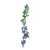
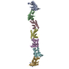
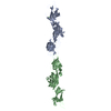
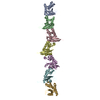
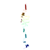
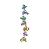
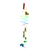
| Title | Cryo-EM map of the FCHSD2 F-BAR domain + SH3-1 | |||||||||
 Map data Map data | FCHSD2 F-BAR SH3-1 domains. | |||||||||
 Sample Sample |
| |||||||||
| Biological species |  Homo sapiens (human) Homo sapiens (human) | |||||||||
| Method | single particle reconstruction / cryo EM / Resolution: 9.2 Å | |||||||||
 Authors Authors | Almeida-Souza L / Frank R / Garcia-Nafria J / Colussi A / Gunawardana N / Johnson CM / Yu M / Howard G / Andrews B / Vallis Y / McMahon HT | |||||||||
 Citation Citation |  Journal: Cell / Year: 2018 Journal: Cell / Year: 2018Title: A Flat BAR Protein Promotes Actin Polymerization at the Base of Clathrin-Coated Pits. Authors: Leonardo Almeida-Souza / Rene A W Frank / Javier García-Nafría / Adeline Colussi / Nushan Gunawardana / Christopher M Johnson / Minmin Yu / Gillian Howard / Byron Andrews / Yvonne Vallis / Harvey T McMahon /  Abstract: Multiple proteins act co-operatively in mammalian clathrin-mediated endocytosis (CME) to generate endocytic vesicles from the plasma membrane. The principles controlling the activation and ...Multiple proteins act co-operatively in mammalian clathrin-mediated endocytosis (CME) to generate endocytic vesicles from the plasma membrane. The principles controlling the activation and organization of the actin cytoskeleton during mammalian CME are, however, not fully understood. Here, we show that the protein FCHSD2 is a major activator of actin polymerization during CME. FCHSD2 deletion leads to decreased ligand uptake caused by slowed pit maturation. FCHSD2 is recruited to endocytic pits by the scaffold protein intersectin via an unusual SH3-SH3 interaction. Here, its flat F-BAR domain binds to the planar region of the plasma membrane surrounding the developing pit forming an annulus. When bound to the membrane, FCHSD2 activates actin polymerization by a mechanism that combines oligomerization and recruitment of N-WASP to PI(4,5)P, thus promoting pit maturation. Our data therefore describe a molecular mechanism for linking spatiotemporally the plasma membrane to a force-generating actin platform guiding endocytic vesicle maturation. | |||||||||
| History |
|
- Structure visualization
| Movie |
 Movie viewer Movie viewer |
|---|---|
| Structure viewer | EM map:  SurfView SurfView Molmil Molmil Jmol/JSmol Jmol/JSmol |
| Supplemental images |
- Downloads & links
-EMDB archive
| Map data |  emd_4371.map.gz emd_4371.map.gz | 36.7 MB |  EMDB map data format EMDB map data format | |
|---|---|---|---|---|
| Header (meta data) |  emd-4371-v30.xml emd-4371-v30.xml emd-4371.xml emd-4371.xml | 12.7 KB 12.7 KB | Display Display |  EMDB header EMDB header |
| Images |  emd_4371.png emd_4371.png | 23 KB | ||
| Archive directory |  http://ftp.pdbj.org/pub/emdb/structures/EMD-4371 http://ftp.pdbj.org/pub/emdb/structures/EMD-4371 ftp://ftp.pdbj.org/pub/emdb/structures/EMD-4371 ftp://ftp.pdbj.org/pub/emdb/structures/EMD-4371 | HTTPS FTP |
-Related structure data
- Links
Links
| EMDB pages |  EMDB (EBI/PDBe) / EMDB (EBI/PDBe) /  EMDataResource EMDataResource |
|---|
- Map
Map
| File |  Download / File: emd_4371.map.gz / Format: CCP4 / Size: 39.5 MB / Type: IMAGE STORED AS FLOATING POINT NUMBER (4 BYTES) Download / File: emd_4371.map.gz / Format: CCP4 / Size: 39.5 MB / Type: IMAGE STORED AS FLOATING POINT NUMBER (4 BYTES) | ||||||||||||||||||||||||||||||||||||||||||||||||||||||||||||
|---|---|---|---|---|---|---|---|---|---|---|---|---|---|---|---|---|---|---|---|---|---|---|---|---|---|---|---|---|---|---|---|---|---|---|---|---|---|---|---|---|---|---|---|---|---|---|---|---|---|---|---|---|---|---|---|---|---|---|---|---|---|
| Annotation | FCHSD2 F-BAR SH3-1 domains. | ||||||||||||||||||||||||||||||||||||||||||||||||||||||||||||
| Projections & slices | Image control
Images are generated by Spider. | ||||||||||||||||||||||||||||||||||||||||||||||||||||||||||||
| Voxel size | X=Y=Z: 1.1 Å | ||||||||||||||||||||||||||||||||||||||||||||||||||||||||||||
| Density |
| ||||||||||||||||||||||||||||||||||||||||||||||||||||||||||||
| Symmetry | Space group: 1 | ||||||||||||||||||||||||||||||||||||||||||||||||||||||||||||
| Details | EMDB XML:
CCP4 map header:
| ||||||||||||||||||||||||||||||||||||||||||||||||||||||||||||
-Supplemental data
- Sample components
-Entire : FCHSD2 F-BAR and SH3-1 domains
| Entire | Name: FCHSD2 F-BAR and SH3-1 domains |
|---|---|
| Components |
|
-Supramolecule #1: FCHSD2 F-BAR and SH3-1 domains
| Supramolecule | Name: FCHSD2 F-BAR and SH3-1 domains / type: organelle_or_cellular_component / ID: 1 / Parent: 0 / Macromolecule list: all |
|---|---|
| Source (natural) | Organism:  Homo sapiens (human) Homo sapiens (human) |
| Molecular weight | Experimental: 142 KDa |
| Recombinant expression | Organism:  |
-Macromolecule #1: FCHSD2 F-BAR + SH3-1
| Macromolecule | Name: FCHSD2 F-BAR + SH3-1 / type: protein_or_peptide / ID: 1 / Enantiomer: LEVO |
|---|---|
| Source (natural) | Organism:  Homo sapiens (human) Homo sapiens (human) |
| Recombinant expression | Organism:  |
| Sequence | String: MQPPPRKVKV TQELKNIQVE QMTKLQAKHQ AECDLLEDMR TFSQKKAAIE REYAQGMQKL ASQYLKRDWP GVKADDRNDY RSMYPVWKSF LEGTMQVAQS RMNICENYKN FISEPARTVR SLKEQQLKRC VDQLTKIQTE LQETVKDLAK GKKKYFETEQ MAHAVREKAD ...String: MQPPPRKVKV TQELKNIQVE QMTKLQAKHQ AECDLLEDMR TFSQKKAAIE REYAQGMQKL ASQYLKRDWP GVKADDRNDY RSMYPVWKSF LEGTMQVAQS RMNICENYKN FISEPARTVR SLKEQQLKRC VDQLTKIQTE LQETVKDLAK GKKKYFETEQ MAHAVREKAD IEAKSKLSLF QSRISLQKAS VKLKARRSEC NSKATHARND YLLTLAAANA HQDRYYQTDL VNIMKALDGN VYDHLKDYLI AFSRTELETC QAVQNTFQFL LENSSKVVRD YNLQLFLQEN AVFHKPQPFQ FQPCDSDTSR QLESETGTTE EHSLNKEARK WATRVAREHK NIVHQQRVLN DLECHGAAVS EQSRAELEQK IDEARENIRK AEIIKLKAEA RLDLLKQIGV SVDTWLKSAM NQVMEELENE RWARPPAVTS NGTLHSLNAD TEREEGEEFE DNMDVFDDSS SSPSGTLRNY PLTCKVVYSY KASQPDELTI EEHEVLEVIE DGDMEDWVKA RNKVGQVGYV PEKYLQFPTS |
-Experimental details
-Structure determination
| Method | cryo EM |
|---|---|
 Processing Processing | single particle reconstruction |
| Aggregation state | particle |
- Sample preparation
| Concentration | 0.15 mg/mL | |||||||||
|---|---|---|---|---|---|---|---|---|---|---|
| Buffer | pH: 7.5 Component:
| |||||||||
| Grid | Model: Quantifoil R1.2/1.3 / Material: GOLD / Mesh: 300 / Pretreatment - Type: GLOW DISCHARGE | |||||||||
| Vitrification | Cryogen name: ETHANE / Chamber humidity: 100 % / Chamber temperature: 277 K / Instrument: FEI VITROBOT MARK III |
- Electron microscopy
| Microscope | FEI TITAN KRIOS |
|---|---|
| Image recording | Film or detector model: GATAN K2 SUMMIT (4k x 4k) / Detector mode: COUNTING / Number real images: 766 / Average electron dose: 40.0 e/Å2 |
| Electron beam | Acceleration voltage: 300 kV / Electron source:  FIELD EMISSION GUN FIELD EMISSION GUN |
| Electron optics | Illumination mode: FLOOD BEAM / Imaging mode: BRIGHT FIELD |
| Sample stage | Specimen holder model: FEI TITAN KRIOS AUTOGRID HOLDER / Cooling holder cryogen: NITROGEN |
| Experimental equipment |  Model: Titan Krios / Image courtesy: FEI Company |
 Movie
Movie Controller





 Z (Sec.)
 Y (Row.)
 X (Col.)


