- EMDB-43607: Human OGG1 bound to a 35-bp DNA with an 8-oxoG in the middle -
+
Open data
ID or keywords:
Loading...
-
Basic information
Entry
Database: EMDB / ID: EMD-43607
Title
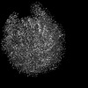
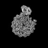
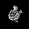
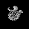
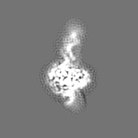
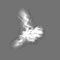
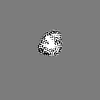
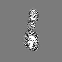
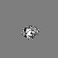
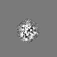
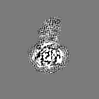
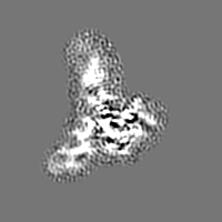
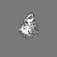
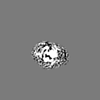
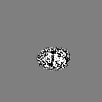
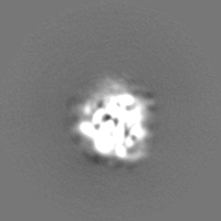
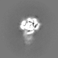
Human OGG1 bound to a 35-bp DNA with an 8-oxoG in the middle
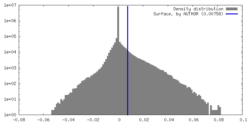
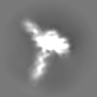
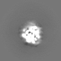
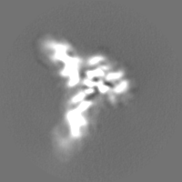
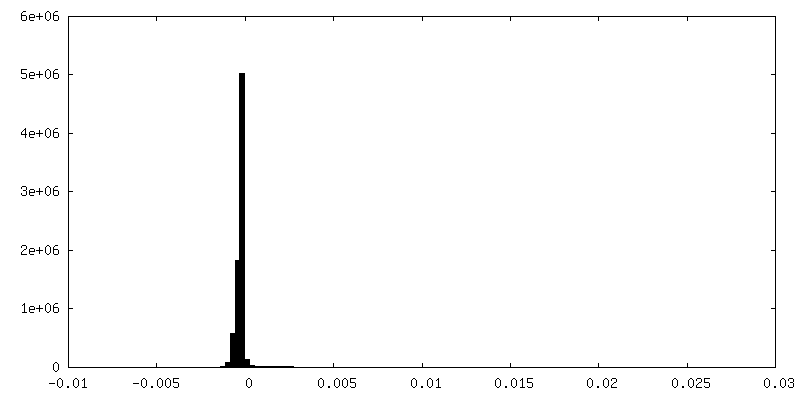
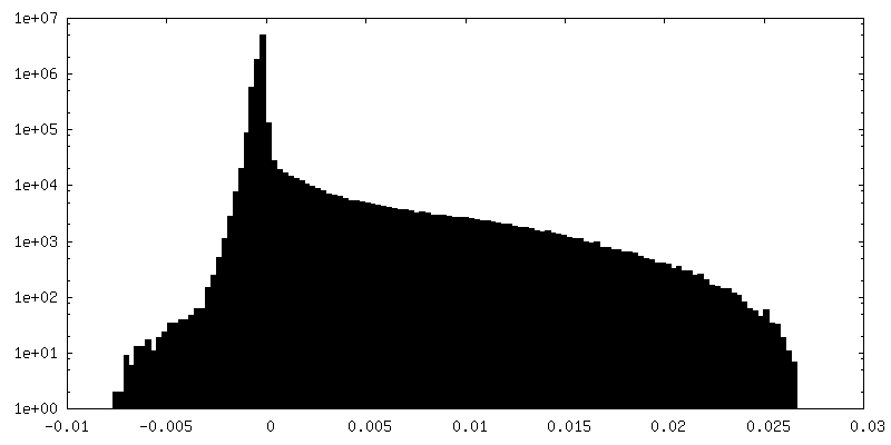
Map data
Human OGG1 binding at 8-oxoG on a 35 bp DNA half map 1
Sample
Complex: Complex of Human OGG1 with a 35 bp DNA duplex containing 8-oxoG
DNA: DNA (35-MER)
DNA: DNA (35-MER)
Protein or peptide: N-glycosylase/DNA lyase
Keywords
Human OGG1 binding at 8-oxoG of a 35 bp DNA duplex / HYDROLASE / LYASE-DNA complex
Function / homology
Function and homology information
Defective OGG1 Substrate Binding / Defective OGG1 Substrate Processing / Defective OGG1 Localization / depurination / negative regulation of double-strand break repair via single-strand annealing / oxidized purine nucleobase lesion DNA N-glycosylase activity / base-excision repair, AP site formation / depyrimidination / 8-oxo-7,8-dihydroguanine DNA N-glycosylase activity / Displacement of DNA glycosylase by APEX1 ...Defective OGG1 Substrate Binding / Defective OGG1 Substrate Processing / Defective OGG1 Localization / depurination / negative regulation of double-strand break repair via single-strand annealing / oxidized purine nucleobase lesion DNA N-glycosylase activity / base-excision repair, AP site formation / depyrimidination / 8-oxo-7,8-dihydroguanine DNA N-glycosylase activity / Displacement of DNA glycosylase by APEX1 / positive regulation of gene expression via chromosomal CpG island demethylation / Hydrolases; Glycosylases; Hydrolysing N-glycosyl compounds / oxidized purine DNA binding / APEX1-Independent Resolution of AP Sites via the Single Nucleotide Replacement Pathway / Recognition and association of DNA glycosylase with site containing an affected purine / Cleavage of the damaged purine / Recognition and association of DNA glycosylase with site containing an affected pyrimidine / Cleavage of the damaged pyrimidine / class I DNA-(apurinic or apyrimidinic site) endonuclease activity / DNA-(apurinic or apyrimidinic site) lyase / nucleotide-excision repair / cellular response to reactive oxygen species / response to radiation / base-excision repair / nuclear matrix / response to oxidative stress / endonuclease activity / microtubule binding / damaged DNA binding / nuclear speck / RNA polymerase II cis-regulatory region sequence-specific DNA binding / mitochondrial matrix / DNA damage response / regulation of DNA-templated transcription / enzyme binding / positive regulation of transcription by RNA polymerase II / protein-containing complex / mitochondrion / DNA binding / nucleoplasm / nucleus / cytosol Similarity search - Function
8-oxoguanine DNA-glycosylase / 8-oxoguanine DNA glycosylase, N-terminal / : / 8-oxoguanine DNA glycosylase, N-terminal domain / HhH-GPD superfamily base excision DNA repair protein / Helix-hairpin-helix, base-excision DNA repair, C-terminal / HhH-GPD domain / endonuclease III / DNA glycosylase Similarity search - Domain/homology
National Institutes of Health/National Cancer Institute (NIH/NCI)
ES011858
United States
National Institutes of Health/National Cancer Institute (NIH/NCI)
GM131754
United States
Citation
Journal: Commun Biol / Year: 2024 Title: Human 8-oxoguanine glycosylase OGG1 binds nucleosome at the dsDNA ends and the super-helical locations. Authors: Qinglong You / Xiang Feng / Yi Cai / Stephen B Baylin / Huilin Li / Abstract: The human glycosylase OGG1 extrudes and excises the oxidized DNA base 8-oxoguanine (8-oxoG) to initiate base excision repair and plays important roles in many pathological conditions such as cancer, ...The human glycosylase OGG1 extrudes and excises the oxidized DNA base 8-oxoguanine (8-oxoG) to initiate base excision repair and plays important roles in many pathological conditions such as cancer, inflammation, and neurodegenerative diseases. Previous structural studies have used a truncated protein and short linear DNA, so it has been unclear how full-length OGG1 operates on longer DNA or on nucleosomes. Here we report cryo-EM structures of human OGG1 bound to a 35-bp long DNA containing an 8-oxoG within an unmethylated Cp-8-oxoG dinucleotide as well as to a nucleosome with an 8-oxoG at super-helical location (SHL)-5. The 8-oxoG in the linear DNA is flipped out by OGG1, consistent with previous crystallographic findings with a 15-bp DNA. OGG1 preferentially binds near dsDNA ends at the nucleosomal entry/exit sites. Such preference may underlie the enzyme's function in DNA double-strand break repair. Unexpectedly, we find that OGG1 bends the nucleosomal entry DNA, flips an undamaged guanine, and binds to internal nucleosomal DNA sites such as SHL-5 and SHL+6. We suggest that the DNA base search mechanism by OGG1 may be chromatin context-dependent and that OGG1 may partner with chromatin remodelers to excise 8-oxoG at the nucleosomal internal sites.
In the structure databanks used in Yorodumi, some data are registered as the other names, "COVID-19 virus" and "2019-nCoV". Here are the details of the virus and the list of structure data.
Jan 31, 2019. EMDB accession codes are about to change! (news from PDBe EMDB page)
EMDB accession codes are about to change! (news from PDBe EMDB page)
The allocation of 4 digits for EMDB accession codes will soon come to an end. Whilst these codes will remain in use, new EMDB accession codes will include an additional digit and will expand incrementally as the available range of codes is exhausted. The current 4-digit format prefixed with “EMD-” (i.e. EMD-XXXX) will advance to a 5-digit format (i.e. EMD-XXXXX), and so on. It is currently estimated that the 4-digit codes will be depleted around Spring 2019, at which point the 5-digit format will come into force.
The EM Navigator/Yorodumi systems omit the EMD- prefix.
Related info.:Q: What is EMD? / ID/Accession-code notation in Yorodumi/EM Navigator
Yorodumi is a browser for structure data from EMDB, PDB, SASBDB, etc.
This page is also the successor to EM Navigator detail page, and also detail information page/front-end page for Omokage search.
The word "yorodu" (or yorozu) is an old Japanese word meaning "ten thousand". "mi" (miru) is to see.
Related info.:EMDB / PDB / SASBDB / Comparison of 3 databanks / Yorodumi Search / Aug 31, 2016. New EM Navigator & Yorodumi / Yorodumi Papers / Jmol/JSmol / Function and homology information / Changes in new EM Navigator and Yorodumi
 Movie
Movie Controller


 Open data
 Basic information
 Map data
 Sample
 Keywords
 Function and homology information
 Homo sapiens (human) / synthetic construct (others)
 Authors
 United States, 2 items
  Citation
 Structure visualization
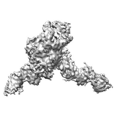
 Downloads & links
 emd_43607.png
 http://ftp.pdbj.org/pub/emdb/structures/EMD-43607


 Z (Sec.)
 Y (Row.)
 X (Col.)


 Sample components
 Processing
 Electron microscopy
 FIELD EMISSION GUN

