Movie
Movie Controller

[English] 日本語
 Yorodumi
- EMDB-4358: Coupling specificity of heterotrimeric Go to the serotonin 5-HT1B... -
+ Open data

- Basic information
| Entry | Database: EMDB / ID: EMD-4358 | |||||||||
|---|---|---|---|---|---|---|---|---|---|---|
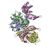
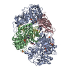
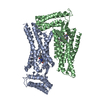
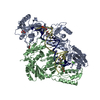
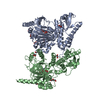
| Title | Coupling specificity of heterotrimeric Go to the serotonin 5-HT1B receptor | |||||||||
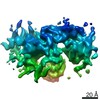
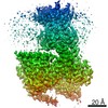
 Map data Map data | Map after RELION "postprocessing" sharpened with a B=-200 and weighted by FSC. | |||||||||
 Sample Sample |
| |||||||||
 Keywords Keywords | G-protein coupled receptor / 5-HT1B / Mini-Go / serotonin / MEMBRANE PROTEIN | |||||||||
| Function / homology |  Function and homology information Function and homology informationnegative regulation of serotonin secretion / serotonergic synapse / Gi/o-coupled serotonin receptor activity / G protein-coupled serotonin receptor complex / regulation of behavior / phospholipase C-activating serotonin receptor signaling pathway / serotonin receptor activity / Serotonin receptors / G protein-coupled serotonin receptor activity / vasoconstriction ...negative regulation of serotonin secretion / serotonergic synapse / Gi/o-coupled serotonin receptor activity / G protein-coupled serotonin receptor complex / regulation of behavior / phospholipase C-activating serotonin receptor signaling pathway / serotonin receptor activity / Serotonin receptors / G protein-coupled serotonin receptor activity / vasoconstriction / neurotransmitter receptor activity / bone remodeling / mu-type opioid receptor binding / : / corticotropin-releasing hormone receptor 1 binding / serotonin binding / G protein-coupled dopamine receptor signaling pathway / cellular response to alkaloid / regulation of heart contraction / parallel fiber to Purkinje cell synapse / G protein-coupled receptor signaling pathway, coupled to cyclic nucleotide second messenger / postsynaptic modulation of chemical synaptic transmission / positive regulation of vascular associated smooth muscle cell proliferation / adenylate cyclase-inhibiting serotonin receptor signaling pathway / G protein-coupled serotonin receptor binding / muscle contraction / locomotory behavior / negative regulation of insulin secretion / cellular response to xenobiotic stimulus / GABA-ergic synapse / G-protein beta/gamma-subunit complex binding / adenylate cyclase-modulating G protein-coupled receptor signaling pathway / adenylate cyclase-inhibiting G protein-coupled receptor signaling pathway / Olfactory Signaling Pathway / Activation of the phototransduction cascade / G protein-coupled acetylcholine receptor signaling pathway / G beta:gamma signalling through PLC beta / Presynaptic function of Kainate receptors / Thromboxane signalling through TP receptor / Activation of G protein gated Potassium channels / Inhibition of voltage gated Ca2+ channels via Gbeta/gamma subunits / G-protein activation / Glucagon signaling in metabolic regulation / Prostacyclin signalling through prostacyclin receptor / G beta:gamma signalling through CDC42 / Synthesis, secretion, and inactivation of Glucagon-like Peptide-1 (GLP-1) / G beta:gamma signalling through BTK / photoreceptor disc membrane / ADP signalling through P2Y purinoceptor 12 / Glucagon-type ligand receptors / Sensory perception of sweet, bitter, and umami (glutamate) taste / Adrenaline,noradrenaline inhibits insulin secretion / Vasopressin regulates renal water homeostasis via Aquaporins / Glucagon-like Peptide-1 (GLP1) regulates insulin secretion / G alpha (z) signalling events / cellular response to catecholamine stimulus / ADP signalling through P2Y purinoceptor 1 / ADORA2B mediated anti-inflammatory cytokines production / G beta:gamma signalling through PI3Kgamma / adenylate cyclase-activating dopamine receptor signaling pathway / Cooperation of PDCL (PhLP1) and TRiC/CCT in G-protein beta folding / GPER1 signaling / cellular response to prostaglandin E stimulus / heterotrimeric G-protein complex / G alpha (12/13) signalling events / Inactivation, recovery and regulation of the phototransduction cascade / G-protein beta-subunit binding / extracellular vesicle / sensory perception of taste / Thrombin signalling through proteinase activated receptors (PARs) / signaling receptor complex adaptor activity / retina development in camera-type eye / cell body / GTPase binding / fibroblast proliferation / G protein activity / presynaptic membrane / Ca2+ pathway / High laminar flow shear stress activates signaling by PIEZO1 and PECAM1:CDH5:KDR in endothelial cells / G alpha (i) signalling events / G alpha (s) signalling events / phospholipase C-activating G protein-coupled receptor signaling pathway / G alpha (q) signalling events / Hydrolases; Acting on acid anhydrides; Acting on GTP to facilitate cellular and subcellular movement / chemical synaptic transmission / Ras protein signal transduction / postsynaptic membrane / Extra-nuclear estrogen signaling / cell population proliferation / G protein-coupled receptor signaling pathway / lysosomal membrane / GTPase activity / synapse / dendrite / GTP binding / protein-containing complex binding / glutamatergic synapse / signal transduction / endoplasmic reticulum / extracellular exosome Similarity search - Function | |||||||||
| Biological species |  Homo sapiens (human) Homo sapiens (human) | |||||||||
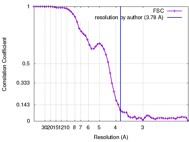
| Method | single particle reconstruction / cryo EM / Resolution: 3.78 Å | |||||||||
 Authors Authors | Garcia-Nafria J / Nehme R / Edwards P / Tate CG | |||||||||
| Funding support |  United Kingdom, 2 items United Kingdom, 2 items
| |||||||||
 Citation Citation | Journal: Nature / Year: 2018 Title: Cryo-EM structure of the serotonin 5-HT receptor coupled to heterotrimeric G. Authors: Javier García-Nafría / Rony Nehmé / Patricia C Edwards / Christopher G Tate / Abstract: G-protein-coupled receptors (GPCRs) form the largest family of receptors encoded by the human genome (around 800 genes). They transduce signals by coupling to a small number of heterotrimeric G ...G-protein-coupled receptors (GPCRs) form the largest family of receptors encoded by the human genome (around 800 genes). They transduce signals by coupling to a small number of heterotrimeric G proteins (16 genes encoding different α-subunits). Each human cell contains several GPCRs and G proteins. The structural determinants of coupling of G to four different GPCRs have been elucidated, but the molecular details of how the other G-protein classes couple to GPCRs are unknown. Here we present the cryo-electron microscopy structure of the serotonin 5-HT receptor (5-HTR) bound to the agonist donitriptan and coupled to an engineered G heterotrimer. In this complex, 5-HTR is in an active state; the intracellular domain of the receptor is in a similar conformation to that observed for the β-adrenoceptor (βAR) or the adenosine A receptor (AR) in complex with G. In contrast to the complexes with G, the gap between the receptor and the Gβ-subunit in the G-5-HTR complex precludes molecular contacts, and the interface between the Gα-subunit of G and the receptor is considerably smaller. These differences are likely to be caused by the differences in the interactions with the C terminus of the G α-subunit. The molecular variations between the interfaces of G and G in complex with GPCRs may contribute substantially to both the specificity of coupling and the kinetics of signalling. | |||||||||
| History |
|
- Structure visualization
| Movie |
Movie viewer |
|---|---|
| Structure viewer | EM map: SurfViewMolmilJmol/JSmol |
| Supplemental images |

- Downloads & links
-EMDB archive
| Map data | emd_4358.map.gz | 5.3 MB | EMDB map data format | |
|---|---|---|---|---|
| Header (meta data) | emd-4358-v30.xmlemd-4358.xml | 19 KB 19 KB | Display Display | EMDB header |
| FSC (resolution estimation) | emd_4358_fsc.xml | 6.3 KB | Display | FSC data file |
| Images | 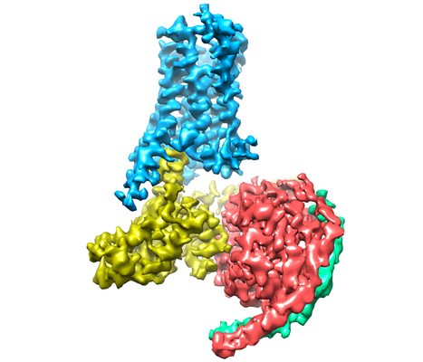 emd_4358.png emd_4358.png | 125.3 KB | ||
| Filedesc metadata | emd-4358.cif.gz | 6.5 KB | ||
| Archive directory |  http://ftp.pdbj.org/pub/emdb/structures/EMD-4358ftp://ftp.pdbj.org/pub/emdb/structures/EMD-4358 http://ftp.pdbj.org/pub/emdb/structures/EMD-4358ftp://ftp.pdbj.org/pub/emdb/structures/EMD-4358 | HTTPS FTP |
-Related structure data
| Related structure data |  6g79MC M: atomic model generated by this map C: citing same article ( |
|---|---|
| Similar structure data | |
| EM raw data | EMPIAR-10308 (Title: Cryo-EM structure of the serotonin 5-HT1B receptor coupled to heterotrimeric Go Data size: 8.0 TB / Data #1: Unaligned movies [micrographs - multiframe] Data #2: Extracted particles [picked particles - multiframe - processed]) |

-Links
| EMDB pages | EMDB (EBI/PDBe) / EMDataResource |
|---|---|
| Related items in Molecule of the Month |


-Map
| File | Download / File: emd_4358.map.gz / Format: CCP4 / Size: 12.9 MB / Type: IMAGE STORED AS FLOATING POINT NUMBER (4 BYTES) | ||||||||||||||||||||||||||||||||||||||||||||||||||||||||||||
|---|---|---|---|---|---|---|---|---|---|---|---|---|---|---|---|---|---|---|---|---|---|---|---|---|---|---|---|---|---|---|---|---|---|---|---|---|---|---|---|---|---|---|---|---|---|---|---|---|---|---|---|---|---|---|---|---|---|---|---|---|---|
| Annotation | Map after RELION "postprocessing" sharpened with a B=-200 and weighted by FSC. | ||||||||||||||||||||||||||||||||||||||||||||||||||||||||||||
| Projections & slices | Image control
Images are generated by Spider. | ||||||||||||||||||||||||||||||||||||||||||||||||||||||||||||
| Voxel size | X=Y=Z: 1.06 Å | ||||||||||||||||||||||||||||||||||||||||||||||||||||||||||||
| Density |
| ||||||||||||||||||||||||||||||||||||||||||||||||||||||||||||
| Symmetry | Space group: 1 | ||||||||||||||||||||||||||||||||||||||||||||||||||||||||||||
| Details | EMDB XML:
CCP4 map header:
| ||||||||||||||||||||||||||||||||||||||||||||||||||||||||||||
 Z (Sec.)
 Y (Row.)
 X (Col.)


-Supplemental data
- Sample components
+Entire : Serotonin 5-HT1B receptor bound to a mini-Go heterotrimer
+Supramolecule #1: Serotonin 5-HT1B receptor bound to a mini-Go heterotrimer
+Supramolecule #2: 5-HT1B receptor
+Supramolecule #3: Beta subunit
+Supramolecule #4: mini-Go
+Supramolecule #5: Gamma subunit
+Macromolecule #1: Guanine nucleotide-binding protein G(I)/G(S)/G(T) subunit beta-1
 Trichoplusia ni (cabbage looper)
+Macromolecule #2: Guanine nucleotide-binding protein G(I)/G(S)/G(O) subunit gamma-2
+Macromolecule #3: Guanine nucleotide-binding protein G(o) subunit alpha

+Macromolecule #4: 5-hydroxytryptamine receptor 1B
+Macromolecule #5: 2-[5-[2-[4-(4-cyanophenyl)piperazin-1-yl]-2-oxidanylidene-ethoxy]...
-Experimental details
-Structure determination
| Method | cryo EM |
|---|---|
 Processing Processing | single particle reconstruction |
| Aggregation state | particle |
-Sample preparation
| Concentration | 2.2 mg/mL | ||||||||||||||||||
|---|---|---|---|---|---|---|---|---|---|---|---|---|---|---|---|---|---|---|---|
| Buffer | pH: 7.5 Component:
| ||||||||||||||||||
| Grid | Model: Quantifoil R1.2/1.3 / Material: GOLD / Mesh: 300 / Pretreatment - Type: GLOW DISCHARGE / Pretreatment - Time: 60 sec. | ||||||||||||||||||
| Vitrification | Cryogen name: ETHANE / Chamber humidity: 100 % / Chamber temperature: 277.15 K / Instrument: FEI VITROBOT MARK IV |
- Electron microscopy
| Microscope | FEI TITAN KRIOS |
|---|---|
| Image recording | Film or detector model: FEI FALCON III (4k x 4k) / Detector mode: COUNTING / Number real images: 5737 / Average exposure time: 60.0 sec. / Average electron dose: 30.0 e/Å2 |
| Electron beam | Acceleration voltage: 300 kV / Electron source:  FIELD EMISSION GUN FIELD EMISSION GUN |
| Electron optics | C2 aperture diameter: 50.0 µm / Illumination mode: FLOOD BEAM / Imaging mode: BRIGHT FIELD / Cs: 2.7 mm |
| Sample stage | Specimen holder model: FEI TITAN KRIOS AUTOGRID HOLDER / Cooling holder cryogen: NITROGEN |
| Experimental equipment |  Model: Titan Krios / Image courtesy: FEI Company |