 Movie
Movie Controller

[English] 日本語
 Yorodumi
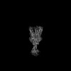
- EMDB-43560: Cryo-EM structure of a type II ZorAB complex from Sulfuricurvum k... -
+ Open data

- Basic information
| Entry | 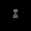 | |||||||||
|---|---|---|---|---|---|---|---|---|---|---|
| Title | Cryo-EM structure of a type II ZorAB complex from Sulfuricurvum kujiense | |||||||||
 Map data Map data | sharpened map used for model refinements | |||||||||
 Sample Sample |
| |||||||||
 Keywords Keywords | phage defense / zorya / phage / membrane protein complex / MEMBRANE PROTEIN | |||||||||
| Function / homology | Motility protein B-like, N-terminal domain / Membrane MotB of proton-channel complex MotA/MotB / : / OmpA-like domain superfamily / membrane / MotA/TolQ/ExbB proton channel domain-containing protein / Motility protein B-like N-terminal domain-containing protein Function and homology information Function and homology information | |||||||||
| Biological species |  Sulfuricurvum kujiense DSM 16994 (bacteria) Sulfuricurvum kujiense DSM 16994 (bacteria) | |||||||||
| Method | single particle reconstruction / cryo EM / Resolution: 2.8 Å | |||||||||
 Authors Authors | Deme JC / Lea SM | |||||||||
| Funding support |  United States, 1 items United States, 1 items
| |||||||||
 Citation Citation | Journal: Nat Commun / Year: 2025 Title: Modularity of Zorya defense systems during phage inhibition. Authors: Giuseppina Mariano / Justin C Deme / Jennifer J Readshaw / Matthew J Grobbelaar / Mackenzie Keenan / Yasmin El-Masri / Lindsay Bamford / Suraj Songra / Tim R Blower / Tracy Palmer / Susan M Lea /  Abstract: Bacteria have evolved an extraordinary diversity of defense systems against bacteriophage (phage) predation. However, the molecular mechanisms underlying these anti-phage systems often remain elusive. ...Bacteria have evolved an extraordinary diversity of defense systems against bacteriophage (phage) predation. However, the molecular mechanisms underlying these anti-phage systems often remain elusive. Here, we provide mechanistic and structural insights into Zorya phage defense systems. Using cryo-EM structural analyses, we show that the Zorya type I and II core components, ZorA and ZorB, assemble in a 5:2 complex that is similar to inner-membrane ion-driven, rotary motors that power flagellar rotation, type 9 secretion, gliding and the Ton nutrient uptake systems. The ZorAB complex has an elongated cytoplasmic tail assembled by bundling the C-termini of the five ZorA subunits. Mutagenesis demonstrates that peptidoglycan binding by the periplasmic domains of ZorB, the structured cytoplasmic tail of ZorA, and ion flow through the motor is important for function in both type I and II systems. Furthermore, we identify ZorE as the effector module of the Zorya II system, possessing nickase activity. Our work reveals the molecular basis of the activity of Zorya systems and highlights the ZorE nickase as crucial for population-wide immunity in the type II system. | |||||||||
| History |
|
- Structure visualization
| Supplemental images |
|---|

- Downloads & links
-EMDB archive
| Map data | emd_43560.map.gz | 398.5 MB | EMDB map data format | |
|---|---|---|---|---|
| Header (meta data) | emd-43560-v30.xmlemd-43560.xml | 20.2 KB 20.2 KB | Display Display | EMDB header |
| FSC (resolution estimation) | emd_43560_fsc.xml | 15.9 KB | Display | FSC data file |
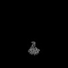
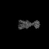
| Images |  emd_43560.png emd_43560.png | 88.4 KB | ||
| Masks | emd_43560_msk_1.map | 421.9 MB | Mask map | |
| Filedesc metadata | emd-43560.cif.gz | 6.4 KB | ||
| Others | emd_43560_additional_1.map.gzemd_43560_half_map_1.map.gzemd_43560_half_map_2.map.gz | 210.7 MB 391 MB 391 MB | ||
| Archive directory |  http://ftp.pdbj.org/pub/emdb/structures/EMD-43560ftp://ftp.pdbj.org/pub/emdb/structures/EMD-43560 http://ftp.pdbj.org/pub/emdb/structures/EMD-43560ftp://ftp.pdbj.org/pub/emdb/structures/EMD-43560 | HTTPS FTP |
-Related structure data
| Related structure data | 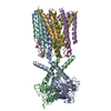 8vviMC 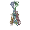 8vvnC M: atomic model generated by this map C: citing same article ( |
|---|---|
| Similar structure data |
-Links
| EMDB pages | EMDB (EBI/PDBe) / EMDataResource |
|---|
-Map
| File | Download / File: emd_43560.map.gz / Format: CCP4 / Size: 421.9 MB / Type: IMAGE STORED AS FLOATING POINT NUMBER (4 BYTES) | ||||||||||||||||||||||||||||||||||||
|---|---|---|---|---|---|---|---|---|---|---|---|---|---|---|---|---|---|---|---|---|---|---|---|---|---|---|---|---|---|---|---|---|---|---|---|---|---|
| Annotation | sharpened map used for model refinements | ||||||||||||||||||||||||||||||||||||
| Projections & slices | Image control
Images are generated by Spider. | ||||||||||||||||||||||||||||||||||||
| Voxel size | X=Y=Z: 0.723 Å | ||||||||||||||||||||||||||||||||||||
| Density |
| ||||||||||||||||||||||||||||||||||||
| Symmetry | Space group: 1 | ||||||||||||||||||||||||||||||||||||
| Details | EMDB XML:
|
 Z (Sec.)
 Y (Row.)
 X (Col.)


-Supplemental data
-Mask #1
| File | emd_43560_msk_1.map | ||||||||||||
|---|---|---|---|---|---|---|---|---|---|---|---|---|---|
| Projections & Slices |
| ||||||||||||
| Density Histograms |


-Additional map: unsharpened map
| File | emd_43560_additional_1.map | ||||||||||||
|---|---|---|---|---|---|---|---|---|---|---|---|---|---|
| Annotation | unsharpened map | ||||||||||||
| Projections & Slices |
| ||||||||||||
| Density Histograms |


-Half map: half map 1
| File | emd_43560_half_map_1.map | ||||||||||||
|---|---|---|---|---|---|---|---|---|---|---|---|---|---|
| Annotation | half map 1 | ||||||||||||
| Projections & Slices |
| ||||||||||||
| Density Histograms |


-Half map: half map 2
| File | emd_43560_half_map_2.map | ||||||||||||
|---|---|---|---|---|---|---|---|---|---|---|---|---|---|
| Annotation | half map 2 | ||||||||||||
| Projections & Slices |
| ||||||||||||
| Density Histograms |


- Sample components
-Entire : ZorAB complex
| Entire | Name: ZorAB complex |
|---|---|
| Components |
|
-Supramolecule #1: ZorAB complex
| Supramolecule | Name: ZorAB complex / type: complex / ID: 1 / Parent: 0 / Macromolecule list: all |
|---|---|
| Source (natural) | Organism: Sulfuricurvum kujiense DSM 16994 (bacteria) |
-Macromolecule #1: MotA/TolQ/ExbB proton channel domain-containing protein
| Macromolecule | Name: MotA/TolQ/ExbB proton channel domain-containing protein type: protein_or_peptide / ID: 1 / Number of copies: 5 / Enantiomer: LEVO |
|---|---|
| Source (natural) | Organism: Sulfuricurvum kujiense DSM 16994 (bacteria) |
| Molecular weight | Theoretical: 42.445297 KDa |
| Recombinant expression | Organism: |
| Sequence | String: MIHNMAYFGV GLITLMFLIF VMNRRNKSIQ ELAPGILITT GIFFTFVGIA IGLVHFNADN VDDSLPTLLN GIKTAFWASA TGVFFALII KILDIFDLTR TNESSAVEGM SIDDIVTYQA KQTDVLVEIL RSIKNMHSSI AAQDDSSLVS QIALLRDDSN K KSDALRQE ...String: MIHNMAYFGV GLITLMFLIF VMNRRNKSIQ ELAPGILITT GIFFTFVGIA IGLVHFNADN VDDSLPTLLN GIKTAFWASA TGVFFALII KILDIFDLTR TNESSAVEGM SIDDIVTYQA KQTDVLVEIL RSIKNMHSSI AAQDDSSLVS QIALLRDDSN K KSDALRQE FRDFAATMAE NNSKIFIEAL KDVIKNFNDK ISEQFGDNFK QLNQAVEKTV IWQENYRNQM AQSIETMTLI AS MLESQAH DYSIIVSNSA EFESHVSAMG RSLEEITFQR EQLQSMIHSL VNFLESASDS LPLIGQKVDD MTERLVKGMN EAT EEVQKQ VTILDHELEI ALKRSLEGLG QQLASLSNKF VQDYTPLTEK LREVVALAAK QR UniProtKB: MotA/TolQ/ExbB proton channel domain-containing protein |
-Macromolecule #2: Motility protein B-like N-terminal domain-containing protein
| Macromolecule | Name: Motility protein B-like N-terminal domain-containing protein type: protein_or_peptide / ID: 2 / Number of copies: 2 / Enantiomer: LEVO |
|---|---|
| Source (natural) | Organism: Sulfuricurvum kujiense DSM 16994 (bacteria) |
| Molecular weight | Theoretical: 31.944371 KDa |
| Recombinant expression | Organism: |
| Sequence | String: MSSLPQKKHH HKEDYWISLS DMMTSLMMLF LLISVIYMIK VQDSVKVPQI YKETTQGLNH ALKKEFDKDL MKWGAVIDKD LTVRFQQPD ILFATGSSAL TPRFKEILDD FFIRYLKIMM SKPFINNIEE IRIEGHTSSM WEGESDRGKA YFKNMTLSQE R TRATLEYI ...String: MSSLPQKKHH HKEDYWISLS DMMTSLMMLF LLISVIYMIK VQDSVKVPQI YKETTQGLNH ALKKEFDKDL MKWGAVIDKD LTVRFQQPD ILFATGSSAL TPRFKEILDD FFIRYLKIMM SKPFINNIEE IRIEGHTSSM WEGESDRGKA YFKNMTLSQE R TRATLEYI MTSDKINLTG EQKEWLMRHF SAIGFSSGHP LTNKGTYLVD GESEDSQLSQ RVEFRVRTNI ERKVADIVEK EN LYFQGQF GSWSHPQFEK GGGSGGGSGG GSWSHPQFEK UniProtKB: Motility protein B-like N-terminal domain-containing protein |
-Experimental details
-Structure determination
| Method | cryo EM |
|---|---|
 Processing Processing | single particle reconstruction |
| Aggregation state | particle |
-Sample preparation
| Buffer | pH: 8 |
|---|---|
| Vitrification | Cryogen name: ETHANE |
- Electron microscopy
| Microscope | TFS KRIOS |
|---|---|
| Image recording | Film or detector model: FEI FALCON IV (4k x 4k) / Average electron dose: 57.0 e/Å2 |
| Electron beam | Acceleration voltage: 300 kV / Electron source:  FIELD EMISSION GUN FIELD EMISSION GUN |
| Electron optics | Illumination mode: FLOOD BEAM / Imaging mode: BRIGHT FIELD / Nominal defocus max: 2.0 µm / Nominal defocus min: 0.5 µm |
| Experimental equipment |  Model: Titan Krios / Image courtesy: FEI Company |

