 ムービー
ムービー コントローラー


+ データを開く

- 基本情報
基本情報
| 登録情報 |  | |||||||||
|---|---|---|---|---|---|---|---|---|---|---|
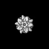
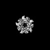
| タイトル | NPM2-H1.8 isolated from Xenopus egg extract | |||||||||
 マップデータ マップデータ | ||||||||||
 試料 試料 |
| |||||||||
 キーワード キーワード | H1 / Xenopus egg extract / MagIC-cryo-EM / Histone chaperone / CHAPERONE | |||||||||
| 機能・相同性 |  機能・相同性情報 機能・相同性情報single fertilization / positive regulation of DNA replication / histone binding / chromatin remodeling / chromatin binding / nucleolus / RNA binding / nucleoplasm / cytoplasm 類似検索 - 分子機能 | |||||||||
| 生物種 | ||||||||||
| 手法 | 単粒子再構成法 / クライオ電子顕微鏡法 / 解像度: 5.0 Å | |||||||||
 データ登録者 データ登録者 | Arimura Y / Funabiki H | |||||||||
| 資金援助 |  米国, 1件 米国, 1件
| |||||||||
 引用 引用 | ジャーナル: Elife / 年: 2025 タイトル: MagIC-Cryo-EM, structural determination on magnetic beads for scarce macromolecules in heterogeneous samples. 著者: Yasuhiro Arimura / Hide A Konishi / Hironori Funabiki / 要旨: Cryo-EM single-particle analyses typically require target macromolecule concentration at 0.05~5.0 mg/ml, which is often difficult to achieve. Here, we devise netic solation and oncentration (MagIC)- ...Cryo-EM single-particle analyses typically require target macromolecule concentration at 0.05~5.0 mg/ml, which is often difficult to achieve. Here, we devise netic solation and oncentration (MagIC)-cryo-EM, a technique enabling direct structural analysis of targets captured on magnetic beads, thereby reducing the targets' concentration requirement to <0.0005 mg/mL. Adapting MagIC-cryo-EM to a Chromatin Immunoprecipitation protocol, we characterized structural variations of the linker histone H1.8-associated nucleosomes that were isolated from interphase and metaphase chromosomes in egg extract. Combining plicated election o xclude ubbish particles (DuSTER), a particle curation method that excludes low signal-to-noise ratio particles, we also resolved the 3D cryo-EM structures of nucleoplasmin NPM2 co-isolated with the linker histone H1.8 and revealed distinct open and closed structural variants. Our study demonstrates the utility of MagIC-cryo-EM for structural analysis of scarce macromolecules in heterogeneous samples and provides structural insights into the cell cycle-regulation of H1.8 association to nucleosomes. #1: ジャーナル: Elife / 年: 2024タイトル: MagIC-Cryo-EM: Structural determination on magnetic beads for scarce macromolecules in heterogeneous samples 著者: Arimura Y / Konishi HA / Funabiki H | |||||||||
| 履歴 |
|
- 構造の表示
構造の表示
| 添付画像 |
|---|

- ダウンロードとリンク
-EMDBアーカイブ
| マップデータ | emd_43238.map.gz | 9.4 MB | EMDBマップデータ形式 | |
|---|---|---|---|---|
| ヘッダ (付随情報) | emd-43238-v30.xmlemd-43238.xml | 19.9 KB 19.9 KB | 表示 表示 | EMDBヘッダ |
| FSC (解像度算出) | emd_43238_fsc.xml | 5.7 KB | 表示 | FSCデータファイル |
| 画像 |  emd_43238.png emd_43238.png | 64.8 KB | ||
| Filedesc metadata | emd-43238.cif.gz | 5.7 KB | ||
| その他 | emd_43238_additional_1.map.gzemd_43238_half_map_1.map.gzemd_43238_half_map_2.map.gz | 18.2 MB 18 MB 18 MB | ||
| アーカイブディレクトリ |  http://ftp.pdbj.org/pub/emdb/structures/EMD-43238ftp://ftp.pdbj.org/pub/emdb/structures/EMD-43238 http://ftp.pdbj.org/pub/emdb/structures/EMD-43238ftp://ftp.pdbj.org/pub/emdb/structures/EMD-43238 | HTTPS FTP |
-関連構造データ


-リンク
| EMDBのページ | EMDB (EBI/PDBe) / EMDataResource |
|---|---|
| 「今月の分子」の関連する項目 |

-マップ
| ファイル | ダウンロード / ファイル: emd_43238.map.gz / 形式: CCP4 / 大きさ: 19.4 MB / タイプ: IMAGE STORED AS FLOATING POINT NUMBER (4 BYTES) | ||||||||||||||||||||||||||||||||||||
|---|---|---|---|---|---|---|---|---|---|---|---|---|---|---|---|---|---|---|---|---|---|---|---|---|---|---|---|---|---|---|---|---|---|---|---|---|---|
| 投影像・断面図 | 画像のコントロール
画像は Spider により作成 | ||||||||||||||||||||||||||||||||||||
| ボクセルのサイズ | X=Y=Z: 1.08 Å | ||||||||||||||||||||||||||||||||||||
| 密度 |
| ||||||||||||||||||||||||||||||||||||
| 対称性 | 空間群: 1 | ||||||||||||||||||||||||||||||||||||
| 詳細 | EMDB XML:
|
 Z (Sec.)
 Y (Row.)
 X (Col.)


-添付データ
-追加マップ: #1
| ファイル | emd_43238_additional_1.map | ||||||||||||
|---|---|---|---|---|---|---|---|---|---|---|---|---|---|
| 投影像・断面図 |
| ||||||||||||
| 密度ヒストグラム |


-ハーフマップ: #1
| ファイル | emd_43238_half_map_1.map | ||||||||||||
|---|---|---|---|---|---|---|---|---|---|---|---|---|---|
| 投影像・断面図 |
| ||||||||||||
| 密度ヒストグラム |


-ハーフマップ: #2
| ファイル | emd_43238_half_map_2.map | ||||||||||||
|---|---|---|---|---|---|---|---|---|---|---|---|---|---|
| 投影像・断面図 |
| ||||||||||||
| 密度ヒストグラム |


- 試料の構成要素
-全体 : NPM2 complexed with H1.8
| 全体 | 名称: NPM2 complexed with H1.8 |
|---|---|
| 要素 |
|
-超分子 #1: NPM2 complexed with H1.8
| 超分子 | 名称: NPM2 complexed with H1.8 / タイプ: cell / ID: 1 / 親要素: 0 / 含まれる分子: all |
|---|---|
| 由来(天然) | 生物種: |
-分子 #1: Nucleoplasmin isoform X1
| 分子 | 名称: Nucleoplasmin isoform X1 / タイプ: protein_or_peptide / ID: 1 / コピー数: 5 / 光学異性体: LEVO |
|---|---|
| 由来(天然) | 生物種: |
| 分子量 | 理論値: 21.949467 KDa |
| 配列 | 文字列: MASTVSNTSK LEKPVSLIWG CELNEQNKTF EFKVEDDEEK CEHQLALRTV CLGDKAKDEF HIVEIVTQEE GAEKSVPIAT LKPSILPMA TMVGIELTPP VTFRLKAGSG PLYISGQHVA MEEDYSWAEE EDEGEAEGEE EEEEEEDQES PPKAVKRPAA T KKAGQAKK ...文字列: MASTVSNTSK LEKPVSLIWG CELNEQNKTF EFKVEDDEEK CEHQLALRTV CLGDKAKDEF HIVEIVTQEE GAEKSVPIAT LKPSILPMA TMVGIELTPP VTFRLKAGSG PLYISGQHVA MEEDYSWAEE EDEGEAEGEE EEEEEEDQES PPKAVKRPAA T KKAGQAKK KLDKEDESSE EDSPTKKGKG AGRGRKPAAK K UniProtKB: Nucleoplasmin |
-実験情報
-構造解析
| 手法 | クライオ電子顕微鏡法 |
|---|---|
 解析 解析 | 単粒子再構成法 |
| 試料の集合状態 | cell |
-試料調製
| 緩衝液 | pH: 7.4 詳細: 10mM HEPES-KOH (pH 7.4), 30 mM KCl, 1 mM EGTA, 10 ng/microL leupeptin, 10 ng/microL pepstatin, 10 ng/microL chymostatin, 1 mM sodium butyrate, 1 mM beta-glycerophosphate, trehalose, and 1,6-hexanediol |
|---|---|
| グリッド | モデル: Quantifoil R1.2/1.3 / 材質: GOLD / メッシュ: 300 / 支持フィルム - 材質: GRAPHENE 詳細: The grid was incubated on the 40 x 20 mm N52 neodymium disc magnets for 5 minutes and vitrified using the Vitrobot Mark IV (FEI) with a 2-second blotting time at room temperature under 100% humidity conditions. |
| 凍結 | 凍結剤: ETHANE |
- 電子顕微鏡法
| 顕微鏡 | FEI TITAN KRIOS |
|---|---|
| 撮影 | #0 - Image recording ID: 1 #0 - フィルム・検出器のモデル: GATAN K3 BIOQUANTUM (6k x 4k) #0 - 平均電子線量: 45.3 e/Å2 / #1 - Image recording ID: 2 #1 - フィルム・検出器のモデル: GATAN K3 BIOQUANTUM (6k x 4k) #1 - 平均電子線量: 51.92 e/Å2 |
| 電子線 | 加速電圧: 300 kV / 電子線源:  FIELD EMISSION GUN FIELD EMISSION GUN |
| 電子光学系 | 照射モード: FLOOD BEAM / 撮影モード: BRIGHT FIELD / 最大 デフォーカス(公称値): 3.5 µm / 最小 デフォーカス(公称値): 1.5 µm |
| 実験機器 |  モデル: Titan Krios / 画像提供: FEI Company |

