Movie
Movie Controller

[English] 日本語
 Yorodumi
- EMDB-41936: CryoEM map of horse spleen apoferritin determined as a reference ... -
+ Open data

- Basic information
| Entry |  | |||||||||
|---|---|---|---|---|---|---|---|---|---|---|
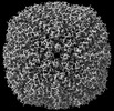
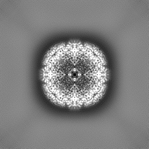
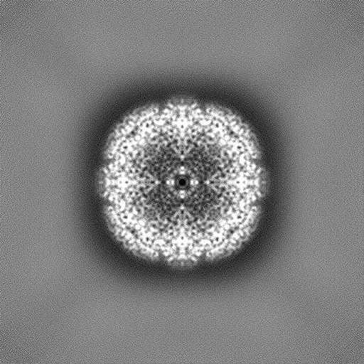
| Title | CryoEM map of horse spleen apoferritin determined as a reference for benchmarking square and rectangular apertures for cryo-EM (Falcon IV round beam) | |||||||||
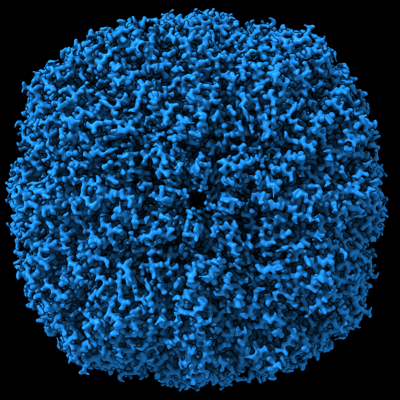
 Map data Map data | Apo-ferritin map reconstructed from dataset recorded using Falcon IV camera and round beam for comparison with square beam | |||||||||
 Sample Sample |
| |||||||||
 Keywords Keywords | Apo-ferritin / METAL BINDING PROTEIN | |||||||||
| Biological species |  | |||||||||
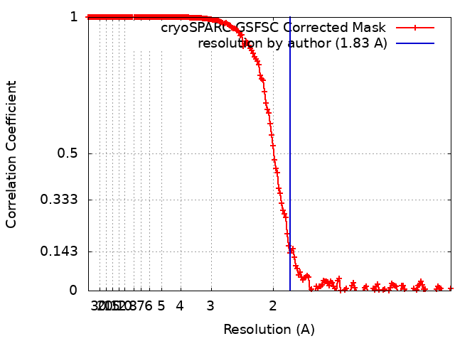
| Method | single particle reconstruction / cryo EM / Resolution: 1.83 Å | |||||||||
 Authors Authors | Brown HG / Hanssen E | |||||||||
| Funding support |  Australia, 1 items Australia, 1 items
| |||||||||
 Citation Citation | Journal: Nat Methods / Year: 2024 Title: Square condenser apertures for square cameras in low-dose transmission electron microscopy. Authors: Hamish G Brown / Dan Smith / Benjamin C Wardle / Eric Hanssen / Abstract: In transmission electron microscopy (TEM), cameras are square or rectangular but beams are round so the circular lobes irradiate adjacent areas, precluding further neighboring acquisition for beam- ...In transmission electron microscopy (TEM), cameras are square or rectangular but beams are round so the circular lobes irradiate adjacent areas, precluding further neighboring acquisition for beam-sensitive samples. We present condenser aperture plates with square and rectangular shapes that improve the efficiency of area usage by 70% and enhance montage imaging for beam-sensitive specimens. We demonstrate the compatibility of these condenser aperture plates with high-resolution cryogenic TEM by reconstructing a 1.8-Å map of equine apo-ferritin. | |||||||||
| History |
|
- Structure visualization
| Supplemental images |
|---|
- Downloads & links
-EMDB archive
| Map data | emd_41936.map.gz | 483.5 MB |  EMDB map data format EMDB map data format | |
|---|---|---|---|---|
| Header (meta data) | emd-41936-v30.xmlemd-41936.xml | 16.6 KB 16.6 KB | Display Display | EMDB header |
| FSC (resolution estimation) | emd_41936_fsc.xml | 16.9 KB | Display | FSC data file |
| Images |  emd_41936.png emd_41936.png | 265.7 KB | ||
| Masks | emd_41936_msk_1.map | 512 MB | Mask map | |
| Filedesc metadata | emd-41936.cif.gz | 4.7 KB | ||
| Others | emd_41936_half_map_1.map.gzemd_41936_half_map_2.map.gz | 474.5 MB 474.5 MB | ||
| Archive directory |  http://ftp.pdbj.org/pub/emdb/structures/EMD-41936ftp://ftp.pdbj.org/pub/emdb/structures/EMD-41936 http://ftp.pdbj.org/pub/emdb/structures/EMD-41936ftp://ftp.pdbj.org/pub/emdb/structures/EMD-41936 | HTTPS FTP |
-Validation report
| Summary document | emd_41936_validation.pdf.gz | 1.1 MB | Display | EMDB validaton report |
|---|---|---|---|---|
| Full document | emd_41936_full_validation.pdf.gz | 1.1 MB | Display | |
| Data in XML | emd_41936_validation.xml.gz | 26.8 KB | Display | |
| Data in CIF | emd_41936_validation.cif.gz | 34.8 KB | Display | |
| Arichive directory | https://ftp.pdbj.org/pub/emdb/validation_reports/EMD-41936ftp://ftp.pdbj.org/pub/emdb/validation_reports/EMD-41936 | HTTPS FTP |
-Related structure data
-Links
| EMDB pages | EMDB (EBI/PDBe) / EMDataResource |
|---|
-Map
| File | Download / File: emd_41936.map.gz / Format: CCP4 / Size: 512 MB / Type: IMAGE STORED AS FLOATING POINT NUMBER (4 BYTES) | ||||||||||||||||||||
|---|---|---|---|---|---|---|---|---|---|---|---|---|---|---|---|---|---|---|---|---|---|
| Annotation | Apo-ferritin map reconstructed from dataset recorded using Falcon IV camera and round beam for comparison with square beam | ||||||||||||||||||||
| Voxel size | X=Y=Z: 0.506 Å | ||||||||||||||||||||
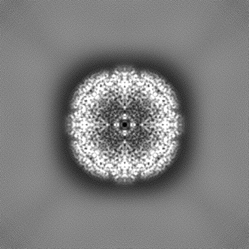
| Density |
| ||||||||||||||||||||
| Symmetry | Space group: 1 | ||||||||||||||||||||
| Details | EMDB XML:
|
-Supplemental data
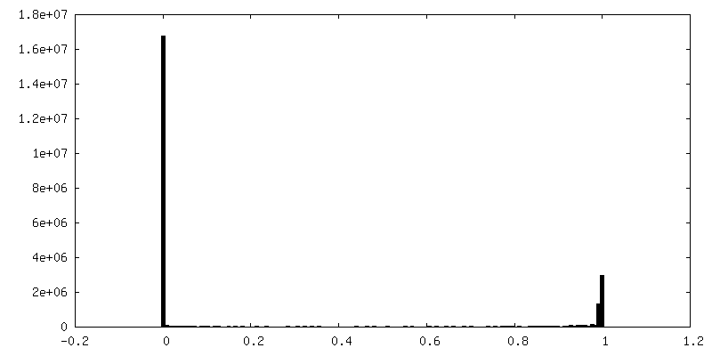
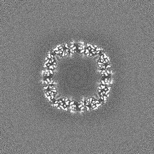
-Mask #1
| File | emd_41936_msk_1.map | ||||||||||||
|---|---|---|---|---|---|---|---|---|---|---|---|---|---|
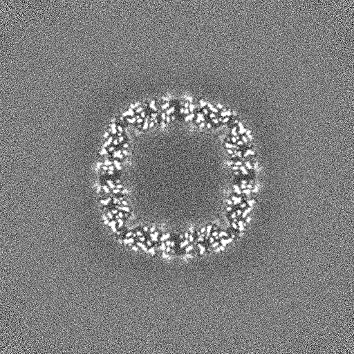
| Projections & Slices |
| ||||||||||||
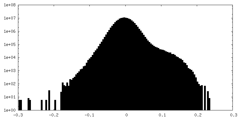
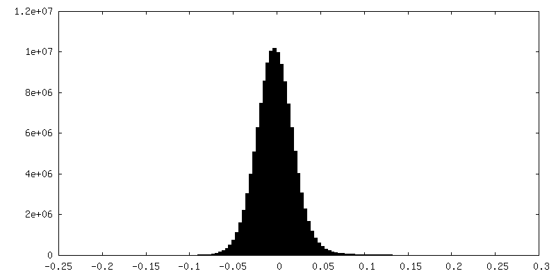
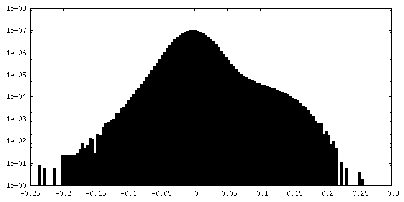
| Density Histograms |
 Z
Z Y
Y X
X


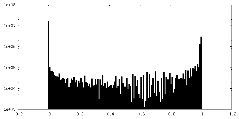
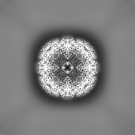
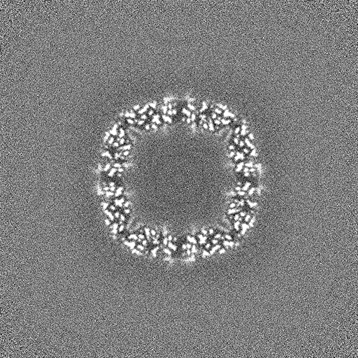
-Half map: Half Map B
| File | emd_41936_half_map_1.map | ||||||||||||
|---|---|---|---|---|---|---|---|---|---|---|---|---|---|
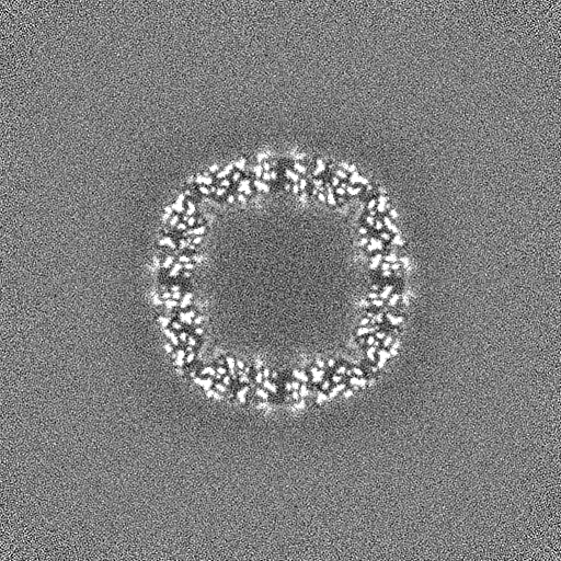
| Annotation | Half Map B | ||||||||||||
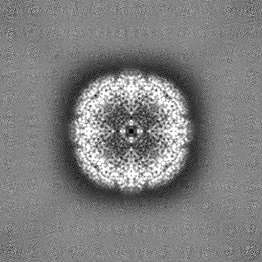
| Projections & Slices |
| ||||||||||||
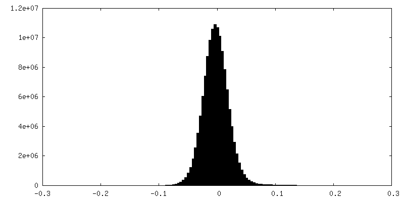
| Density Histograms |
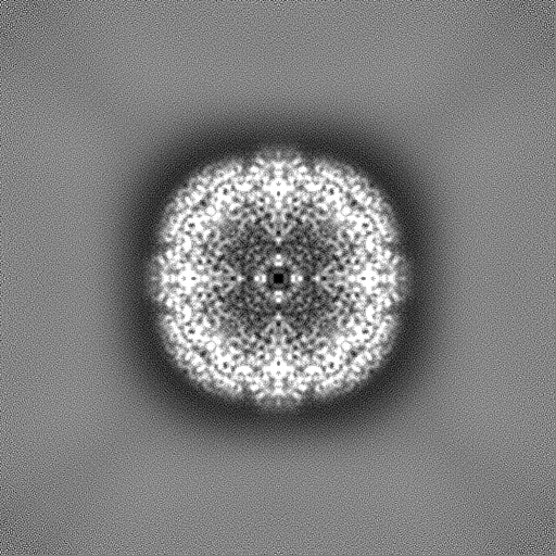
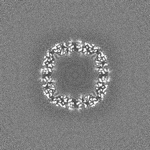
-Half map: Half Map A
| File | emd_41936_half_map_2.map | ||||||||||||
|---|---|---|---|---|---|---|---|---|---|---|---|---|---|
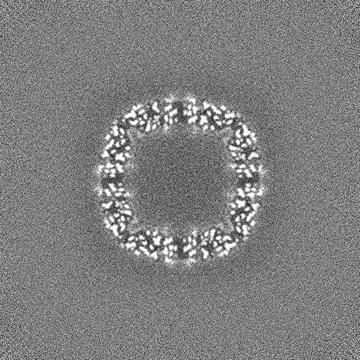
| Annotation | Half Map A | ||||||||||||
| Projections & Slices |
| ||||||||||||
| Density Histograms |
- Sample components
-Entire : Apoferritin from equine spleen
| Entire | Name: Apoferritin from equine spleen |
|---|---|
| Components |
|
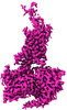
-Supramolecule #1: Apoferritin from equine spleen
| Supramolecule | Name: Apoferritin from equine spleen / type: complex / ID: 1 / Parent: 0 / Macromolecule list: #1 / Details: Purchased from Sigma Aldrich product number A3641 |
|---|---|
| Source (natural) | Organism: |
| Molecular weight | Theoretical: 481.2 KDa |
-Experimental details
-Structure determination
| Method | cryo EM |
|---|---|
 Processing Processing | single particle reconstruction |
| Aggregation state | particle |
-Sample preparation
| Concentration | 35 mg/mL |
|---|---|
| Buffer | pH: 7.4 / Component - Formula: PBS |
| Grid | Model: Quantifoil R1.2/1.3 / Material: GOLD / Mesh: 400 / Support film - Material: GOLD / Support film - topology: HOLEY ARRAY / Pretreatment - Type: GLOW DISCHARGE / Pretreatment - Time: 180 sec. / Pretreatment - Atmosphere: AIR / Details: Quorum Glocube |
| Vitrification | Cryogen name: ETHANE / Chamber humidity: 95 % / Chamber temperature: 295 K / Instrument: FEI VITROBOT MARK IV |
| Details | Solution purchased from Sigma Aldrich and no further purification |
- Electron microscopy
| Microscope | FEI TITAN KRIOS |
|---|---|
| Specialist optics | Energy filter - Name: GIF Bioquantum / Energy filter - Slit width: 10 eV |
| Image recording | Film or detector model: GATAN K3 BIOQUANTUM (6k x 4k) / Digitization - Dimensions - Width: 5760 pixel / Digitization - Dimensions - Height: 4092 pixel / Number real images: 3111 / Average electron dose: 50.0 e/Å2 |
| Electron beam | Acceleration voltage: 300 kV / Electron source:  FIELD EMISSION GUN FIELD EMISSION GUN |
| Electron optics | C2 aperture diameter: 50.0 µm / Illumination mode: FLOOD BEAM / Imaging mode: BRIGHT FIELD / Cs: 2.7 mm / Nominal defocus max: 1.4000000000000001 µm / Nominal defocus min: 0.6 µm |
| Sample stage | Specimen holder model: FEI TITAN KRIOS AUTOGRID HOLDER / Cooling holder cryogen: NITROGEN |
| Experimental equipment |  Model: Titan Krios / Image courtesy: FEI Company |