 Movie
Movie Controller


+ Open data

- Basic information
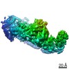
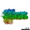
| Entry | Database: EMDB / ID: EMD-4188 | |||||||||
|---|---|---|---|---|---|---|---|---|---|---|
| Title | Cryo-EM structure of microtubule co-polymerized with SSNA1. | |||||||||
 Map data Map data | ||||||||||
 Sample Sample |
| |||||||||
| Biological species | Chlamydo | |||||||||
| Method | helical reconstruction / cryo EM / Resolution: 6.1 Å | |||||||||
 Authors Authors | Basnet N / Mizuno N | |||||||||
 Citation Citation | Journal: Nat Cell Biol / Year: 2018 Title: Direct induction of microtubule branching by microtubule nucleation factor SSNA1. Authors: Nirakar Basnet / Hana Nedozralova / Alvaro H Crevenna / Satish Bodakuntla / Thomas Schlichthaerle / Michael Taschner / Giovanni Cardone / Carsten Janke / Ralf Jungmann / Maria M Magiera / ...Authors: Nirakar Basnet / Hana Nedozralova / Alvaro H Crevenna / Satish Bodakuntla / Thomas Schlichthaerle / Michael Taschner / Giovanni Cardone / Carsten Janke / Ralf Jungmann / Maria M Magiera / Christian Biertümpfel / Naoko Mizuno /     Abstract: Microtubules are central elements of the eukaryotic cytoskeleton that often function as part of branched networks. Current models for branching include nucleation of new microtubules from severed ...Microtubules are central elements of the eukaryotic cytoskeleton that often function as part of branched networks. Current models for branching include nucleation of new microtubules from severed microtubule seeds or from γ-tubulin recruited to the side of a pre-existing microtubule. Here, we found that microtubules can be directly remodelled into branched structures by the microtubule-remodelling factor SSNA1 (also known as NA14 or DIP13). The branching activity of SSNA1 relies on its ability to self-assemble into fibrils in a head-to-tail fashion. SSNA1 fibrils guide protofilaments of a microtubule to split apart to form daughter microtubules. We further found that SSNA1 localizes at axon branching sites and has a key role in neuronal development. SSNA1 mutants that abolish microtubule branching in vitro also fail to promote axon development and branching when overexpressed in neurons. We have, therefore, discovered a mechanism for microtubule branching and implicated its role in neuronal development. | |||||||||
| History |
|
- Structure visualization
| Movie |
 Movie viewer Movie viewer |
|---|---|
| Structure viewer | EM map: SurfViewMolmilJmol/JSmol |
| Supplemental images |

- Downloads & links
-EMDB archive
| Map data | emd_4188.map.gz | 3.4 MB | EMDB map data format | |
|---|---|---|---|---|
| Header (meta data) | emd-4188-v30.xmlemd-4188.xml | 6.6 KB 6.6 KB | Display Display | EMDB header |
| Images | 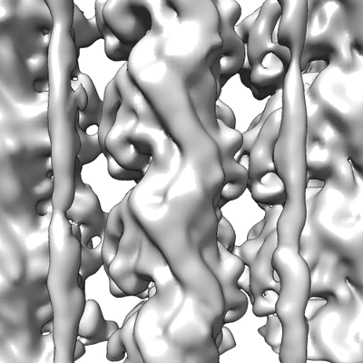 emd_4188.png emd_4188.png | 119.2 KB | ||
| Archive directory |  http://ftp.pdbj.org/pub/emdb/structures/EMD-4188ftp://ftp.pdbj.org/pub/emdb/structures/EMD-4188 http://ftp.pdbj.org/pub/emdb/structures/EMD-4188ftp://ftp.pdbj.org/pub/emdb/structures/EMD-4188 | HTTPS FTP |
-Related structure data
| Similar structure data |
|---|


-Links
| EMDB pages | EMDB (EBI/PDBe) / EMDataResource |
|---|
-Map
| File | Download / File: emd_4188.map.gz / Format: CCP4 / Size: 23 MB / Type: IMAGE STORED AS FLOATING POINT NUMBER (4 BYTES) | ||||||||||||||||||||||||||||||||||||||||||||||||||||||||||||||||||||
|---|---|---|---|---|---|---|---|---|---|---|---|---|---|---|---|---|---|---|---|---|---|---|---|---|---|---|---|---|---|---|---|---|---|---|---|---|---|---|---|---|---|---|---|---|---|---|---|---|---|---|---|---|---|---|---|---|---|---|---|---|---|---|---|---|---|---|---|---|---|
| Projections & slices | Image control
Images are generated by Spider. generated in cubic-lattice coordinate | ||||||||||||||||||||||||||||||||||||||||||||||||||||||||||||||||||||
| Voxel size | X=Y=Z: 1.34 Å | ||||||||||||||||||||||||||||||||||||||||||||||||||||||||||||||||||||
| Density |
| ||||||||||||||||||||||||||||||||||||||||||||||||||||||||||||||||||||
| Symmetry | Space group: 1 | ||||||||||||||||||||||||||||||||||||||||||||||||||||||||||||||||||||
| Details | EMDB XML:
CCP4 map header:
| ||||||||||||||||||||||||||||||||||||||||||||||||||||||||||||||||||||
 Z (Sec.)
 Y (Row.)
 X (Col.)


-Supplemental data
- Sample components
-Entire : SSNA1 in complex with porcine microtubules
| Entire | Name: SSNA1 in complex with porcine microtubules |
|---|---|
| Components |
|
-Supramolecule #1: SSNA1 in complex with porcine microtubules
| Supramolecule | Name: SSNA1 in complex with porcine microtubules / type: complex / ID: 1 / Parent: 0 |
|---|---|
| Source (natural) | Organism: Chlamydo |
-Experimental details
-Structure determination
| Method | cryo EM |
|---|---|
 Processing Processing | helical reconstruction |
| Aggregation state | helical array |
-Sample preparation
| Concentration | 1.5 mg/mL |
|---|---|
| Buffer | pH: 6.8 / Details: 80mM PIPES, 1 mM EGTA, 1 mM MgCl2 |
| Vitrification | Cryogen name: ETHANE / Chamber humidity: 95 % |
- Electron microscopy
| Microscope | FEI TITAN KRIOS |
|---|---|
| Image recording | Film or detector model: GATAN K2 SUMMIT (4k x 4k) / Average electron dose: 34.08 e/Å2 |
| Electron beam | Acceleration voltage: 300 kV / Electron source:  FIELD EMISSION GUN FIELD EMISSION GUN |
| Electron optics | Illumination mode: FLOOD BEAM / Imaging mode: BRIGHT FIELD |
| Experimental equipment |  Model: Titan Krios / Image courtesy: FEI Company |
-Image processing
| Final reconstruction | Applied symmetry - Helical parameters - Δz: 9.37308 Å Applied symmetry - Helical parameters - Δ&Phi: 27.692 ° Applied symmetry - Helical parameters - Axial symmetry: C1 (asymmetric) Resolution.type: BY AUTHOR / Resolution: 6.1 Å / Resolution method: FSC 0.143 CUT-OFF / Number images used: 30625 |
|---|---|
| Final angle assignment | Type: NOT APPLICABLE |
