Movie
Movie Controller

[English] 日本語
 Yorodumi
- EMDB-41807: QUS bounds mGlu5 in complex with Nb43, non uniform refinement on ... -
+ Open data

- Basic information
| Entry |  | |||||||||
|---|---|---|---|---|---|---|---|---|---|---|
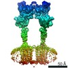
| Title | QUS bounds mGlu5 in complex with Nb43, non uniform refinement on the whole complex | |||||||||
 Map data Map data | sharpened map of QUS bound inactive mGlu5 in complex with Nb43, non uniform refinement on the whole complex | |||||||||
 Sample Sample |
| |||||||||
 Keywords Keywords | GPCR / SIGNALING PROTEIN | |||||||||
| Biological species |  Homo sapiens (human) Homo sapiens (human) | |||||||||
| Method | single particle reconstruction / cryo EM / Resolution: 3.33 Å | |||||||||
 Authors Authors | Krishna Kumar K / Wang H / Kobilka BK | |||||||||
| Funding support |  United States, 1 items United States, 1 items
| |||||||||
 Citation Citation | Journal: Nature / Year: 2024 Title: Stepwise activation of a metabotropic glutamate receptor. Authors: Kaavya Krishna Kumar / Haoqing Wang / Chris Habrian / Naomi R Latorraca / Jun Xu / Evan S O'Brien / Chensong Zhang / Elizabeth Montabana / Antoine Koehl / Susan Marqusee / Ehud Y Isacoff / Brian K Kobilka / Abstract: Metabotropic glutamate receptors belong to a family of G protein-coupled receptors that are obligate dimers and possess a large extracellular ligand-binding domain that is linked via a cysteine-rich ...Metabotropic glutamate receptors belong to a family of G protein-coupled receptors that are obligate dimers and possess a large extracellular ligand-binding domain that is linked via a cysteine-rich domain to their 7-transmembrane domain. Upon activation, these receptors undergo a large conformational change to transmit the ligand binding signal from the extracellular ligand-binding domain to the G protein-coupling 7-transmembrane domain. In this manuscript, we propose a model for a sequential, multistep activation mechanism of metabotropic glutamate receptor subtype 5. We present a series of structures in lipid nanodiscs, from inactive to fully active, including agonist-bound intermediate states. Further, using bulk and single-molecule fluorescence imaging, we reveal distinct receptor conformations upon allosteric modulator and G protein binding. | |||||||||
| History |
|
- Structure visualization
| Supplemental images |
|---|

- Downloads & links
-EMDB archive
| Map data | emd_41807.map.gz | 141.7 MB |  EMDB map data format EMDB map data format | |
|---|---|---|---|---|
| Header (meta data) | emd-41807-v30.xmlemd-41807.xml | 13.3 KB 13.3 KB | Display Display | EMDB header |
| Images |  emd_41807.png emd_41807.png | 54.1 KB | ||
| Filedesc metadata | emd-41807.cif.gz | 4 KB | ||
| Others | emd_41807_half_map_1.map.gzemd_41807_half_map_2.map.gz | 139 MB 139 MB | ||
| Archive directory |  http://ftp.pdbj.org/pub/emdb/structures/EMD-41807ftp://ftp.pdbj.org/pub/emdb/structures/EMD-41807 http://ftp.pdbj.org/pub/emdb/structures/EMD-41807ftp://ftp.pdbj.org/pub/emdb/structures/EMD-41807 | HTTPS FTP |
-Related structure data


-Links
| EMDB pages | EMDB (EBI/PDBe) / EMDataResource |
|---|
-Map
| File | Download / File: emd_41807.map.gz / Format: CCP4 / Size: 149.9 MB / Type: IMAGE STORED AS FLOATING POINT NUMBER (4 BYTES) | ||||||||||||||||||||||||||||||||||||
|---|---|---|---|---|---|---|---|---|---|---|---|---|---|---|---|---|---|---|---|---|---|---|---|---|---|---|---|---|---|---|---|---|---|---|---|---|---|
| Annotation | sharpened map of QUS bound inactive mGlu5 in complex with Nb43, non uniform refinement on the whole complex | ||||||||||||||||||||||||||||||||||||
| Projections & slices | Image control
Images are generated by Spider. | ||||||||||||||||||||||||||||||||||||
| Voxel size | X=Y=Z: 1.111 Å | ||||||||||||||||||||||||||||||||||||
| Density |
| ||||||||||||||||||||||||||||||||||||
| Symmetry | Space group: 1 | ||||||||||||||||||||||||||||||||||||
| Details | EMDB XML:
|
 Z (Sec.)
 Y (Row.)
 X (Col.)


-Supplemental data
-Half map: half map of QUS bound inactive mGlu5 in...
| File | emd_41807_half_map_1.map | ||||||||||||
|---|---|---|---|---|---|---|---|---|---|---|---|---|---|
| Annotation | half map of QUS bound inactive mGlu5 in complex with Nb43, non uniform refinement on the whole complex | ||||||||||||
| Projections & Slices |
| ||||||||||||
| Density Histograms |


-Half map: half map of QUS bound inactive mGlu5 in...
| File | emd_41807_half_map_2.map | ||||||||||||
|---|---|---|---|---|---|---|---|---|---|---|---|---|---|
| Annotation | half map of QUS bound inactive mGlu5 in complex with Nb43, non uniform refinement on the whole complex | ||||||||||||
| Projections & Slices |
| ||||||||||||
| Density Histograms |


- Sample components
-Entire : Metabotropic glutamate receptor 5 in complex with Quis
| Entire | Name: Metabotropic glutamate receptor 5 in complex with Quis |
|---|---|
| Components |
|
-Supramolecule #1: Metabotropic glutamate receptor 5 in complex with Quis
| Supramolecule | Name: Metabotropic glutamate receptor 5 in complex with Quis type: organelle_or_cellular_component / ID: 1 / Parent: 0 / Macromolecule list: #1-#2 |
|---|---|
| Source (natural) | Organism: Homo sapiens (human) |
-Experimental details
-Structure determination
| Method | cryo EM |
|---|---|
 Processing Processing | single particle reconstruction |
| Aggregation state | particle |
-Sample preparation
| Buffer | pH: 7.4 |
|---|---|
| Vitrification | Cryogen name: ETHANE |
- Electron microscopy
| Microscope | FEI TITAN KRIOS |
|---|---|
| Image recording | Film or detector model: GATAN K3 BIOQUANTUM (6k x 4k) / Average electron dose: 50.0 e/Å2 |
| Electron beam | Acceleration voltage: 300 kV / Electron source:  FIELD EMISSION GUN FIELD EMISSION GUN |
| Electron optics | Illumination mode: FLOOD BEAM / Imaging mode: BRIGHT FIELD / Nominal defocus max: 2.0 µm / Nominal defocus min: 0.7000000000000001 µm |
| Experimental equipment |  Model: Titan Krios / Image courtesy: FEI Company |