+データを開く
-基本情報
登録情報 | データベース: EMDB / ID: EMD-4166 | |||||||||
---|---|---|---|---|---|---|---|---|---|---|
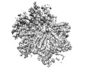
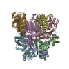
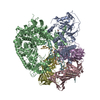
タイトル | Cryo-EM structure of TRIP13 in complex with ATP gamma S, p31comet, C-Mad2 and Cdc20 | |||||||||
マップデータ | TRIP13:p31-Substrate map | |||||||||
試料 |
| |||||||||
機能・相同性 | 機能・相同性情報 deactivation of mitotic spindle assembly checkpoint / metaphase/anaphase transition of cell cycle / mitotic spindle assembly checkpoint MAD1-MAD2 complex / metaphase/anaphase transition of meiosis I / Inhibition of the proteolytic activity of APC/C required for the onset of anaphase by mitotic spindle checkpoint components / mitotic checkpoint complex / positive regulation of anaphase-promoting complex-dependent catabolic process / meiotic recombination checkpoint signaling / positive regulation of mitotic cell cycle spindle assembly checkpoint / regulation of meiotic nuclear division ...deactivation of mitotic spindle assembly checkpoint / metaphase/anaphase transition of cell cycle / mitotic spindle assembly checkpoint MAD1-MAD2 complex / metaphase/anaphase transition of meiosis I / Inhibition of the proteolytic activity of APC/C required for the onset of anaphase by mitotic spindle checkpoint components / mitotic checkpoint complex / positive regulation of anaphase-promoting complex-dependent catabolic process / meiotic recombination checkpoint signaling / positive regulation of mitotic cell cycle spindle assembly checkpoint / regulation of meiotic nuclear division / establishment of centrosome localization / positive regulation of synapse maturation / Conversion from APC/C:Cdc20 to APC/C:Cdh1 in late anaphase / Inactivation of APC/C via direct inhibition of the APC/C complex / APC/C:Cdc20 mediated degradation of mitotic proteins / synaptonemal complex assembly / Phosphorylation of Emi1 / anaphase-promoting complex / regulation of meiotic cell cycle / anaphase-promoting complex-dependent catabolic process / positive regulation of synaptic plasticity / regulation of exit from mitosis / anaphase-promoting complex binding / nuclear pore nuclear basket / positive regulation of mitotic metaphase/anaphase transition / ubiquitin ligase activator activity / positive regulation of ubiquitin protein ligase activity / reciprocal meiotic recombination / oocyte maturation / female meiosis I / negative regulation of ubiquitin protein ligase activity / mitotic sister chromatid cohesion / oogenesis / mitotic spindle assembly checkpoint signaling / male meiosis I / establishment of mitotic spindle orientation / Regulation of APC/C activators between G1/S and early anaphase / mitotic sister chromatid segregation / negative regulation of mitotic cell cycle / spermatid development / mitotic spindle assembly / ubiquitin-like ligase-substrate adaptor activity / Amplification of signal from unattached kinetochores via a MAD2 inhibitory signal / Mitotic Prometaphase / EML4 and NUDC in mitotic spindle formation / regulation of mitotic cell cycle / Resolution of Sister Chromatid Cohesion / APC/C:Cdc20 mediated degradation of Cyclin B / APC-Cdc20 mediated degradation of Nek2A / male germ cell nucleus / APC/C:Cdc20 mediated degradation of Securin / SCF-beta-TrCP mediated degradation of Emi1 / RHO GTPases Activate Formins / Cdc20:Phospho-APC/C mediated degradation of Cyclin A / transcription coregulator activity / APC/C:Cdh1 mediated degradation of Cdc20 and other APC/C:Cdh1 targeted proteins in late mitosis/early G1 / negative regulation of protein catabolic process / mitotic spindle / kinetochore / spindle pole / spindle / Separation of Sister Chromatids / double-strand break repair / Antigen processing: Ubiquitination & Proteasome degradation / chromosome / nervous system development / spermatogenesis / nuclear membrane / transcription by RNA polymerase II / cell differentiation / Ub-specific processing proteases / protein ubiquitination / cell division / centrosome / negative regulation of apoptotic process / nucleolus / perinuclear region of cytoplasm / protein homodimerization activity / ATP hydrolysis activity / nucleoplasm / ATP binding / identical protein binding / nucleus / cytosol / cytoplasm 類似検索 - 分子機能 | |||||||||
生物種 | Homo sapiens (ヒト) | |||||||||
手法 | 単粒子再構成法 / クライオ電子顕微鏡法 / 解像度: 4.6 Å | |||||||||
データ登録者 | Alfieri C / Chang L / Barford D | |||||||||
資金援助 | 英国, 1件
| |||||||||
引用 | ジャーナル: Nature / 年: 2018 タイトル: Mechanism for remodelling of the cell cycle checkpoint protein MAD2 by the ATPase TRIP13. 著者: Claudio Alfieri / Leifu Chang / David Barford / 要旨: The maintenance of genome stability during mitosis is coordinated by the spindle assembly checkpoint (SAC) through its effector the mitotic checkpoint complex (MCC), an inhibitor of the anaphase- ...The maintenance of genome stability during mitosis is coordinated by the spindle assembly checkpoint (SAC) through its effector the mitotic checkpoint complex (MCC), an inhibitor of the anaphase-promoting complex (APC/C, also known as the cyclosome). Unattached kinetochores control MCC assembly by catalysing a change in the topology of the β-sheet of MAD2 (an MCC subunit), thereby generating the active closed MAD2 (C-MAD2) conformer. Disassembly of free MCC, which is required for SAC inactivation and chromosome segregation, is an ATP-dependent process driven by the AAA+ ATPase TRIP13. In combination with p31, an SAC antagonist, TRIP13 remodels C-MAD2 into inactive open MAD2 (O-MAD2). Here, we present a mechanism that explains how TRIP13-p31 disassembles the MCC. Cryo-electron microscopy structures of the TRIP13-p31-C-MAD2-CDC20 complex reveal that p31 recruits C-MAD2 to a defined site on the TRIP13 hexameric ring, positioning the N terminus of C-MAD2 (MAD2) to insert into the axial pore of TRIP13 and distorting the TRIP13 ring to initiate remodelling. Molecular modelling suggests that by gripping MAD2 within its axial pore, TRIP13 couples sequential ATP-driven translocation of its hexameric ring along MAD2 to push upwards on, and simultaneously rotate, the globular domains of the p31-C-MAD2 complex. This unwinds a region of the αA helix of C-MAD2 that is required to stabilize the C-MAD2 β-sheet, thus destabilizing C-MAD2 in favour of O-MAD2 and dissociating MAD2 from p31. Our study provides insights into how specific substrates are recruited to AAA+ ATPases through adaptor proteins and suggests a model of how translocation through the axial pore of AAA+ ATPases is coupled to protein remodelling. | |||||||||
履歴 |
|
-構造の表示
ムービー |
ムービービューア |
---|---|
構造ビューア | EMマップ: SurfViewMolmilJmol/JSmol |
添付画像 |
-ダウンロードとリンク
-EMDBアーカイブ
マップデータ | emd_4166.map.gz | 1.9 MB | EMDBマップデータ形式 | |
---|---|---|---|---|
ヘッダ (付随情報) | emd-4166-v30.xml emd-4166.xml | 26 KB 26 KB | 表示 表示 | EMDBヘッダ |
画像 | emd_4166.png | 77.5 KB | ||
その他 | emd_4166_additional_1.map.gz emd_4166_additional_2.map.gz emd_4166_additional_3.map.gz emd_4166_additional_4.map.gz emd_4166_additional_5.map.gz | 1.8 MB 2 MB 1.9 MB 1.9 MB 1.3 MB | ||
アーカイブディレクトリ | http://ftp.pdbj.org/pub/emdb/structures/EMD-4166 ftp://ftp.pdbj.org/pub/emdb/structures/EMD-4166 | HTTPS FTP |
-検証レポート
文書・要旨 | emd_4166_validation.pdf.gz | 235.4 KB | 表示 | EMDB検証レポート |
---|---|---|---|---|
文書・詳細版 | emd_4166_full_validation.pdf.gz | 234.5 KB | 表示 | |
XML形式データ | emd_4166_validation.xml.gz | 5.3 KB | 表示 | |
アーカイブディレクトリ | https://ftp.pdbj.org/pub/emdb/validation_reports/EMD-4166 ftp://ftp.pdbj.org/pub/emdb/validation_reports/EMD-4166 | HTTPS FTP |
-関連構造データ
-リンク
EMDBのページ | EMDB (EBI/PDBe) / EMDataResource |
---|---|
「今月の分子」の関連する項目 |
-マップ
ファイル | ダウンロード / ファイル: emd_4166.map.gz / 形式: CCP4 / 大きさ: 10.5 MB / タイプ: IMAGE STORED AS FLOATING POINT NUMBER (4 BYTES) | ||||||||||||||||||||||||||||||||||||||||||||||||||||||||||||
---|---|---|---|---|---|---|---|---|---|---|---|---|---|---|---|---|---|---|---|---|---|---|---|---|---|---|---|---|---|---|---|---|---|---|---|---|---|---|---|---|---|---|---|---|---|---|---|---|---|---|---|---|---|---|---|---|---|---|---|---|---|
注釈 | TRIP13:p31-Substrate map | ||||||||||||||||||||||||||||||||||||||||||||||||||||||||||||
投影像・断面図 | 画像のコントロール
画像は Spider により作成 | ||||||||||||||||||||||||||||||||||||||||||||||||||||||||||||
ボクセルのサイズ | X=Y=Z: 1.43 Å | ||||||||||||||||||||||||||||||||||||||||||||||||||||||||||||
密度 |
| ||||||||||||||||||||||||||||||||||||||||||||||||||||||||||||
対称性 | 空間群: 1 | ||||||||||||||||||||||||||||||||||||||||||||||||||||||||||||
詳細 | EMDB XML:
CCP4マップ ヘッダ情報:
|
-添付データ
-追加マップ: TRIP13 apo map
ファイル | emd_4166_additional_1.map | ||||||||||||
---|---|---|---|---|---|---|---|---|---|---|---|---|---|
注釈 | TRIP13_apo map | ||||||||||||
投影像・断面図 |
| ||||||||||||
密度ヒストグラム |
-追加マップ: TRIP13:p31-Substrate basal state map
ファイル | emd_4166_additional_2.map | ||||||||||||
---|---|---|---|---|---|---|---|---|---|---|---|---|---|
注釈 | TRIP13:p31-Substrate_basal_state map | ||||||||||||
投影像・断面図 |
| ||||||||||||
密度ヒストグラム |
-追加マップ: TRIP13:p31-Substrate Activated state map
ファイル | emd_4166_additional_3.map | ||||||||||||
---|---|---|---|---|---|---|---|---|---|---|---|---|---|
注釈 | TRIP13:p31-Substrate_Activated_state map | ||||||||||||
投影像・断面図 |
| ||||||||||||
密度ヒストグラム |
-追加マップ: TRIP13:p31-Substrate All Particles map
ファイル | emd_4166_additional_4.map | ||||||||||||
---|---|---|---|---|---|---|---|---|---|---|---|---|---|
注釈 | TRIP13:p31-Substrate_All_Particles map | ||||||||||||
投影像・断面図 |
| ||||||||||||
密度ヒストグラム |
-追加マップ: TRIP13 A,B,C and D monomers map
ファイル | emd_4166_additional_5.map | ||||||||||||
---|---|---|---|---|---|---|---|---|---|---|---|---|---|
注釈 | TRIP13_A,B,C and D monomers map | ||||||||||||
投影像・断面図 |
| ||||||||||||
密度ヒストグラム |
-試料の構成要素
-全体 : TRIP13 hexamer in complex with ATP gamma S, p31comet, C-Mad2 and Cdc20
全体 | 名称: TRIP13 hexamer in complex with ATP gamma S, p31comet, C-Mad2 and Cdc20 |
---|---|
要素 |
|
-超分子 #1: TRIP13 hexamer in complex with ATP gamma S, p31comet, C-Mad2 and Cdc20
超分子 | 名称: TRIP13 hexamer in complex with ATP gamma S, p31comet, C-Mad2 and Cdc20 タイプ: complex / ID: 1 / 親要素: 0 / 含まれる分子: #1-#4 |
---|---|
分子量 | 理論値: 400 kDa/nm |
-超分子 #2: MAD2L1-binding protein, Pachytene checkpoint protein 2 homolog
超分子 | 名称: MAD2L1-binding protein, Pachytene checkpoint protein 2 homolog タイプ: complex / ID: 2 / 親要素: 1 / 含まれる分子: #1-#2 |
---|---|
由来(天然) | 生物種: Homo sapiens (ヒト) |
組換発現 | 生物種: Escherichia coli (大腸菌) |
-超分子 #3: Cell division cycle protein 20 homolog, Mitotic spindle assembly ...
超分子 | 名称: Cell division cycle protein 20 homolog, Mitotic spindle assembly checkpoint protein MAD2A タイプ: complex / ID: 3 / 親要素: 1 / 含まれる分子: #3-#4 |
---|---|
由来(天然) | 生物種: Homo sapiens (ヒト) |
組換発現 | 生物種: Trichoplusia ni (イラクサキンウワバ) |
-分子 #1: Pachytene checkpoint protein 2 homolog
分子 | 名称: Pachytene checkpoint protein 2 homolog / タイプ: protein_or_peptide / ID: 1 / コピー数: 6 / 光学異性体: LEVO |
---|---|
由来(天然) | 生物種: Homo sapiens (ヒト) |
分子量 | 理論値: 48.606664 KDa |
組換発現 | 生物種: Escherichia coli (大腸菌) |
配列 | 文字列: MDEAVGDLKQ ALPCVAESPT VHVEVHQRGS STAKKEDINL SVRKLLNRHN IVFGDYTWTE FDEPFLTRNV QSVSIIDTEL KVKDSQPID LSACTVALHI FQLNEDGPSS ENLEEETENI IAANHWVLPA AEFHGLWDSL VYDVEVKSHL LDYVMTTLLF S DKNVNSNL ...文字列: MDEAVGDLKQ ALPCVAESPT VHVEVHQRGS STAKKEDINL SVRKLLNRHN IVFGDYTWTE FDEPFLTRNV QSVSIIDTEL KVKDSQPID LSACTVALHI FQLNEDGPSS ENLEEETENI IAANHWVLPA AEFHGLWDSL VYDVEVKSHL LDYVMTTLLF S DKNVNSNL ITWNRVVLLH GPPGTGKTSL CKALAQKLTI RLSSRYRYGQ LIEINSHSLF SKWFSESGKL VTKMFQKIQD LI DDKDALV FVLIDQVESL TAARNACRAG TEPSDAIRVV NAVLTQIDQI KRHSNVVILT TSNITEKIDV AFVDRADIKQ YIG PPSAAA IFKIYLSCLE ELMKCQIIYP RQQLLTLREL EMIGFIENNV SKLSLLLNDI SRKSEGLSGR VLRKLPFLAH ALYV QAPTV TIEGFLQALS LAVDKQFEER KKLAAYI |
-分子 #2: MAD2L1-binding protein
分子 | 名称: MAD2L1-binding protein / タイプ: protein_or_peptide / ID: 2 / コピー数: 1 / 光学異性体: LEVO |
---|---|
由来(天然) | 生物種: Homo sapiens (ヒト) |
分子量 | 理論値: 31.086646 KDa |
組換発現 | 生物種: Escherichia coli (大腸菌) |
配列 | 文字列: MAAPEAEVLS SAAVPDLEWY EKSEETHASQ IELLETSSTQ EPLNASEAFC PRDCMVPVVF PGPVSQEGCC QFTCELLKHI MYQRQQLPL PYEQLKHFYR KPSPQAEEML KKKPRATTEV SSRKCQQALA ELESVLSHLE DFFARTLVPR VLILLGGNAL S PKEFYELD ...文字列: MAAPEAEVLS SAAVPDLEWY EKSEETHASQ IELLETSSTQ EPLNASEAFC PRDCMVPVVF PGPVSQEGCC QFTCELLKHI MYQRQQLPL PYEQLKHFYR KPSPQAEEML KKKPRATTEV SSRKCQQALA ELESVLSHLE DFFARTLVPR VLILLGGNAL S PKEFYELD LSLLAPYSVD QSLSTAACLR RLFRAIFMAD AFSELQAPPL MGTVVMAQGH RNCGEDWFRP KLNYRVPSRG HK LTVTLSC GRPSIRTTAW EDYIWFQAPV TFKGFRE |
-分子 #3: Cell division cycle protein 20 homolog
分子 | 名称: Cell division cycle protein 20 homolog / タイプ: protein_or_peptide / ID: 3 / コピー数: 1 / 光学異性体: LEVO |
---|---|
由来(天然) | 生物種: Homo sapiens (ヒト) |
分子量 | 理論値: 54.796508 KDa |
組換発現 | 生物種: Trichoplusia ni (イラクサキンウワバ) |
配列 | 文字列: MAQFAFESDL HSLLQLDAPI PNAPPARWQR KAKEAAGPAP SPMRAANRSH SAGRTPGRTP GKSSSKVQTT PSKPGGDRYI PHRSAAQME VASFLLSKEN QPENSQTPTK KEHQKAWALN LNGFDVEEAK ILRLSGKPQN APEGYQNRLK VLYSQKATPG S SRKTCRYI ...文字列: MAQFAFESDL HSLLQLDAPI PNAPPARWQR KAKEAAGPAP SPMRAANRSH SAGRTPGRTP GKSSSKVQTT PSKPGGDRYI PHRSAAQME VASFLLSKEN QPENSQTPTK KEHQKAWALN LNGFDVEEAK ILRLSGKPQN APEGYQNRLK VLYSQKATPG S SRKTCRYI PSLPDRILDA PEIRNDYYLN LVDWSSGNVL AVALDNSVYL WSASSGDILQ LLQMEQPGEY ISSVAWIKEG NY LAVGTSS AEVQLWDVQQ QKRLRNMTSH SARVGSLSWN SYILSSGSRS GHIHHHDVRV AEHHVATLSG HSQEVCGLRW APD GRHLAS GGNDNLVNVW PSAPGEGGWV PLQTFTQHQG AVKAVAWCPW QSNVLATGGG TSDRHIRIWN VCSGACLSAV DAHS QVCSI LWSPHYKELI SGHGFAQNQL VIWKYPTMAK VAELKGHTSR VLSLTMSPDG ATVASAAADE TLRLWRCFEL DPARR RERE KASAAKSSLI HQGIR |
-分子 #4: Mitotic spindle assembly checkpoint protein MAD2A
分子 | 名称: Mitotic spindle assembly checkpoint protein MAD2A / タイプ: protein_or_peptide / ID: 4 / コピー数: 1 / 光学異性体: LEVO |
---|---|
由来(天然) | 生物種: Homo sapiens (ヒト) |
分子量 | 理論値: 23.533883 KDa |
組換発現 | 生物種: Trichoplusia ni (イラクサキンウワバ) |
配列 | 文字列: MALQLSREQG ITLRGSAEIV AEFFSFGINS ILYQRGIYPS ETFTRVQKYG LTLLVTTDLE LIKYLNNVVE QLKDWLYKCS VQKLVVVIS NIESGEVLER WQFDIECDKT AKDDSAPREK SQKAIQDEIR SVIRQITATV TFLPLLEVSC SFDLLIYTDK D LVVPEKWE ...文字列: MALQLSREQG ITLRGSAEIV AEFFSFGINS ILYQRGIYPS ETFTRVQKYG LTLLVTTDLE LIKYLNNVVE QLKDWLYKCS VQKLVVVIS NIESGEVLER WQFDIECDKT AKDDSAPREK SQKAIQDEIR SVIRQITATV TFLPLLEVSC SFDLLIYTDK D LVVPEKWE ESGPQFITNS EEVRLRSFTT TIHKVNSMVA YKIPVND |
-分子 #5: PHOSPHOTHIOPHOSPHORIC ACID-ADENYLATE ESTER
分子 | 名称: PHOSPHOTHIOPHOSPHORIC ACID-ADENYLATE ESTER / タイプ: ligand / ID: 5 / コピー数: 5 / 式: AGS |
---|---|
分子量 | 理論値: 523.247 Da |
Chemical component information | ChemComp-AGS: |
-実験情報
-構造解析
手法 | クライオ電子顕微鏡法 |
---|---|
解析 | 単粒子再構成法 |
試料の集合状態 | particle |
-試料調製
濃度 | 0.3 mg/mL |
---|---|
緩衝液 | pH: 7.5 |
凍結 | 凍結剤: ETHANE |
-電子顕微鏡法
顕微鏡 | FEI TITAN KRIOS |
---|---|
撮影 | フィルム・検出器のモデル: GATAN K2 SUMMIT (4k x 4k) 平均電子線量: 40.0 e/Å2 |
電子線 | 加速電圧: 300 kV / 電子線源: FIELD EMISSION GUN |
電子光学系 | 照射モード: FLOOD BEAM / 撮影モード: BRIGHT FIELD |
実験機器 | モデル: Titan Krios / 画像提供: FEI Company |
-画像解析
最終 再構成 | 想定した対称性 - 点群: C1 (非対称) / 解像度のタイプ: BY AUTHOR / 解像度: 4.6 Å / 解像度の算出法: FSC 0.143 CUT-OFF / ソフトウェア - 名称: RELION (ver. 2.1-beta-1) / 使用した粒子像数: 354157 |
---|---|
初期 角度割当 | タイプ: PROJECTION MATCHING |
最終 角度割当 | タイプ: PROJECTION MATCHING |