+ Open data
- Basic information
| Entry |  | |||||||||
|---|---|---|---|---|---|---|---|---|---|---|
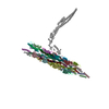
| Title | Outer Mat-T4P complex | |||||||||
 Map data Map data | ||||||||||
 Sample Sample |
| |||||||||
 Keywords Keywords | Acinetobacter / SsRNA phage virus / T4P / VIRUS | |||||||||
| Function / homology |  Function and homology information Function and homology informationvirion attachment to host cell pilus / protein secretion by the type II secretion system / type II protein secretion system complex / pilus / virion component / cell adhesion / membrane Similarity search - Function | |||||||||
| Biological species |  Acinetobacter phage AP205 (virus) / Acinetobacter phage AP205 (virus) /  Acinetobacter genomosp. 16BJ (bacteria) Acinetobacter genomosp. 16BJ (bacteria) | |||||||||
| Method | single particle reconstruction / cryo EM / Resolution: 8.55 Å | |||||||||
 Authors Authors | Meng R / Xing Z / Thongchol J / Zhang J | |||||||||
| Funding support |  United States, 1 items United States, 1 items
| |||||||||
 Citation Citation |  Journal: Nat Commun / Year: 2024 Journal: Nat Commun / Year: 2024Title: Structural basis of Acinetobacter type IV pili targeting by an RNA virus. Authors: Ran Meng / Zhongliang Xing / Jeng-Yih Chang / Zihao Yu / Jirapat Thongchol / Wen Xiao / Yuhang Wang / Karthik Chamakura / Zhiqi Zeng / Fengbin Wang / Ry Young / Lanying Zeng / Junjie Zhang /  Abstract: Acinetobacters pose a significant threat to human health, especially those with weakened immune systems. Type IV pili of acinetobacters play crucial roles in virulence and antibiotic resistance. ...Acinetobacters pose a significant threat to human health, especially those with weakened immune systems. Type IV pili of acinetobacters play crucial roles in virulence and antibiotic resistance. Single-stranded RNA bacteriophages target the bacterial retractile pili, including type IV. Our study delves into the interaction between Acinetobacter phage AP205 and type IV pili. Using cryo-electron microscopy, we solve structures of the AP205 virion with an asymmetric dimer of maturation proteins, the native Acinetobacter type IV pili bearing a distinct post-translational pilin cleavage, and the pili-bound AP205 showing its maturation proteins adapted to pilin modifications, allowing each phage to bind to one or two pili. Leveraging these results, we develop a 20-kilodalton AP205-derived protein scaffold targeting type IV pili in situ, with potential for research and diagnostics. | |||||||||
| History |
|
- Structure visualization
| Supplemental images |
|---|
- Downloads & links
-EMDB archive
| Map data |  emd_41635.map.gz emd_41635.map.gz | 81 MB |  EMDB map data format EMDB map data format | |
|---|---|---|---|---|
| Header (meta data) |  emd-41635-v30.xml emd-41635-v30.xml emd-41635.xml emd-41635.xml | 18.7 KB 18.7 KB | Display Display |  EMDB header EMDB header |
| FSC (resolution estimation) |  emd_41635_fsc.xml emd_41635_fsc.xml | 11.7 KB | Display |  FSC data file FSC data file |
| Images |  emd_41635.png emd_41635.png | 42.9 KB | ||
| Filedesc metadata |  emd-41635.cif.gz emd-41635.cif.gz | 6.1 KB | ||
| Others |  emd_41635_half_map_1.map.gz emd_41635_half_map_1.map.gz emd_41635_half_map_2.map.gz emd_41635_half_map_2.map.gz | 151.6 MB 151.6 MB | ||
| Archive directory |  http://ftp.pdbj.org/pub/emdb/structures/EMD-41635 http://ftp.pdbj.org/pub/emdb/structures/EMD-41635 ftp://ftp.pdbj.org/pub/emdb/structures/EMD-41635 ftp://ftp.pdbj.org/pub/emdb/structures/EMD-41635 | HTTPS FTP |
-Validation report
| Summary document |  emd_41635_validation.pdf.gz emd_41635_validation.pdf.gz | 883 KB | Display |  EMDB validaton report EMDB validaton report |
|---|---|---|---|---|
| Full document |  emd_41635_full_validation.pdf.gz emd_41635_full_validation.pdf.gz | 882.6 KB | Display | |
| Data in XML |  emd_41635_validation.xml.gz emd_41635_validation.xml.gz | 20.1 KB | Display | |
| Data in CIF |  emd_41635_validation.cif.gz emd_41635_validation.cif.gz | 26.4 KB | Display | |
| Arichive directory |  https://ftp.pdbj.org/pub/emdb/validation_reports/EMD-41635 https://ftp.pdbj.org/pub/emdb/validation_reports/EMD-41635 ftp://ftp.pdbj.org/pub/emdb/validation_reports/EMD-41635 ftp://ftp.pdbj.org/pub/emdb/validation_reports/EMD-41635 | HTTPS FTP |
-Related structure data
| Related structure data |  8tvaMC  8tobC  8tocC  8tv9C  8tw2C  8twcC C: citing same article ( M: atomic model generated by this map |
|---|---|
| Similar structure data | Similarity search - Function & homology  F&H Search F&H Search |
- Links
Links
| EMDB pages |  EMDB (EBI/PDBe) / EMDB (EBI/PDBe) /  EMDataResource EMDataResource |
|---|---|
| Related items in Molecule of the Month |
- Map
Map
| File |  Download / File: emd_41635.map.gz / Format: CCP4 / Size: 163.6 MB / Type: IMAGE STORED AS FLOATING POINT NUMBER (4 BYTES) Download / File: emd_41635.map.gz / Format: CCP4 / Size: 163.6 MB / Type: IMAGE STORED AS FLOATING POINT NUMBER (4 BYTES) | ||||||||||||||||||||||||||||||||||||
|---|---|---|---|---|---|---|---|---|---|---|---|---|---|---|---|---|---|---|---|---|---|---|---|---|---|---|---|---|---|---|---|---|---|---|---|---|---|
| Projections & slices | Image control
Images are generated by Spider. | ||||||||||||||||||||||||||||||||||||
| Voxel size | X=Y=Z: 2.12 Å | ||||||||||||||||||||||||||||||||||||
| Density |
| ||||||||||||||||||||||||||||||||||||
| Symmetry | Space group: 1 | ||||||||||||||||||||||||||||||||||||
| Details | EMDB XML:
|
-Supplemental data
-Half map: #2
| File | emd_41635_half_map_1.map | ||||||||||||
|---|---|---|---|---|---|---|---|---|---|---|---|---|---|
| Projections & Slices |
| ||||||||||||
| Density Histograms |
-Half map: #1
| File | emd_41635_half_map_2.map | ||||||||||||
|---|---|---|---|---|---|---|---|---|---|---|---|---|---|
| Projections & Slices |
| ||||||||||||
| Density Histograms |
- Sample components
-Entire : Acinetobacter phage AP205
| Entire | Name:  Acinetobacter phage AP205 (virus) Acinetobacter phage AP205 (virus) |
|---|---|
| Components |
|
-Supramolecule #1: Acinetobacter phage AP205
| Supramolecule | Name: Acinetobacter phage AP205 / type: virus / ID: 1 / Parent: 0 / Macromolecule list: all / NCBI-ID: 154784 / Sci species name: Acinetobacter phage AP205 / Virus type: VIRION / Virus isolate: SPECIES / Virus enveloped: Yes / Virus empty: No |
|---|---|
| Host (natural) | Organism:  Acinetobacter genomosp. 16BJ (bacteria) Acinetobacter genomosp. 16BJ (bacteria) |
| Molecular weight | Theoretical: 378 KDa |
| Virus shell | Shell ID: 1 / Name: Coat Capsid / Diameter: 290.0 Å / T number (triangulation number): 3 |
-Macromolecule #1: Maturation protein
| Macromolecule | Name: Maturation protein / type: protein_or_peptide / ID: 1 / Number of copies: 1 / Enantiomer: LEVO |
|---|---|
| Source (natural) | Organism:  Acinetobacter phage AP205 (virus) Acinetobacter phage AP205 (virus) |
| Molecular weight | Theoretical: 61.063852 KDa |
| Sequence | String: MNMYKWVPES IRDSGEGQPS YSNNGDYAPS GPWVAAGIHT MPQSLRDSMR NSIMVTAQAR RDVIGPEWGP DGRFTGYASV IGTPDPKPA DIVNKFTVER RPVSNGNFQQ RVKAGDIVVA PYTSDGKITV KLVAGQKDIS STPDYDYRID SSLASSAGFV V AGERWYYT ...String: MNMYKWVPES IRDSGEGQPS YSNNGDYAPS GPWVAAGIHT MPQSLRDSMR NSIMVTAQAR RDVIGPEWGP DGRFTGYASV IGTPDPKPA DIVNKFTVER RPVSNGNFQQ RVKAGDIVVA PYTSDGKITV KLVAGQKDIS STPDYDYRID SSLASSAGFV V AGERWYYT KRHFIIPRYF QNWRMRRRKY VTGWVMPTFY SPKEIFNRLK DSLVPDTGLV TQVWADNNTK RMDFLTAMAE IP QTLSSFL DALGYLGSLI KDFKRRRFFL NKAHQRIRNK LGVSFAERRS QIVSKYDRKI ASARKPAIIV KLRQRKEKAL KAL DKMRVR EEKKMIREFA TQAASLWLSF RYEIMPLYYQ SQDVLDVIAN STSEFMTSRD FVAKAINIGI PLEWNLDQEN LVSQ PRHNV MVKSKLSPEN NIGKTLSVNP FTTAWELLTL SFVVDWFVNF GDVIAGFTGG YSDDSGATAS WRFDDKKVFH LKNIP SAMV IVDINFYTRQ VIDPRLCGGL AFSPKLNLFR YLDAMSLSWN RSRLKISRAT UniProtKB: Maturation protein |
-Macromolecule #2: Fimbrial protein
| Macromolecule | Name: Fimbrial protein / type: protein_or_peptide / ID: 2 / Number of copies: 20 / Enantiomer: LEVO |
|---|---|
| Source (natural) | Organism:  Acinetobacter genomosp. 16BJ (bacteria) Acinetobacter genomosp. 16BJ (bacteria) |
| Molecular weight | Theoretical: 7.470747 KDa |
| Sequence | String: TLIELMIVVA IIGILAAIAI PQYQNYIAKS QVSRVMSETG SLKTVIETCI LDGKTAANCE LGWTNSNLLG UniProtKB: Fimbrial protein |
-Macromolecule #3: Fimbrial protein
| Macromolecule | Name: Fimbrial protein / type: protein_or_peptide / ID: 3 / Number of copies: 20 / Enantiomer: LEVO |
|---|---|
| Source (natural) | Organism:  Acinetobacter genomosp. 16BJ (bacteria) Acinetobacter genomosp. 16BJ (bacteria) |
| Molecular weight | Theoretical: 6.999778 KDa |
| Sequence | String: STAAVTGQTG LTITYPASAT ESAAIQGTFG NSAAIKIKNQ TLTWTRTPEG AWSCATTVEA KFKPAGCAS UniProtKB: Fimbrial protein |
-Experimental details
-Structure determination
| Method | cryo EM |
|---|---|
 Processing Processing | single particle reconstruction |
| Aggregation state | particle |
- Sample preparation
| Buffer | pH: 8 Component:
Details: 20mM Tris-HCl, 150mM NaCl, pH 8.0 | |||||||||
|---|---|---|---|---|---|---|---|---|---|---|
| Vitrification | Cryogen name: ETHANE / Chamber humidity: 100 % / Chamber temperature: 277 K / Instrument: FEI VITROBOT MARK III / Details: 3uL sample applied to a 300-mesh 2/1 copper grid. | |||||||||
| Details | AP205 virion particle |
- Electron microscopy
| Microscope | FEI TITAN KRIOS |
|---|---|
| Image recording | Film or detector model: GATAN K3 BIOQUANTUM (6k x 4k) / Detector mode: COUNTING / Average electron dose: 50.0 e/Å2 |
| Electron beam | Acceleration voltage: 300 kV / Electron source:  FIELD EMISSION GUN FIELD EMISSION GUN |
| Electron optics | Illumination mode: FLOOD BEAM / Imaging mode: BRIGHT FIELD / Cs: 2.7 mm / Nominal defocus max: 3.5 µm / Nominal defocus min: 1.0 µm / Nominal magnification: 130000 |
| Sample stage | Cooling holder cryogen: NITROGEN |
| Experimental equipment |  Model: Titan Krios / Image courtesy: FEI Company |
+ Image processing
-Atomic model buiding 1
| Refinement | Protocol: RIGID BODY FIT |
|---|---|
| Output model |  PDB-8tva: |
 Movie
Movie Controller


 Z (Sec.)
 Y (Row.)
 X (Col.)


