 Movie
Movie Controller


+ Open data

- Basic information
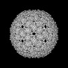
| Entry | Database: PDB / ID: 8twc | ||||||
|---|---|---|---|---|---|---|---|
| Title | Acinetobacter phage AP205 T=3 VLP | ||||||
 Components Components | Coat protein | ||||||
 Keywords Keywords | VIRUS LIKE PARTICLE / Acinetobacter / SsRNA phage virus / VIRUS / VLP | ||||||
| Function / homology | : / AP205 coat protein / viral capsid / Coat protein Function and homology information Function and homology information | ||||||
| Biological species |  Acinetobacter phage AP205 (virus) Acinetobacter phage AP205 (virus) | ||||||
| Method | ELECTRON MICROSCOPY / single particle reconstruction / cryo EM / Resolution: 3 Å | ||||||
 Authors Authors | Meng, R. / Xing, Z. / Zhang, J. | ||||||
| Funding support |  United States, 1items United States, 1items
| ||||||
 Citation Citation | Journal: Nat Commun / Year: 2024 Title: Structural basis of Acinetobacter type IV pili targeting by an RNA virus. Authors: Ran Meng / Zhongliang Xing / Jeng-Yih Chang / Zihao Yu / Jirapat Thongchol / Wen Xiao / Yuhang Wang / Karthik Chamakura / Zhiqi Zeng / Fengbin Wang / Ry Young / Lanying Zeng / Junjie Zhang / Abstract: Acinetobacters pose a significant threat to human health, especially those with weakened immune systems. Type IV pili of acinetobacters play crucial roles in virulence and antibiotic resistance. ...Acinetobacters pose a significant threat to human health, especially those with weakened immune systems. Type IV pili of acinetobacters play crucial roles in virulence and antibiotic resistance. Single-stranded RNA bacteriophages target the bacterial retractile pili, including type IV. Our study delves into the interaction between Acinetobacter phage AP205 and type IV pili. Using cryo-electron microscopy, we solve structures of the AP205 virion with an asymmetric dimer of maturation proteins, the native Acinetobacter type IV pili bearing a distinct post-translational pilin cleavage, and the pili-bound AP205 showing its maturation proteins adapted to pilin modifications, allowing each phage to bind to one or two pili. Leveraging these results, we develop a 20-kilodalton AP205-derived protein scaffold targeting type IV pili in situ, with potential for research and diagnostics. | ||||||
| History |
|
- Structure visualization
| Structure viewer | Molecule: MolmilJmol/JSmol |
|---|
- Downloads & links
-Download
| PDBx/mmCIF format | 8twc.cif.gz | 3.5 MB | Display | PDBx/mmCIF format |
|---|---|---|---|---|
| PDB format | pdb8twc.ent.gz | Display | PDB format | |
| PDBx/mmJSON format | 8twc.json.gz | Tree view | PDBx/mmJSON format | |
| Others |  Other downloads Other downloads |
-Validation report
| Arichive directory | https://data.pdbj.org/pub/pdb/validation_reports/tw/8twcftp://data.pdbj.org/pub/pdb/validation_reports/tw/8twc | HTTPS FTP |
|---|
-Related structure data
| Related structure data | 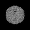 41666MC  8tobC  8tocC  8tv9C  8tvaC  8tw2C C: citing same article ( M: map data used to model this data |
|---|---|
| Similar structure data |


-Links
 PDBj
PDBj- Assembly
| Deposited unit | 
|
|---|---|
| 1 |
|
-Components
| #1: Protein | Mass: 13820.569 Da / Num. of mol.: 180 / Source method: isolated from a natural source / Source: (natural) Acinetobacter phage AP205 (virus) / References: UniProt: Q9AZ42Has protein modification | Y | |
|---|
-Experimental details
-Experiment
| Experiment | Method: ELECTRON MICROSCOPY |
|---|---|
| EM experiment | Aggregation state: PARTICLE / 3D reconstruction method: single particle reconstruction |
- Sample preparation
| Component | Name: Acinetobacter phage AP205 / Type: VIRUS Details: amplified and purified from infected Acinetobacter GP16 cells. Entity ID: all / Source: NATURAL | |||||||||||||||
|---|---|---|---|---|---|---|---|---|---|---|---|---|---|---|---|---|
| Molecular weight | Value: 2.48 MDa / Experimental value: NO | |||||||||||||||
| Source (natural) | Organism: Acinetobacter phage AP205 (virus) | |||||||||||||||
| Details of virus | Empty: NO / Enveloped: YES / Isolate: SPECIES / Type: VIRION | |||||||||||||||
| Natural host | Organism: Acinetobacter genomosp. 16BJ | |||||||||||||||
| Virus shell | Name: Coat / Diameter: 300 nm / Triangulation number (T number): 3 | |||||||||||||||
| Buffer solution | pH: 8 / Details: 20mM Tris-HCl, 150mM NaCl, pH 8.0 | |||||||||||||||
| Buffer component |
| |||||||||||||||
| Specimen | Embedding applied: NO / Shadowing applied: NO / Staining applied: NO / Vitrification applied: YES / Details: AP205 virion particle | |||||||||||||||
| Vitrification | Instrument: FEI VITROBOT MARK III / Cryogen name: ETHANE / Humidity: 100 % / Chamber temperature: 277 K / Details: 3uL sample applied to a 300-mesh 2/1 copper grid |
- Electron microscopy imaging
| Experimental equipment |  Model: Titan Krios / Image courtesy: FEI Company |
|---|---|
| Microscopy | Model: FEI TITAN KRIOS |
| Electron gun | Electron source:  FIELD EMISSION GUN / Accelerating voltage: 300 kV / Illumination mode: FLOOD BEAM FIELD EMISSION GUN / Accelerating voltage: 300 kV / Illumination mode: FLOOD BEAM |
| Electron lens | Mode: BRIGHT FIELD / Nominal magnification: 130000 X / Nominal defocus max: 3500 nm / Nominal defocus min: 1000 nm / Cs: 2.7 mm |
| Specimen holder | Cryogen: NITROGEN |
| Image recording | Electron dose: 50 e/Å2 / Detector mode: COUNTING / Film or detector model: GATAN K3 BIOQUANTUM (6k x 4k) |
- Processing
| EM software |
| ||||||||||||||||||||||||
|---|---|---|---|---|---|---|---|---|---|---|---|---|---|---|---|---|---|---|---|---|---|---|---|---|---|
| CTF correction | Type: NONE | ||||||||||||||||||||||||
| 3D reconstruction | Resolution: 3 Å / Resolution method: FSC 0.143 CUT-OFF / Num. of particles: 10000 / Symmetry type: POINT | ||||||||||||||||||||||||
| Atomic model building | Protocol: RIGID BODY FIT | ||||||||||||||||||||||||
| Refine LS restraints |
|
