 ムービー
ムービー コントローラー


+ データを開く

- 基本情報
基本情報
| 登録情報 | データベース: EMDB / ID: EMD-4000 | |||||||||
|---|---|---|---|---|---|---|---|---|---|---|
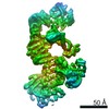
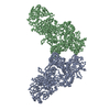
| タイトル | Subtomographic reconstruction of a single VP4 spike from Halorubrum pleomorphic virus 1. | |||||||||
 マップデータ マップデータ | None | |||||||||
 試料 試料 |
| |||||||||
| 生物種 |  Halorubrum pleomorphic virus 1 (ウイルス) Halorubrum pleomorphic virus 1 (ウイルス) | |||||||||
| 手法 | サブトモグラム平均法 / クライオ電子顕微鏡法 / 解像度: 40.0 Å | |||||||||
 データ登録者 データ登録者 | Butcher SJ / Liljeroos L / Manole V | |||||||||
 引用 引用 | ジャーナル: J Virol / 年: 2012 タイトル: Virion architecture unifies globally distributed pleolipoviruses infecting halophilic archaea. 著者: Maija K Pietilä / Nina S Atanasova / Violeta Manole / Lassi Liljeroos / Sarah J Butcher / Hanna M Oksanen / Dennis H Bamford /  要旨: Our understanding of the third domain of life, Archaea, has greatly increased since its establishment some 20 years ago. The increasing information on archaea has also brought their viruses into the ...Our understanding of the third domain of life, Archaea, has greatly increased since its establishment some 20 years ago. The increasing information on archaea has also brought their viruses into the limelight. Today, about 100 archaeal viruses are known, which is a low number compared to the numbers of characterized bacterial or eukaryotic viruses. Here, we have performed a comparative biological and structural study of seven pleomorphic viruses infecting extremely halophilic archaea. The pleomorphic nature of this novel virion type was established by sedimentation analysis and cryo-electron microscopy. These nonlytic viruses form virions characterized by a lipid vesicle enclosing the genome, without any nucleoproteins. The viral lipids are unselectively acquired from host cell membranes. The virions contain two to three major structural proteins, which either are embedded in the membrane or form spikes distributed randomly on the external membrane surface. Thus, the most important step during virion assembly is most likely the interaction of the membrane proteins with the genome. The interaction can be driven by single-stranded or double-stranded DNA, resulting in the virions having similar architectures but different genome types. Based on our comparative study, these viruses probably form a novel group, which we define as pleolipoviruses. | |||||||||
| 履歴 |
|
- 構造の表示
構造の表示
| ムービー |
 ムービービューア ムービービューア |
|---|---|
| 構造ビューア | EMマップ: SurfViewMolmilJmol/JSmol |
| 添付画像 |

- ダウンロードとリンク
-EMDBアーカイブ
| マップデータ | emd_4000.map.gz | 89.8 KB | EMDBマップデータ形式 | |
|---|---|---|---|---|
| ヘッダ (付随情報) | emd-4000-v30.xmlemd-4000.xml | 13.8 KB 13.8 KB | 表示 表示 | EMDBヘッダ |
| 画像 |  emd_4000.png emd_4000.png | 55 KB | ||
| アーカイブディレクトリ |  http://ftp.pdbj.org/pub/emdb/structures/EMD-4000ftp://ftp.pdbj.org/pub/emdb/structures/EMD-4000 http://ftp.pdbj.org/pub/emdb/structures/EMD-4000ftp://ftp.pdbj.org/pub/emdb/structures/EMD-4000 | HTTPS FTP |
-関連構造データ


-リンク
| EMDBのページ | EMDB (EBI/PDBe) / EMDataResource |
|---|
-マップ
| ファイル | ダウンロード / ファイル: emd_4000.map.gz / 形式: CCP4 / 大きさ: 106.4 KB / タイプ: IMAGE STORED AS FLOATING POINT NUMBER (4 BYTES) | ||||||||||||||||||||||||||||||||||||||||||||||||||||||||||||
|---|---|---|---|---|---|---|---|---|---|---|---|---|---|---|---|---|---|---|---|---|---|---|---|---|---|---|---|---|---|---|---|---|---|---|---|---|---|---|---|---|---|---|---|---|---|---|---|---|---|---|---|---|---|---|---|---|---|---|---|---|---|
| 注釈 | None | ||||||||||||||||||||||||||||||||||||||||||||||||||||||||||||
| 投影像・断面図 | 画像のコントロール
画像は Spider により作成 | ||||||||||||||||||||||||||||||||||||||||||||||||||||||||||||
| ボクセルのサイズ | X=Y=Z: 11.5 Å | ||||||||||||||||||||||||||||||||||||||||||||||||||||||||||||
| 密度 |
| ||||||||||||||||||||||||||||||||||||||||||||||||||||||||||||
| 対称性 | 空間群: 1 | ||||||||||||||||||||||||||||||||||||||||||||||||||||||||||||
| 詳細 | EMDB XML:
CCP4マップ ヘッダ情報:
| ||||||||||||||||||||||||||||||||||||||||||||||||||||||||||||
 Z (Sec.)
 Y (Row.)
 X (Col.)


-添付データ
- 試料の構成要素
-全体 : Halorubrum pleomorphic virus 1
| 全体 | 名称: Halorubrum pleomorphic virus 1 (ウイルス) |
|---|---|
| 要素 |
|
-超分子 #1: Halorubrum pleomorphic virus 1
| 超分子 | 名称: Halorubrum pleomorphic virus 1 / タイプ: virus / ID: 1 / 親要素: 0 / 含まれる分子: all / NCBI-ID: 634168 / 生物種: Halorubrum pleomorphic virus 1 / ウイルスタイプ: VIRION / ウイルス・単離状態: SPECIES / ウイルス・エンベロープ: Yes / ウイルス・中空状態: No |
|---|---|
| 宿主 | 生物種:  Halorubrum sp. PV6 (古細菌) Halorubrum sp. PV6 (古細菌) |
-分子 #1: VP4
| 分子 | 名称: VP4 / タイプ: protein_or_peptide / ID: 1 / 光学異性体: LEVO |
|---|---|
| 由来(天然) | 生物種: Halorubrum pleomorphic virus 1 (ウイルス) |
| 配列 | 文字列: msvnrssiks llmvfmivs s sllapvgg aa adefrtp aas dtspea gecs nlddf imfls vgri nadscs rqa yvdaavq dm kdsdanqt k vdiysaaag vkggsetwaa pydnylndt e siawmkae sa iaqsyse ges kteakv aaka aiady ...文字列: msvnrssiks llmvfmivs s sllapvgg aa adefrtp aas dtspea gecs nlddf imfls vgri nadscs rqa yvdaavq dm kdsdanqt k vdiysaaag vkggsetwaa pydnylndt e siawmkae sa iaqsyse ges kteakv aaka aiady yatkq knli eqwnfa naq mftlreq ar medgisrn y vepayrnve ktnspdysla ysnttveks l vdgttvnt tg vsmdvtv qht tvsdva tvss gpvra gkynn qyne wkatyy sws vepasps qd tlyavhfq p yadrwqriv dmngalqsea dnfvnatwd d ydtgqina sd vlsanta mse ygvrsg sese glwrs taals mmgy dtpnln nsg mmtveyk nv qhtgllma k napngswqv nttyntsnid gpvfmatte g tkldfadg ee ftivgmt akd gtavns tqtt kyryk tantt elle vqnqli elr qeiedre pe aggffgsg s tdtmlvgll alagvlllaq snnrggrr |
-実験情報
-構造解析
| 手法 | クライオ電子顕微鏡法 |
|---|---|
 解析 解析 | サブトモグラム平均法 |
| 試料の集合状態 | particle |
-試料調製
| 緩衝液 | pH: 7.5 構成要素:
| |||||||||||||||
|---|---|---|---|---|---|---|---|---|---|---|---|---|---|---|---|---|
| グリッド | モデル: Quantifoil R 2/2 / 材質: COPPER / 支持フィルム - 材質: CARBON / 支持フィルム - トポロジー: HOLEY ARRAY / 前処理 - タイプ: GLOW DISCHARGE / 前処理 - 雰囲気: AIR / 前処理 - 気圧: 101.325 kPa / 詳細: Almost at sea level | |||||||||||||||
| 凍結 | 凍結剤: ETHANE / 装置: HOMEMADE PLUNGER | |||||||||||||||
| 詳細 | The sample was monodisperse |
- 電子顕微鏡法
| 顕微鏡 | FEI TECNAI F20 |
|---|---|
| 温度 | 最低: 77.0 K / 最高: 77.0 K |
| 撮影 | フィルム・検出器のモデル: GATAN ULTRASCAN 4000 (4k x 4k) デジタル化 - サイズ - 横: 4080 pixel / デジタル化 - サイズ - 縦: 4080 pixel / デジタル化 - サンプリング間隔: 15.0 µm / 平均電子線量: 2.0 e/Å2 |
| 電子線 | 加速電圧: 200 kV / 電子線源:  FIELD EMISSION GUN FIELD EMISSION GUN |
| 電子光学系 | 照射モード: FLOOD BEAM / 撮影モード: BRIGHT FIELD / Cs: 2.0 mm / 最大 デフォーカス(公称値): 6.0 µm / 最小 デフォーカス(公称値): 6.0 µm / 倍率(公称値): 26000 |
| 試料ステージ | 試料ホルダーモデル: GATAN 626 SINGLE TILT LIQUID NITROGEN CRYO TRANSFER HOLDER ホルダー冷却材: NITROGEN |
| 実験機器 |  モデル: Tecnai F20 / 画像提供: FEI Company |
-画像解析
| 最終 再構成 | 想定した対称性 - 点群: C1 (非対称) / アルゴリズム: BACK PROJECTION / 解像度のタイプ: BY AUTHOR / 解像度: 40.0 Å / 解像度の算出法: OTHER / ソフトウェア - 名称: IMOD 詳細: R-weighted back-projection. The resolution was not estimated quantitatively for this structure, however, I estimate it qualitatively to be around 4 nm. 使用したサブトモグラム数: 1019 |
|---|---|
| 抽出 | トモグラム数: 17 / 使用した粒子像数: 3588 / 手法: volumes picked interactively / ソフトウェア - 名称: JSUBTOMO ソフトウェア - 詳細: Particles were iteratively centered, first against a spherical model of a 17.3-nm radius and then against their own 3D radial density profile, without any symmetry ...ソフトウェア - 詳細: Particles were iteratively centered, first against a spherical model of a 17.3-nm radius and then against their own 3D radial density profile, without any symmetry imposed. These particles were all roughly spherical and were assumed to have the membrane at a similar radius in subsequent steps. This enabled the automatic selection of 3,588 membrane subvolumes from all over the surface of the centered particles, on a regular grid, irrespective of whether or not the volume contained spikes, in a box of 30 by 30 by 30 pixels. A surface normal in each location defined the orientation of each subvolume relative to the viral surface. 詳細: 78 initial subtomograms each contained one virus particle were first extractd. These were further dissected to recover 3588 subtomograms containing spikes. |
| CTF補正 | 詳細: Data were truncated to the first zero of the CTF |
| 最終 角度割当 | タイプ: OTHER / ソフトウェア - 名称: IMOD 詳細: The tilt angles are recorded in the microscope software and utilised in the |
