 ムービー
ムービー コントローラー


+ データを開く

- 基本情報
基本情報
| 登録情報 | データベース: EMDB / ID: EMD-3977 | |||||||||
|---|---|---|---|---|---|---|---|---|---|---|
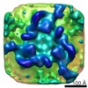
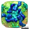
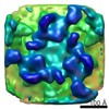
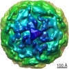
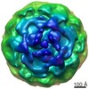
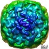
| タイトル | Cryo-electron tomogram of the Chlamydomonas reinhardtii Golgi apparatus | |||||||||
 マップデータ マップデータ | Cryo-electron tomogram of Chlamydomonas reinhardtii Golgi apparatus | |||||||||
 試料 試料 |
| |||||||||
| 生物種 |   Chlamydomonas reinhardtii (クラミドモナス) Chlamydomonas reinhardtii (クラミドモナス) | |||||||||
| 手法 | 電子線トモグラフィー法 / クライオ電子顕微鏡法 | |||||||||
 データ登録者 データ登録者 | Bykov YS / Schaffer M / Dodonova SO / Albert S / Plitzko JM / Baumeister W / Engel BD / Briggs JAG | |||||||||
 引用 引用 | ジャーナル: Elife / 年: 2017 タイトル: The structure of the COPI coat determined within the cell. 著者: Yury S Bykov / Miroslava Schaffer / Svetlana O Dodonova / Sahradha Albert / Jürgen M Plitzko / Wolfgang Baumeister / Benjamin D Engel / John Ag Briggs /   要旨: COPI-coated vesicles mediate trafficking within the Golgi apparatus and from the Golgi to the endoplasmic reticulum. The structures of membrane protein coats, including COPI, have been extensively ...COPI-coated vesicles mediate trafficking within the Golgi apparatus and from the Golgi to the endoplasmic reticulum. The structures of membrane protein coats, including COPI, have been extensively studied with reconstitution systems using purified components. Previously we have determined a complete structural model of the reconstituted COPI coat (Dodonova et al., 2017). Here, we applied cryo-focused ion beam milling, cryo-electron tomography and subtomogram averaging to determine the native structure of the COPI coat within vitrified cells. The native algal structure resembles the mammalian structure, but additionally reveals cargo bound beneath β'-COP. We find that all coat components disassemble simultaneously and relatively rapidly after budding. Structural analysis , maintaining Golgi topology, shows that vesicles change their size, membrane thickness, and cargo content as they progress from to , but the structure of the coat machinery remains constant. | |||||||||
| 履歴 |
|
- 構造の表示
構造の表示
| ムービー |
 ムービービューア ムービービューア |
|---|---|
| 構造ビューア | EMマップ: SurfViewMolmilJmol/JSmol |
| 添付画像 |

- ダウンロードとリンク
-EMDBアーカイブ
| マップデータ | emd_3977.map.gz | 599.9 MB | EMDBマップデータ形式 | |
|---|---|---|---|---|
| ヘッダ (付随情報) | emd-3977-v30.xmlemd-3977.xml | 10.9 KB 10.9 KB | 表示 表示 | EMDBヘッダ |
| 画像 | 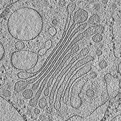 emd_3977.png emd_3977.png | 151 KB | ||
| アーカイブディレクトリ |  http://ftp.pdbj.org/pub/emdb/structures/EMD-3977ftp://ftp.pdbj.org/pub/emdb/structures/EMD-3977 http://ftp.pdbj.org/pub/emdb/structures/EMD-3977ftp://ftp.pdbj.org/pub/emdb/structures/EMD-3977 | HTTPS FTP |
-検証レポート
| 文書・要旨 | emd_3977_validation.pdf.gz | 174 KB | 表示 | EMDB検証レポート |
|---|---|---|---|---|
| 文書・詳細版 | emd_3977_full_validation.pdf.gz | 173.2 KB | 表示 | |
| XML形式データ | emd_3977_validation.xml.gz | 3.9 KB | 表示 | |
| アーカイブディレクトリ | https://ftp.pdbj.org/pub/emdb/validation_reports/EMD-3977ftp://ftp.pdbj.org/pub/emdb/validation_reports/EMD-3977 | HTTPS FTP |
-関連構造データ
-リンク
| EMDBのページ | EMDB (EBI/PDBe) / EMDataResource |
|---|
-マップ
| ファイル | ダウンロード / ファイル: emd_3977.map.gz / 形式: CCP4 / 大きさ: 762.2 MB / タイプ: IMAGE STORED AS SIGNED INTEGER (2 BYTES) | ||||||||||||||||||||||||||||||||||||||||||||||||||||||||||||
|---|---|---|---|---|---|---|---|---|---|---|---|---|---|---|---|---|---|---|---|---|---|---|---|---|---|---|---|---|---|---|---|---|---|---|---|---|---|---|---|---|---|---|---|---|---|---|---|---|---|---|---|---|---|---|---|---|---|---|---|---|---|
| 注釈 | Cryo-electron tomogram of Chlamydomonas reinhardtii Golgi apparatus | ||||||||||||||||||||||||||||||||||||||||||||||||||||||||||||
| 投影像・断面図 | 画像のコントロール
画像は Spider により作成 これらの図は立方格子座標系で作成されたものです | ||||||||||||||||||||||||||||||||||||||||||||||||||||||||||||
| ボクセルのサイズ | X=Y=Z: 13.68 Å | ||||||||||||||||||||||||||||||||||||||||||||||||||||||||||||
| 密度 |
| ||||||||||||||||||||||||||||||||||||||||||||||||||||||||||||
| 対称性 | 空間群: 1 | ||||||||||||||||||||||||||||||||||||||||||||||||||||||||||||
| 詳細 | EMDB XML:
CCP4マップ ヘッダ情報:
| ||||||||||||||||||||||||||||||||||||||||||||||||||||||||||||
 Z (Sec.)
 Y (Row.)
 X (Col.)


-添付データ
- 試料の構成要素
-全体 : Whole Chlamydomonas cells
| 全体 | 名称: Whole Chlamydomonas cells |
|---|---|
| 要素 |
|
-超分子 #1: Whole Chlamydomonas cells
| 超分子 | 名称: Whole Chlamydomonas cells / タイプ: organelle_or_cellular_component / ID: 1 / 親要素: 0 詳細: Grown suspended in TAP media, with normal atmosphere aeration and constant light |
|---|---|
| 由来(天然) | 生物種: Chlamydomonas reinhardtii (クラミドモナス) 株: mat3-4 / Organelle: Golgi |
-実験情報
-構造解析
| 手法 | クライオ電子顕微鏡法 |
|---|---|
 解析 解析 | 電子線トモグラフィー法 |
| 試料の集合状態 | cell |
-試料調製
| 緩衝液 | pH: 7 / 詳細: TAP media |
|---|---|
| グリッド | モデル: Quantifoil R2/1 / 材質: COPPER / メッシュ: 200 / 前処理 - タイプ: GLOW DISCHARGE / 前処理 - 雰囲気: AIR |
| 凍結 | 凍結剤: ETHANE-PROPANE / チャンバー内湿度: 90 % / 装置: FEI VITROBOT MARK IV 詳細: Blotted from the back side for 10 seconds with 10 blot force before plunging. |
| 詳細 | The cells were frozen onto grids, then thinned using cryo-focused ion beam milling. |
| 切片作成 | 集束イオンビーム - 装置: OTHER / 集束イオンビーム - イオン: OTHER / 集束イオンビーム - 電圧: 30 kV / 集束イオンビーム - 電流: 0.03 nA / 集束イオンビーム - Dose rate: 2 / 集束イオンビーム - 時間: 2400 sec. / 集束イオンビーム - 温度: 80 K / 集束イオンビーム - Initial thickness: 5000 / 集束イオンビーム - 最終 厚さ: 240 集束イオンビーム - 詳細: Starting curent: 0.5 nA, Final current: 0.03 nA. The value given for _emd_sectioning_focused_ion_beam.instrument is FEI Scios DB-FIB. This is not in a list of ...集束イオンビーム - 詳細: Starting curent: 0.5 nA, Final current: 0.03 nA. The value given for _emd_sectioning_focused_ion_beam.instrument is FEI Scios DB-FIB. This is not in a list of allowed values set(['DB235', 'OTHER']) so OTHER is written into the XML file. |
- 電子顕微鏡法
| 顕微鏡 | FEI TITAN KRIOS |
|---|---|
| 特殊光学系 | エネルギーフィルター - 名称: GIF Quantum LS エネルギーフィルター - エネルギー下限: 0 eV エネルギーフィルター - エネルギー上限: 20 eV |
| 撮影 | フィルム・検出器のモデル: GATAN K2 SUMMIT (4k x 4k) 検出モード: COUNTING / デジタル化 - 画像ごとのフレーム数: 1-12 / 平均露光時間: 1.5 sec. / 平均電子線量: 1.5 e/Å2 |
| 電子線 | 加速電圧: 300 kV / 電子線源:  FIELD EMISSION GUN FIELD EMISSION GUN |
| 電子光学系 | C2レンズ絞り径: 70.0 µm / 照射モード: FLOOD BEAM / 撮影モード: BRIGHT FIELD / Cs: 2.7 mm / 最大 デフォーカス(公称値): 6.0 µm / 最小 デフォーカス(公称値): 4.0 µm / 倍率(公称値): 42000 |
| 試料ステージ | 試料ホルダーモデル: FEI TITAN KRIOS AUTOGRID HOLDER ホルダー冷却材: NITROGEN |
| 実験機器 |  モデル: Titan Krios / 画像提供: FEI Company |
-画像解析
| 最終 再構成 | アルゴリズム: BACK PROJECTION / ソフトウェア - 名称: IMOD 詳細: Each of the images in the tilt series was low-pass filtered according to the electron-dose acquired by the sample (Grant and Grigorieff, 2015). This is a bin4 (twice binned) tomogram. Three ...詳細: Each of the images in the tilt series was low-pass filtered according to the electron-dose acquired by the sample (Grant and Grigorieff, 2015). This is a bin4 (twice binned) tomogram. Three last tilts (56, 58, and 60 degrees) were excluded from the reconstruction. 使用した粒子像数: 55 |
|---|---|
| CTF補正 | ソフトウェア: (名称: CTFFIND, CTFPHASEFLIP) |
