- EMDB-39612: canine immunoproteasome 20S subunit in complex with compound 1 -
+
Open data
ID or keywords:
Loading...
-
Basic information
Entry
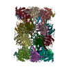
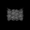
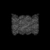
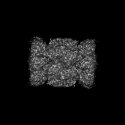
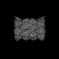
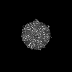
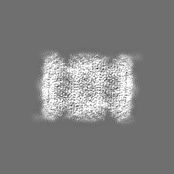
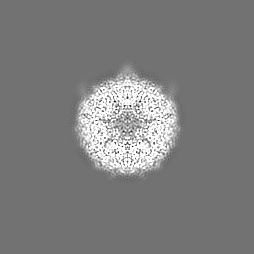
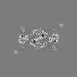
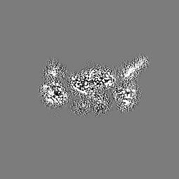
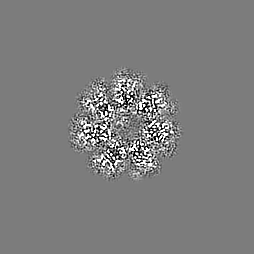
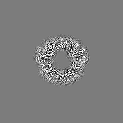
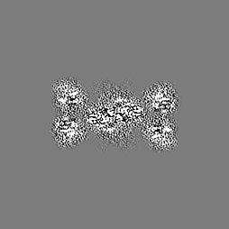
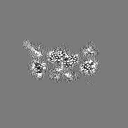
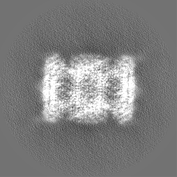
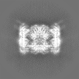
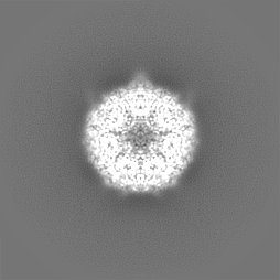
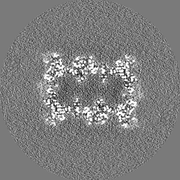
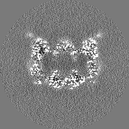
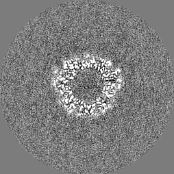
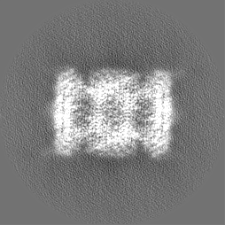
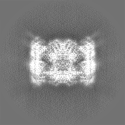
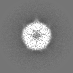
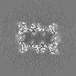
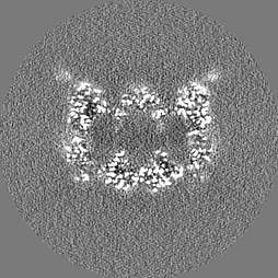
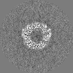
Database: EMDB / ID: EMD-39612
Title
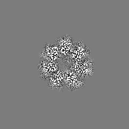
canine immunoproteasome 20S subunit in complex with compound 1
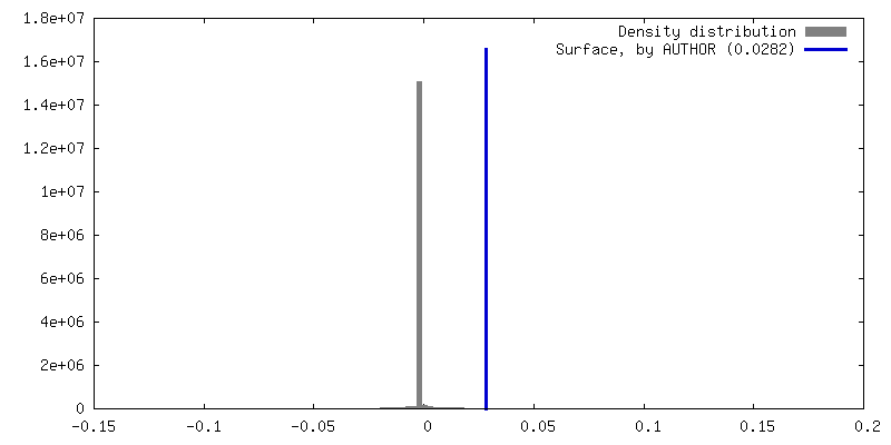
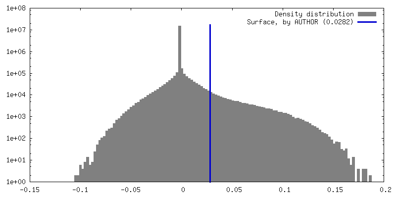
Map data
Sample
Complex: 20S proteasome
Protein or peptide: x 14 types
Ligand: x 2 types
Keywords
inhibitor / complex / proteasome / hydrolase
Function / homology
Function and homology information
Regulation of ornithine decarboxylase (ODC) / Proteasome assembly / Cross-presentation of soluble exogenous antigens (endosomes) / Autodegradation of Cdh1 by Cdh1:APC/C / APC/C:Cdc20 mediated degradation of Securin / Ubiquitin-Mediated Degradation of Phosphorylated Cdc25A / Ubiquitin-dependent degradation of Cyclin D / AUF1 (hnRNP D0) binds and destabilizes mRNA / Cdc20:Phospho-APC/C mediated degradation of Cyclin A / SCF(Skp2)-mediated degradation of p27/p21 ...Regulation of ornithine decarboxylase (ODC) / Proteasome assembly / Cross-presentation of soluble exogenous antigens (endosomes) / Autodegradation of Cdh1 by Cdh1:APC/C / APC/C:Cdc20 mediated degradation of Securin / Ubiquitin-Mediated Degradation of Phosphorylated Cdc25A / Ubiquitin-dependent degradation of Cyclin D / AUF1 (hnRNP D0) binds and destabilizes mRNA / Cdc20:Phospho-APC/C mediated degradation of Cyclin A / SCF(Skp2)-mediated degradation of p27/p21 / Assembly of the pre-replicative complex / CDK-mediated phosphorylation and removal of Cdc6 / Autodegradation of the E3 ubiquitin ligase COP1 / G2/M Checkpoints / Degradation of AXIN / APC/C:Cdh1 mediated degradation of Cdc20 and other APC/C:Cdh1 targeted proteins in late mitosis/early G1 / Asymmetric localization of PCP proteins / Regulation of RUNX3 expression and activity / Regulation of RAS by GAPs / Regulation of PTEN stability and activity / Regulation of RUNX2 expression and activity / Degradation of GLI1 by the proteasome / UCH proteinases / FBXL7 down-regulates AURKA during mitotic entry and in early mitosis / Degradation of DVL / Orc1 removal from chromatin / GSK3B and BTRC:CUL1-mediated-degradation of NFE2L2 / Dectin-1 mediated noncanonical NF-kB signaling / NIK-->noncanonical NF-kB signaling / Hedgehog ligand biogenesis / TNFR2 non-canonical NF-kB pathway / Oxygen-dependent proline hydroxylation of Hypoxia-inducible Factor Alpha / Hedgehog 'on' state / Degradation of beta-catenin by the destruction complex / Activation of NF-kappaB in B cells / The role of GTSE1 in G2/M progression after G2 checkpoint / FCERI mediated NF-kB activation / CLEC7A (Dectin-1) signaling / Interleukin-1 signaling / RUNX1 regulates transcription of genes involved in differentiation of HSCs / Downstream TCR signaling / Separation of Sister Chromatids / MAPK6/MAPK4 signaling / GLI3 is processed to GLI3R by the proteasome / ABC-family proteins mediated transport / Neddylation / Ub-specific processing proteases / KEAP1-NFE2L2 pathway / Antigen processing: Ubiquitination & Proteasome degradation / proteasome core complex / myofibril / immune system process / NF-kappaB binding / proteasome endopeptidase complex / proteasome core complex, beta-subunit complex / threonine-type endopeptidase activity / proteasome core complex, alpha-subunit complex / proteasomal protein catabolic process / skeletal muscle tissue development / Neutrophil degranulation / proteasome complex / proteolysis involved in protein catabolic process / sarcomere / lipopolysaccharide binding / negative regulation of inflammatory response to antigenic stimulus / P-body / protein catabolic process / : / response to virus / nuclear matrix / positive regulation of NF-kappaB transcription factor activity / peptidase activity / ubiquitin-dependent protein catabolic process / endopeptidase activity / response to oxidative stress / proteasome-mediated ubiquitin-dependent protein catabolic process / ciliary basal body / nuclear body / cilium / ribosome / centrosome / ubiquitin protein ligase binding / mitochondrion / proteolysis / RNA binding / nucleoplasm / identical protein binding / cytosol / cytoplasm Similarity search - Function
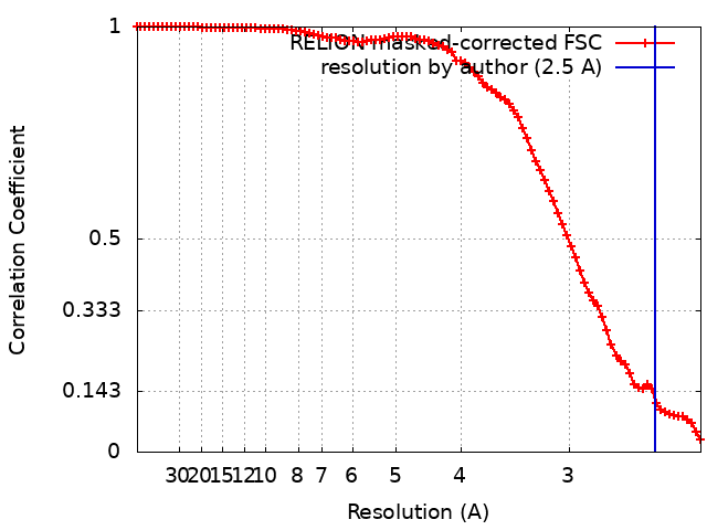
Journal: Bioorg Med Chem / Year: 2024 Title: Optimization of α-amido boronic acids via cryo-electron microscopy analysis: Discovery of a novel highly selective immunoproteasome subunit LMP7 (β5i)/LMP2 (β1i) dual inhibitor. Authors: Yuuki Arai / Hiroaki Shitama / Masahito Yamagishi / Satoshi Ono / Akiko Kashima / Masahiro Hiraizumi / Naoki Tsuda / Koushirou Katayama / Kouji Tanaka / Yuzo Koda / Sayuka Kato / Kei Sakata ...Authors: Yuuki Arai / Hiroaki Shitama / Masahito Yamagishi / Satoshi Ono / Akiko Kashima / Masahiro Hiraizumi / Naoki Tsuda / Koushirou Katayama / Kouji Tanaka / Yuzo Koda / Sayuka Kato / Kei Sakata / Osamu Nureki / Hiroshi Miyazaki / Abstract: The immunoproteasome subunit LMP7 (β5i)/LMP2 (β1i) dual blockade has been reported to suppress B cell differentiation and activation, suggesting that the dual inhibition of LMP7/LMP2 is a promising ...The immunoproteasome subunit LMP7 (β5i)/LMP2 (β1i) dual blockade has been reported to suppress B cell differentiation and activation, suggesting that the dual inhibition of LMP7/LMP2 is a promising approach for treating autoimmune diseases. In contrast, the inhibition of the constitutive proteasome subunit β5c correlates with cytotoxicity against non-immune cells. Therefore, LMP7/LMP2 dual inhibitors with high selectivity over β5c may be desirable for treating autoimmune diseases. In this study, we present the optimization and discovery of α-amido boronic acids using cryo-electron microscopy (cryo-EM). The exploitation of structural differences between the proteasome subunits led to the identification of a highly selective LMP7/LMP2 dual inhibitor 19. Molecular dynamics simulation based on cryo-EM structures of the proteasome subunits complexed with 19 explained the inhibitory activity profile. In mice immunized with 4-hydroxy-3-nitrophenylacetyl conjugated to ovalbumin, results indicate that 19 is orally bioavailable and shows promise as potential treatment for autoimmune diseases.
In the structure databanks used in Yorodumi, some data are registered as the other names, "COVID-19 virus" and "2019-nCoV". Here are the details of the virus and the list of structure data.
Jan 31, 2019. EMDB accession codes are about to change! (news from PDBe EMDB page)
EMDB accession codes are about to change! (news from PDBe EMDB page)
The allocation of 4 digits for EMDB accession codes will soon come to an end. Whilst these codes will remain in use, new EMDB accession codes will include an additional digit and will expand incrementally as the available range of codes is exhausted. The current 4-digit format prefixed with “EMD-” (i.e. EMD-XXXX) will advance to a 5-digit format (i.e. EMD-XXXXX), and so on. It is currently estimated that the 4-digit codes will be depleted around Spring 2019, at which point the 5-digit format will come into force.
The EM Navigator/Yorodumi systems omit the EMD- prefix.
Related info.:Q: What is EMD? / ID/Accession-code notation in Yorodumi/EM Navigator
Yorodumi is a browser for structure data from EMDB, PDB, SASBDB, etc.
This page is also the successor to EM Navigator detail page, and also detail information page/front-end page for Omokage search.
The word "yorodu" (or yorozu) is an old Japanese word meaning "ten thousand". "mi" (miru) is to see.
Related info.:EMDB / PDB / SASBDB / Comparison of 3 databanks / Yorodumi Search / Aug 31, 2016. New EM Navigator & Yorodumi / Yorodumi Papers / Jmol/JSmol / Function and homology information / Changes in new EM Navigator and Yorodumi
 Movie
Movie Controller


 Open data
 Basic information
 Map data
 Sample
 Keywords
 Function and homology information
 Authors
 Citation
 Structure visualization
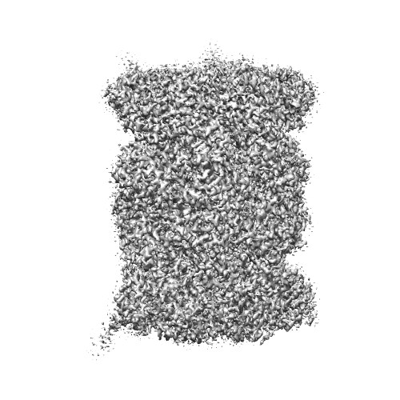
 Downloads & links
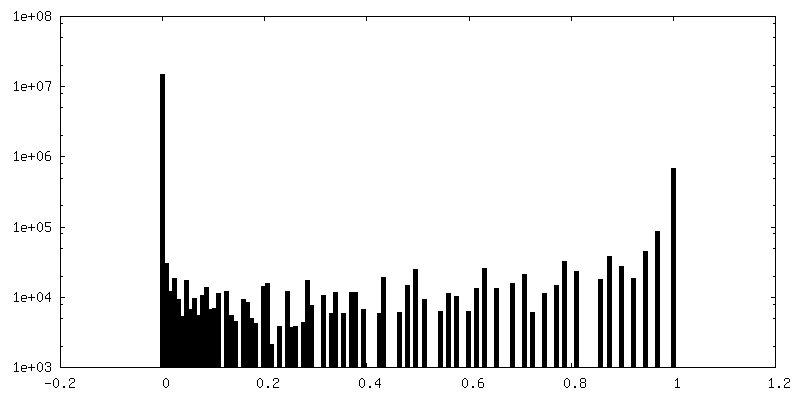
 emd_39612.png
 http://ftp.pdbj.org/pub/emdb/structures/EMD-39612


 Z (Sec.)
 Y (Row.)
 X (Col.)


 Sample components
 Processing
 Electron microscopy
 FIELD EMISSION GUN