 Movie
Movie Controller

[English] 日本語
 Yorodumi
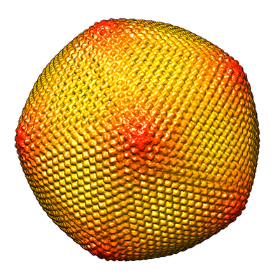
- EMDB-3868: Cryo-EM single particle reconstruction of Melbournevirus particle. -
+ Open data

- Basic information
| Entry | Database: EMDB / ID: EMD-3868 | |||||||||
|---|---|---|---|---|---|---|---|---|---|---|
| Title | Cryo-EM single particle reconstruction of Melbournevirus particle. | |||||||||
 Map data Map data | Melbournevirus capsid(applied inner mask) | |||||||||
 Sample Sample | Melbournevirus particle != Melbournevirus Melbournevirus particle
| |||||||||
| Biological species |  Melbournevirus Melbournevirus | |||||||||
| Method | single particle reconstruction / cryo EM / Resolution: 26.3 Å | |||||||||
 Authors Authors | Okamoto K / Miyazaki N / Reddy KNH / Hantke FM / Maia RNCF / Larsson SDD / Abergel C / Claverie JM / Hajdu J / Murata K / Svenda M | |||||||||
 Citation Citation | Journal: Virology / Year: 2018 Title: Cryo-EM structure of a Marseilleviridae virus particle reveals a large internal microassembly. Authors: Kenta Okamoto / Naoyuki Miyazaki / Hemanth K N Reddy / Max F Hantke / Filipe R N C Maia / Daniel S D Larsson / Chantal Abergel / Jean-Michel Claverie / Janos Hajdu / Kazuyoshi Murata / Martin Svenda /     Abstract: Nucleocytoplasmic large DNA viruses (NCLDVs) blur the line between viruses and cells. Melbournevirus (MelV, family Marseilleviridae) belongs to a new family of NCLDVs. Here we present an electron ...Nucleocytoplasmic large DNA viruses (NCLDVs) blur the line between viruses and cells. Melbournevirus (MelV, family Marseilleviridae) belongs to a new family of NCLDVs. Here we present an electron cryo-microscopy structure of the MelV particle, with the large triangulation number T = 309 constructed by 3080 pseudo-hexagonal capsomers. The most distinct feature of the particle is a large and dense body (LDB) consistently found inside all particles. Electron cryo-tomography of 147 particles shows that the LDB is preferentially located in proximity to the probable lipid bilayer. The LDB is 30 nm in size and its density matches that of a genome/protein complex. The observed LDB reinforces the structural complexity of MelV, setting it apart from other NCLDVs. | |||||||||
| History |
|
- Structure visualization
| Movie |
 Movie viewer Movie viewer |
|---|---|
| Structure viewer | EM map: SurfViewMolmilJmol/JSmol |
| Supplemental images |
- Downloads & links
-EMDB archive
| Map data | emd_3868.map.gz | 644.2 MB | EMDB map data format | |
|---|---|---|---|---|
| Header (meta data) | emd-3868-v30.xmlemd-3868.xml | 9.9 KB 9.9 KB | Display Display | EMDB header |
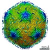
| Images |  emd_3868.png emd_3868.png | 236.8 KB | ||
| Archive directory |  http://ftp.pdbj.org/pub/emdb/structures/EMD-3868ftp://ftp.pdbj.org/pub/emdb/structures/EMD-3868 http://ftp.pdbj.org/pub/emdb/structures/EMD-3868ftp://ftp.pdbj.org/pub/emdb/structures/EMD-3868 | HTTPS FTP |
-Related structure data
| Similar structure data |
|---|


-Links
| EMDB pages | EMDB (EBI/PDBe) / EMDataResource |
|---|
-Map
| File | Download / File: emd_3868.map.gz / Format: CCP4 / Size: 2.3 MB / Type: IMAGE STORED AS FLOATING POINT NUMBER (4 BYTES) | ||||||||||||||||||||||||||||||||||||||||||||||||||||||||||||
|---|---|---|---|---|---|---|---|---|---|---|---|---|---|---|---|---|---|---|---|---|---|---|---|---|---|---|---|---|---|---|---|---|---|---|---|---|---|---|---|---|---|---|---|---|---|---|---|---|---|---|---|---|---|---|---|---|---|---|---|---|---|
| Annotation | Melbournevirus capsid(applied inner mask) | ||||||||||||||||||||||||||||||||||||||||||||||||||||||||||||
| Projections & slices | Image control
Images are generated by Spider. | ||||||||||||||||||||||||||||||||||||||||||||||||||||||||||||
| Voxel size | X=Y=Z: 3.31 Å | ||||||||||||||||||||||||||||||||||||||||||||||||||||||||||||
| Density |
| ||||||||||||||||||||||||||||||||||||||||||||||||||||||||||||
| Symmetry | Space group: 1 | ||||||||||||||||||||||||||||||||||||||||||||||||||||||||||||
| Details | EMDB XML:
CCP4 map header:
| ||||||||||||||||||||||||||||||||||||||||||||||||||||||||||||
 Z (Sec.)
 Y (Row.)
 X (Col.)


-Supplemental data
- Sample components
-Entire : Melbournevirus particle
| Entire | Name: Melbournevirus particle |
|---|---|
| Components |
|
-Supramolecule #1: Melbournevirus
| Supramolecule | Name: Melbournevirus / type: virus / ID: 1 / Parent: 0 / NCBI-ID: 1560514 / Sci species name: Melbournevirus / Virus type: VIRION / Virus isolate: SPECIES / Virus enveloped: No / Virus empty: No |
|---|---|
| Host (natural) | Organism:  Acanthamoeba (eukaryote) Acanthamoeba (eukaryote) |
| Virus shell | Shell ID: 1 / Name: Capsid / Diameter: 2320.0 Å / T number (triangulation number): 309 |
-Experimental details
-Structure determination
| Method | cryo EM |
|---|---|
 Processing Processing | single particle reconstruction |
| Aggregation state | particle |
-Sample preparation
| Buffer | pH: 7.4 |
|---|---|
| Grid | Model: R1.2/1.3 Quantifoil Micro Tools GmbH / Material: COPPER / Support film - Material: CARBON / Support film - topology: HOLEY / Pretreatment - Type: PLASMA CLEANING |
| Vitrification | Cryogen name: ETHANE |
- Electron microscopy
| Microscope | JEOL 2200FS |
|---|---|
| Image recording | Film or detector model: TVIPS TEMCAM-F415 (4k x 4k) / Average electron dose: 20.0 e/Å2 |
| Electron beam | Acceleration voltage: 200 kV / Electron source:  FIELD EMISSION GUN FIELD EMISSION GUN |
| Electron optics | Illumination mode: FLOOD BEAM / Imaging mode: BRIGHT FIELD / Cs: 4.2 mm / Nominal defocus max: 5.0 µm / Nominal defocus min: 1.0 µm |
| Sample stage | Specimen holder model: JEOL / Cooling holder cryogen: NITROGEN |
