Movie
Movie Controller


+ Open data

- Basic information
| Entry |  | |||||||||
|---|---|---|---|---|---|---|---|---|---|---|
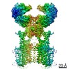
| Title | The focused map of ACE2-SIT1 bound with tiagabine | |||||||||
 Map data Map data | ||||||||||
 Sample Sample |
| |||||||||
 Keywords Keywords | Transporter / inhibitors / TRANSPORT PROTEIN / TRANSPORT PROTEIN-HYDROLASE complex | |||||||||
| Biological species |  Homo sapiens (human) Homo sapiens (human) | |||||||||
| Method | single particle reconstruction / cryo EM / Resolution: 3.8 Å | |||||||||
 Authors Authors | Yan R / Dai L / Hu Z / Zhang T | |||||||||
| Funding support |  China, 1 items China, 1 items
| |||||||||
 Citation Citation | Journal: J Biol Chem / Year: 2024 Title: Cryo-EM structure of ACE2-SIT1 in complex with tiagabine. Authors: Angelika Bröer / Ziwei Hu / Jędrzej Kukułowicz / Aditya Yadav / Ting Zhang / Lu Dai / Marek Bajda / Renhong Yan / Stefan Bröer /   Abstract: The pharmacology of amino acid transporters in the SLC6 family is poorly developed compared to that of the neurotransmitter transporters. To identify new inhibitors of the proline transporter SIT1 ...The pharmacology of amino acid transporters in the SLC6 family is poorly developed compared to that of the neurotransmitter transporters. To identify new inhibitors of the proline transporter SIT1 (SLC6A20), its expression in Xenopus laevis oocytes was optimized. Trafficking of SIT1 was augmented by co-expression of angiotensin-converting enzyme 2 (ACE2) in oocytes but there was no strict requirement for co-expression of ACE2. A pharmacophore-guided screen identified tiagabine as a potent non-competitive inhibitor of SIT1. To understand its binding mode, we determined the cryo-electron microscopy (cryo-EM) structure of ACE2-SIT1 bound with tiagabine. The inhibitor binds close to the orthosteric proline binding site, but due to its size extends into the cytosolic vestibule. This causes the transporter to adopt an inward-open conformation, in which the intracellular gate is blocked. This study provides the first structural insight into inhibition of SIT1 and generates tools for a better understanding of the ACE2-SIT1 complex. These findings may have significance for SARS-CoV-2 binding to its receptor ACE2 in human lung alveolar cells where SIT1 and ACE2 are functionally expressed. | |||||||||
| History |
|
- Structure visualization
| Supplemental images |
|---|

- Downloads & links
-EMDB archive
| Map data | emd_37868.map.gz | 59.7 MB |  EMDB map data format EMDB map data format | |
|---|---|---|---|---|
| Header (meta data) | emd-37868-v30.xmlemd-37868.xml | 12 KB 12 KB | Display Display | EMDB header |
| Images |  emd_37868.png emd_37868.png | 17.4 KB | ||
| Filedesc metadata | emd-37868.cif.gz | 3.7 KB | ||
| Others | emd_37868_half_map_1.map.gzemd_37868_half_map_2.map.gz | 58.5 MB 58.5 MB | ||
| Archive directory |  http://ftp.pdbj.org/pub/emdb/structures/EMD-37868ftp://ftp.pdbj.org/pub/emdb/structures/EMD-37868 http://ftp.pdbj.org/pub/emdb/structures/EMD-37868ftp://ftp.pdbj.org/pub/emdb/structures/EMD-37868 | HTTPS FTP |
-Validation report
| Summary document | emd_37868_validation.pdf.gz | 719.3 KB | Display | EMDB validaton report |
|---|---|---|---|---|
| Full document | emd_37868_full_validation.pdf.gz | 718.9 KB | Display | |
| Data in XML | emd_37868_validation.xml.gz | 12 KB | Display | |
| Data in CIF | emd_37868_validation.cif.gz | 14.1 KB | Display | |
| Arichive directory | https://ftp.pdbj.org/pub/emdb/validation_reports/EMD-37868ftp://ftp.pdbj.org/pub/emdb/validation_reports/EMD-37868 | HTTPS FTP |
-Related structure data

-Links
| EMDB pages | EMDB (EBI/PDBe) / EMDataResource |
|---|
-Map
| File | Download / File: emd_37868.map.gz / Format: CCP4 / Size: 64 MB / Type: IMAGE STORED AS FLOATING POINT NUMBER (4 BYTES) | ||||||||||||||||||||||||||||||||||||
|---|---|---|---|---|---|---|---|---|---|---|---|---|---|---|---|---|---|---|---|---|---|---|---|---|---|---|---|---|---|---|---|---|---|---|---|---|---|
| Projections & slices | Image control
Images are generated by Spider. | ||||||||||||||||||||||||||||||||||||
| Voxel size | X=Y=Z: 1.095 Å | ||||||||||||||||||||||||||||||||||||
| Density |
| ||||||||||||||||||||||||||||||||||||
| Symmetry | Space group: 1 | ||||||||||||||||||||||||||||||||||||
| Details | EMDB XML:
|
 Z (Sec.)
 Y (Row.)
 X (Col.)


-Supplemental data
-Half map: #1
| File | emd_37868_half_map_1.map | ||||||||||||
|---|---|---|---|---|---|---|---|---|---|---|---|---|---|
| Projections & Slices |
| ||||||||||||
| Density Histograms |


-Half map: #2
| File | emd_37868_half_map_2.map | ||||||||||||
|---|---|---|---|---|---|---|---|---|---|---|---|---|---|
| Projections & Slices |
| ||||||||||||
| Density Histograms |


- Sample components
-Entire : ACE2-SIT1 compelx with Tiagabine
| Entire | Name: ACE2-SIT1 compelx with Tiagabine |
|---|---|
| Components |
|
-Supramolecule #1: ACE2-SIT1 compelx with Tiagabine
| Supramolecule | Name: ACE2-SIT1 compelx with Tiagabine / type: complex / ID: 1 / Parent: 0 / Macromolecule list: #1-#2 |
|---|---|
| Source (natural) | Organism: Homo sapiens (human) |
-Experimental details
-Structure determination
| Method | cryo EM |
|---|---|
 Processing Processing | single particle reconstruction |
| Aggregation state | particle |
-Sample preparation
| Buffer | pH: 7.5 |
|---|---|
| Vitrification | Cryogen name: NITROGEN |
- Electron microscopy
| Microscope | FEI TITAN KRIOS |
|---|---|
| Image recording | Film or detector model: GATAN K3 BIOQUANTUM (6k x 4k) / Average electron dose: 1.059 e/Å2 |
| Electron beam | Acceleration voltage: 300 kV / Electron source:  FIELD EMISSION GUN FIELD EMISSION GUN |
| Electron optics | Illumination mode: FLOOD BEAM / Imaging mode: BRIGHT FIELD / Nominal defocus max: 0.8 µm / Nominal defocus min: 0.7000000000000001 µm |
| Experimental equipment |  Model: Titan Krios / Image courtesy: FEI Company |
-Image processing
| Startup model | Type of model: EMDB MAP EMDB ID: |
|---|---|
| Final reconstruction | Resolution.type: BY AUTHOR / Resolution: 3.8 Å / Resolution method: FSC 0.143 CUT-OFF / Number images used: 193320 |
| Initial angle assignment | Type: COMMON LINE |
| Final angle assignment | Type: COMMON LINE |