 Movie
Movie Controller

[English] 日本語
 Yorodumi
- EMDB-3762: Sub-tomogram average of the cytosolic yeast ribosome associated w... -
+ Open data

- Basic information
| Entry | Database: EMDB / ID: EMD-3762 | |||||||||
|---|---|---|---|---|---|---|---|---|---|---|
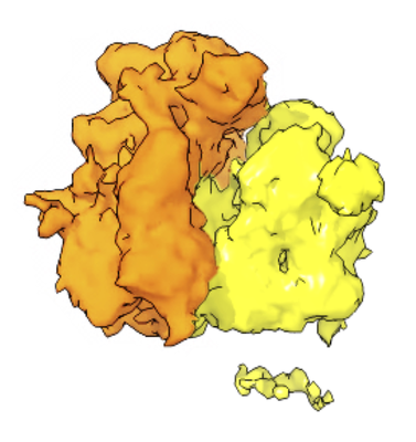
| Title | Sub-tomogram average of the cytosolic yeast ribosome associated with mitochondria | |||||||||
 Map data Map data | Sub-tomogram average of the cytosolic yeast ribosome bound to mitochondria | |||||||||
 Sample Sample |
| |||||||||
| Biological species |  | |||||||||
| Method | subtomogram averaging / cryo EM / Resolution: 38.0 Å | |||||||||
 Authors Authors | Gold VAM / Chroscicki P / Bragoszewski P / Chacinska A | |||||||||
 Citation Citation | Journal: EMBO Rep / Year: 2017 Title: Visualization of cytosolic ribosomes on the surface of mitochondria by electron cryo-tomography. Authors: Vicki Am Gold / Piotr Chroscicki / Piotr Bragoszewski / Agnieszka Chacinska /    Abstract: We employed electron cryo-tomography to visualize cytosolic ribosomes on the surface of mitochondria. Translation-arrested ribosomes reveal the clustered organization of the TOM complex, ...We employed electron cryo-tomography to visualize cytosolic ribosomes on the surface of mitochondria. Translation-arrested ribosomes reveal the clustered organization of the TOM complex, corroborating earlier reports of localized translation. Ribosomes are shown to interact specifically with the TOM complex, and nascent chain binding is crucial for ribosome recruitment and stabilization. Ribosomes are bound to the membrane in discrete clusters, often in the vicinity of the crista junctions. This interaction highlights how protein synthesis may be coupled with transport. Our work provides unique insights into the spatial organization of cytosolic ribosomes on mitochondria. | |||||||||
| History |
|
- Structure visualization
| Movie |
 Movie viewer Movie viewer |
|---|---|
| Structure viewer | EM map: SurfViewMolmilJmol/JSmol |
| Supplemental images |
- Downloads & links
-EMDB archive
| Map data | emd_3762.map.gz | 1.4 MB | EMDB map data format | |
|---|---|---|---|---|
| Header (meta data) | emd-3762-v30.xmlemd-3762.xml | 8.1 KB 8.1 KB | Display Display | EMDB header |
| Images |  emd_3762.png emd_3762.png | 113.9 KB | ||
| Archive directory |  http://ftp.pdbj.org/pub/emdb/structures/EMD-3762ftp://ftp.pdbj.org/pub/emdb/structures/EMD-3762 http://ftp.pdbj.org/pub/emdb/structures/EMD-3762ftp://ftp.pdbj.org/pub/emdb/structures/EMD-3762 | HTTPS FTP |
-Related structure data
-Links
| EMDB pages | EMDB (EBI/PDBe) / EMDataResource |
|---|---|
| Related items in Molecule of the Month |

-Map
| File | Download / File: emd_3762.map.gz / Format: CCP4 / Size: 2 MB / Type: IMAGE STORED AS FLOATING POINT NUMBER (4 BYTES) | ||||||||||||||||||||||||||||||||||||||||||||||||||||||||||||
|---|---|---|---|---|---|---|---|---|---|---|---|---|---|---|---|---|---|---|---|---|---|---|---|---|---|---|---|---|---|---|---|---|---|---|---|---|---|---|---|---|---|---|---|---|---|---|---|---|---|---|---|---|---|---|---|---|---|---|---|---|---|
| Annotation | Sub-tomogram average of the cytosolic yeast ribosome bound to mitochondria | ||||||||||||||||||||||||||||||||||||||||||||||||||||||||||||
| Projections & slices | Image control
Images are generated by Spider. | ||||||||||||||||||||||||||||||||||||||||||||||||||||||||||||
| Voxel size | X=Y=Z: 6.7 Å | ||||||||||||||||||||||||||||||||||||||||||||||||||||||||||||
| Density |
| ||||||||||||||||||||||||||||||||||||||||||||||||||||||||||||
| Symmetry | Space group: 1 | ||||||||||||||||||||||||||||||||||||||||||||||||||||||||||||
| Details | EMDB XML:
CCP4 map header:
| ||||||||||||||||||||||||||||||||||||||||||||||||||||||||||||
 Z (Sec.)
 Y (Row.)
 X (Col.)


-Supplemental data
- Sample components
-Entire : S. cerevisiae cytosolic ribosome associated with mitochondria
| Entire | Name: S. cerevisiae cytosolic ribosome associated with mitochondria |
|---|---|
| Components |
|
-Supramolecule #1: S. cerevisiae cytosolic ribosome associated with mitochondria
| Supramolecule | Name: S. cerevisiae cytosolic ribosome associated with mitochondria type: complex / ID: 1 / Parent: 0 |
|---|---|
| Source (natural) | Organism: |
-Experimental details
-Structure determination
| Method | cryo EM |
|---|---|
 Processing Processing | subtomogram averaging |
| Aggregation state | particle |
-Sample preparation
| Buffer | pH: 7.2 |
|---|---|
| Vitrification | Cryogen name: ETHANE Details: blotted for ~4 s on one side in a humidified atmosphere. |
- Electron microscopy
| Microscope | FEI TITAN KRIOS |
|---|---|
| Image recording | Film or detector model: GATAN K2 SUMMIT (4k x 4k) / Detector mode: COUNTING / Average electron dose: 2.0 e/Å2 |
| Electron beam | Acceleration voltage: 300 kV / Electron source:  FIELD EMISSION GUN FIELD EMISSION GUN |
| Electron optics | Illumination mode: FLOOD BEAM / Imaging mode: BRIGHT FIELD / Nominal defocus max: 8.0 µm / Nominal defocus min: 5.0 µm |
| Experimental equipment |  Model: Titan Krios / Image courtesy: FEI Company |
-Image processing
| Final reconstruction | Applied symmetry - Point group: C1 (asymmetric) / Resolution.type: BY AUTHOR / Resolution: 38.0 Å / Resolution method: FSC 0.5 CUT-OFF / Software - Name: IMOD (ver. 4.8.46) / Number subtomograms used: 1215 |
|---|---|
| Extraction | Number tomograms: 23 / Number images used: 1215 |
| CTF correction | Software - Name: IMOD (ver. 4.8.46) |
| Final angle assignment | Type: NOT APPLICABLE |
