 Movie
Movie Controller

[English] 日本語
 Yorodumi
- EMDB-37334: Consensus cryo-EM map of human 26S RP (Ed state) bound to K11/K48... -
+ Open data

- Basic information
| Entry |  | |||||||||||||||
|---|---|---|---|---|---|---|---|---|---|---|---|---|---|---|---|---|
| Title | Consensus cryo-EM map of human 26S RP (Ed state) bound to K11/K48-branched ubiquitin (Ub) chain composed of four Ub. | |||||||||||||||
 Map data Map data | Consensus cryo-EM map of human 26S RP (Ed state) bound to K11/K48-branched ubiquitin (Ub) chain composed of four Ub. | |||||||||||||||
 Sample Sample |
| |||||||||||||||
 Keywords Keywords | protein degradation / macromolecular complex / ubiquitin-proteasome system / CYTOSOLIC PROTEIN | |||||||||||||||
| Biological species |  Homo sapiens (human) / Homo sapiens (human) /  | |||||||||||||||
| Method | single particle reconstruction / cryo EM / Resolution: 3.8 Å | |||||||||||||||
 Authors Authors | Hsu STD / Draczkowski P / Wang YS | |||||||||||||||
| Funding support |  Taiwan, 4 items Taiwan, 4 items
| |||||||||||||||
 Citation Citation | Journal: Nat Commun / Year: 2025 Title: Structural basis of K11/K48-branched ubiquitin chain recognition by the human 26S proteasome. Authors: Piotr Draczkowski / Szu-Ni Chen / Ting Chen / Yong-Sheng Wang / Hsin-An Shih / Jessica Y C Huang / Ming-Chieh Tsai / Shu-Yu Lin / Steven Lin / Rosa Viner / Yuan-Chih Chang / Kuen-Phon Wu / ...Authors: Piotr Draczkowski / Szu-Ni Chen / Ting Chen / Yong-Sheng Wang / Hsin-An Shih / Jessica Y C Huang / Ming-Chieh Tsai / Shu-Yu Lin / Steven Lin / Rosa Viner / Yuan-Chih Chang / Kuen-Phon Wu / Shang-Te Danny Hsu /   Abstract: Beyond the canonical K48-linked homotypic polyubiquitination for proteasome-targeted proteolysis, K11/K48-branched ubiquitin (Ub) chains are involved in fast-tracking protein turnover during cell ...Beyond the canonical K48-linked homotypic polyubiquitination for proteasome-targeted proteolysis, K11/K48-branched ubiquitin (Ub) chains are involved in fast-tracking protein turnover during cell cycle progression and proteotoxic stress. Here, we report cryo-EM structures of human 26S proteasome in a complex with a K11/K48-branched Ub chain. The structures revealed a multivalent substrate recognition mechanism involving a hitherto unknown K11-linked Ub binding site at the groove formed by RPN2 and RPN10 in addition to the canonical K48-linkage binding site formed by RPN10 and RPT4/5 coiled-coil. Additionally, RPN2 recognizes an alternating K11-K48-linkage through a conserved motif similar to the K48-specific T1 binding site of RPN1. The insights gleaned from these structures explain the molecular mechanism underlying the recognition of the K11/K48-branched Ub as a priority signal in the ubiquitin-mediated proteasomal degradation. | |||||||||||||||
| History |
|
- Structure visualization
| Supplemental images |
|---|

- Downloads & links
-EMDB archive
| Map data | emd_37334.map.gz | 122.4 MB |  EMDB map data format EMDB map data format | |
|---|---|---|---|---|
| Header (meta data) | emd-37334-v30.xmlemd-37334.xml | 31.4 KB 31.4 KB | Display Display | EMDB header |
| FSC (resolution estimation) | emd_37334_fsc.xml | 13.2 KB | Display | FSC data file |
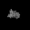
| Images |  emd_37334.png emd_37334.png | 99.3 KB | ||
| Filedesc metadata | emd-37334.cif.gz | 5.5 KB | ||
| Others | emd_37334_half_map_1.map.gzemd_37334_half_map_2.map.gz | 226.3 MB 226.3 MB | ||
| Archive directory |  http://ftp.pdbj.org/pub/emdb/structures/EMD-37334ftp://ftp.pdbj.org/pub/emdb/structures/EMD-37334 http://ftp.pdbj.org/pub/emdb/structures/EMD-37334ftp://ftp.pdbj.org/pub/emdb/structures/EMD-37334 | HTTPS FTP |
-Related structure data


-Links
| EMDB pages | EMDB (EBI/PDBe) / EMDataResource |
|---|
-Map
| File | Download / File: emd_37334.map.gz / Format: CCP4 / Size: 244.1 MB / Type: IMAGE STORED AS FLOATING POINT NUMBER (4 BYTES) | ||||||||||||||||||||||||||||||||||||
|---|---|---|---|---|---|---|---|---|---|---|---|---|---|---|---|---|---|---|---|---|---|---|---|---|---|---|---|---|---|---|---|---|---|---|---|---|---|
| Annotation | Consensus cryo-EM map of human 26S RP (Ed state) bound to K11/K48-branched ubiquitin (Ub) chain composed of four Ub. | ||||||||||||||||||||||||||||||||||||
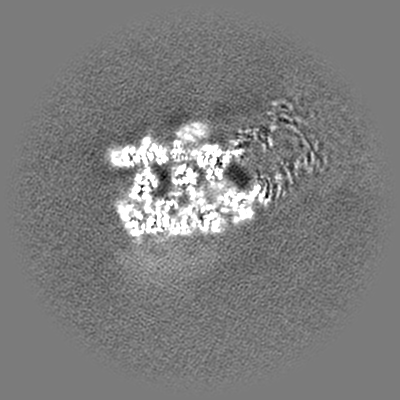
| Projections & slices | Image control
Images are generated by Spider. | ||||||||||||||||||||||||||||||||||||
| Voxel size | X=Y=Z: 1.4 Å | ||||||||||||||||||||||||||||||||||||
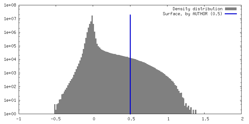
| Density |
| ||||||||||||||||||||||||||||||||||||
| Symmetry | Space group: 1 | ||||||||||||||||||||||||||||||||||||
| Details | EMDB XML:
|
 Z (Sec.)
 Y (Row.)
 X (Col.)


-Supplemental data
-Half map: Consensus cryo-EM half map of human 26S RP...
| File | emd_37334_half_map_1.map | ||||||||||||
|---|---|---|---|---|---|---|---|---|---|---|---|---|---|
| Annotation | Consensus cryo-EM half map of human 26S RP (Ed state) bound to K11/K48-branched ubiquitin (Ub) chain composed of four Ub. | ||||||||||||
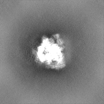
| Projections & Slices |
| ||||||||||||
| Density Histograms |


-Half map: Consensus cryo-EM half map of human 26S RP...
| File | emd_37334_half_map_2.map | ||||||||||||
|---|---|---|---|---|---|---|---|---|---|---|---|---|---|
| Annotation | Consensus cryo-EM half map of human 26S RP (Ed state) bound to K11/K48-branched ubiquitin (Ub) chain composed of four Ub. | ||||||||||||
| Projections & Slices |
| ||||||||||||
| Density Histograms |


- Sample components
-Entire : Human 26S proteasome
| Entire | Name: Human 26S proteasome |
|---|---|
| Components |
|
-Supramolecule #1: Human 26S proteasome
| Supramolecule | Name: Human 26S proteasome / type: complex / ID: 1 / Parent: 0 / Macromolecule list: #1-#27 Details: Images of double-capped (RP-CP-RP) 26S proteasome particles were split in half. After combining with subset of single-capped (RP-CP) complexes the signal in particle images was partially ...Details: Images of double-capped (RP-CP-RP) 26S proteasome particles were split in half. After combining with subset of single-capped (RP-CP) complexes the signal in particle images was partially subtracted leaving only the signal of 19S RP together with alpha subunits of the 20S CP proteasomal subcomplex. |
|---|---|
| Source (natural) | Organism: Homo sapiens (human) |
| Molecular weight | Theoretical: 2.5 MDa |
-Supramolecule #2: 19S regulatory particle (RP) of human 26S proteasome
| Supramolecule | Name: 19S regulatory particle (RP) of human 26S proteasome / type: complex / ID: 2 / Parent: 1 / Macromolecule list: #1-#5, #13-#19, #21-#26 |
|---|---|
| Source (natural) | Organism: Homo sapiens (human) |
-Supramolecule #3: AAA+-ATPase of 19S regulatory particle (RP) of human 26S proteasome
| Supramolecule | Name: AAA+-ATPase of 19S regulatory particle (RP) of human 26S proteasome type: complex / ID: 3 / Parent: 2 / Macromolecule list: #1-#5, #21 |
|---|
-Supramolecule #4: alpha subunits of the 20S core particle (CP) of human 26S proteasome
| Supramolecule | Name: alpha subunits of the 20S core particle (CP) of human 26S proteasome type: complex / ID: 4 / Parent: 1 / Macromolecule list: #6-#12 |
|---|---|
| Source (natural) | Organism: Homo sapiens (human) |
-Supramolecule #5: Ubiquitinated substrate
| Supramolecule | Name: Ubiquitinated substrate / type: complex / ID: 5 / Parent: 1 / Macromolecule list: #20, #27 Details: Sic1_PY conjugated with K11/K48-branched ubiquitin chain. |
|---|
-Supramolecule #6: K11/K48-branched ubiquitin (Ub) chain composed of four Ub.
| Supramolecule | Name: K11/K48-branched ubiquitin (Ub) chain composed of four Ub. type: complex / ID: 6 / Parent: 5 / Macromolecule list: #27 |
|---|---|
| Source (natural) | Organism: Homo sapiens (human) |
-Supramolecule #7: Sic1_PY substrate polypeptide
| Supramolecule | Name: Sic1_PY substrate polypeptide / type: complex / ID: 7 / Parent: 5 / Macromolecule list: #20 |
|---|---|
| Source (natural) | Organism: |
-Experimental details
-Structure determination
| Method | cryo EM |
|---|---|
 Processing Processing | single particle reconstruction |
| Aggregation state | particle |
-Sample preparation
| Concentration | 1 mg/mL | |||||||||||||||||||||
|---|---|---|---|---|---|---|---|---|---|---|---|---|---|---|---|---|---|---|---|---|---|---|
| Buffer | pH: 7.6 Component:
| |||||||||||||||||||||
| Grid | Model: Quantifoil R1.2/1.3 / Material: COPPER / Mesh: 300 / Pretreatment - Type: GLOW DISCHARGE / Pretreatment - Time: 30 sec. / Pretreatment - Atmosphere: AIR / Details: 20 mA | |||||||||||||||||||||
| Vitrification | Cryogen name: ETHANE / Chamber humidity: 100 % / Chamber temperature: 277.15 K / Instrument: FEI VITROBOT MARK IV / Details: incubation time= 3 s blotting time= 2.5 s. | |||||||||||||||||||||
| Details | The complex was additionally supplemented with an excess of preformed and SEC-purified RPN13:UCHL5 complex. |
- Electron microscopy
| Microscope | FEI TITAN KRIOS |
|---|---|
| Specialist optics | Energy filter - Slit width: 20 eV |
| Software | Name: EPU |
| Image recording | Film or detector model: GATAN K3 (6k x 4k) / Number grids imaged: 3 / Number real images: 15242 / Average exposure time: 1.8 sec. / Average electron dose: 49.0 e/Å2 |
| Electron beam | Acceleration voltage: 300 kV / Electron source:  FIELD EMISSION GUN FIELD EMISSION GUN |
| Electron optics | Illumination mode: FLOOD BEAM / Imaging mode: BRIGHT FIELD / Nominal defocus max: 1.8 µm / Nominal defocus min: 1.2 µm / Nominal magnification: 70000 |
| Sample stage | Specimen holder model: FEI TITAN KRIOS AUTOGRID HOLDER / Cooling holder cryogen: NITROGEN |
| Experimental equipment |  Model: Titan Krios / Image courtesy: FEI Company |

