 Movie
Movie Controller

[English] 日本語
 Yorodumi
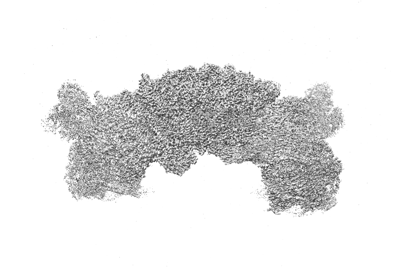
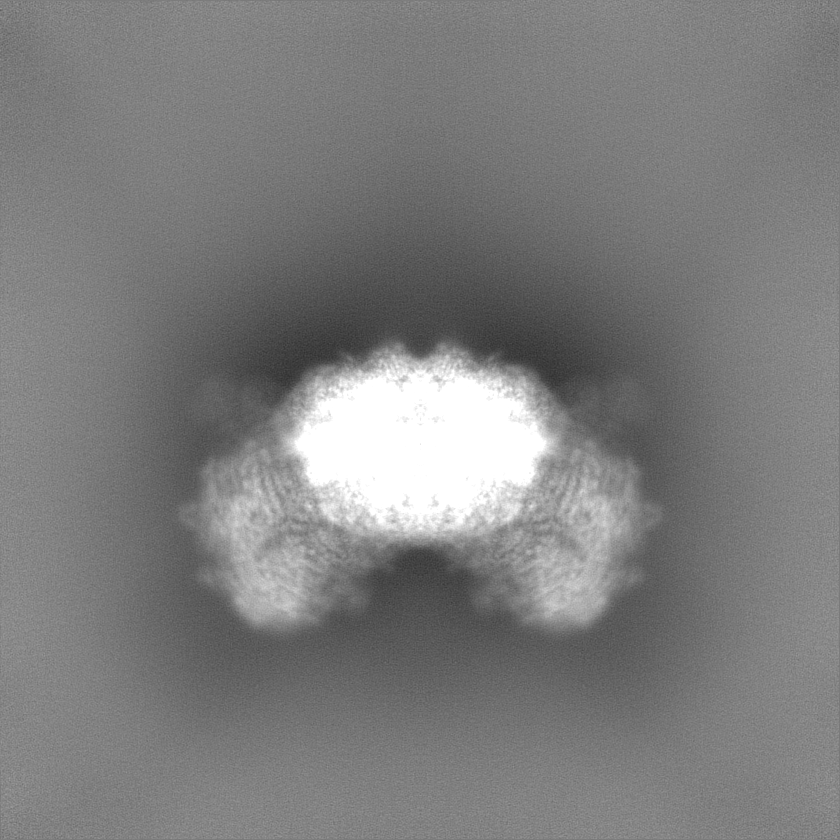
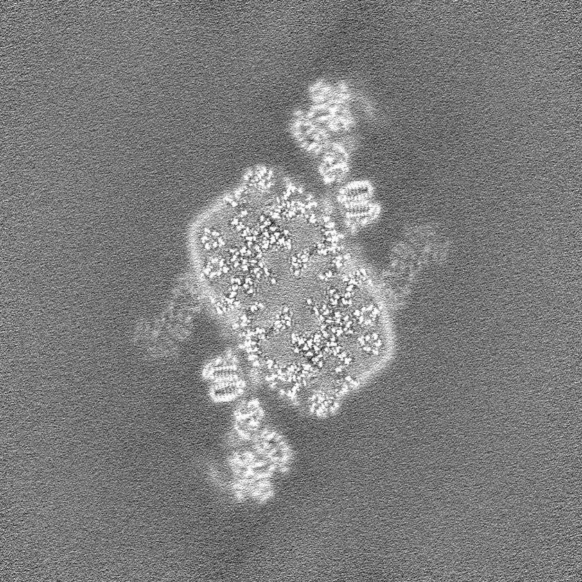
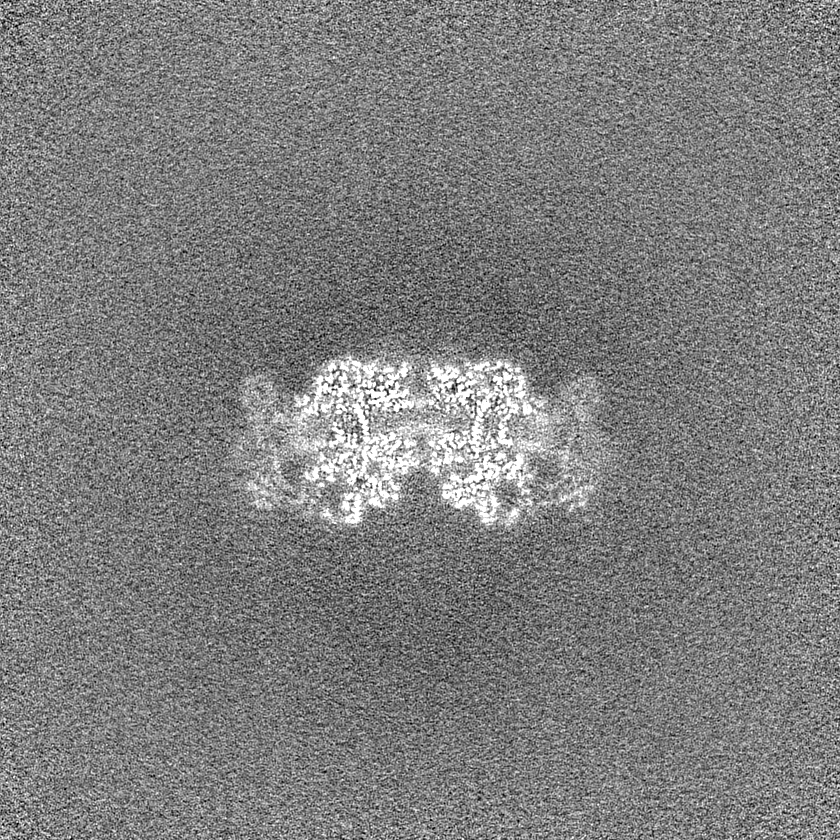
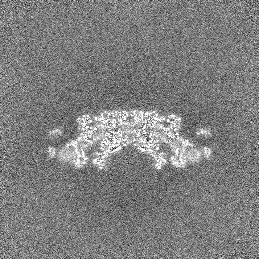
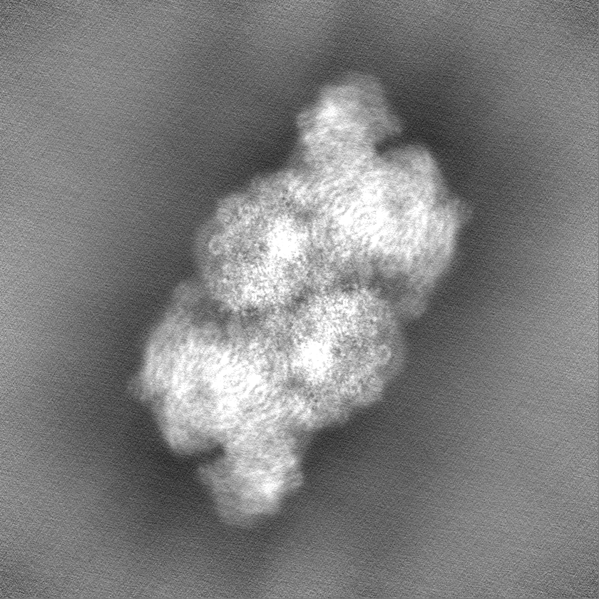
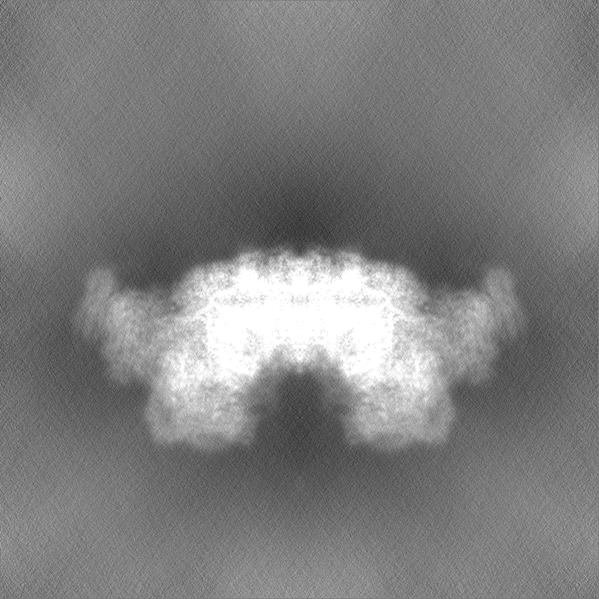
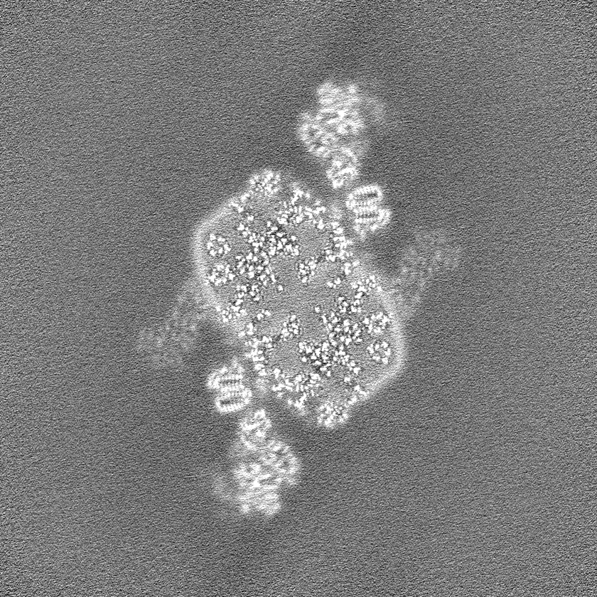
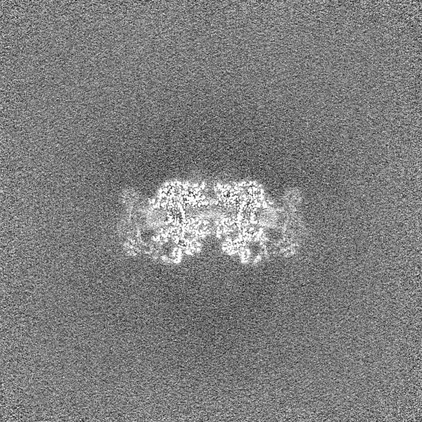
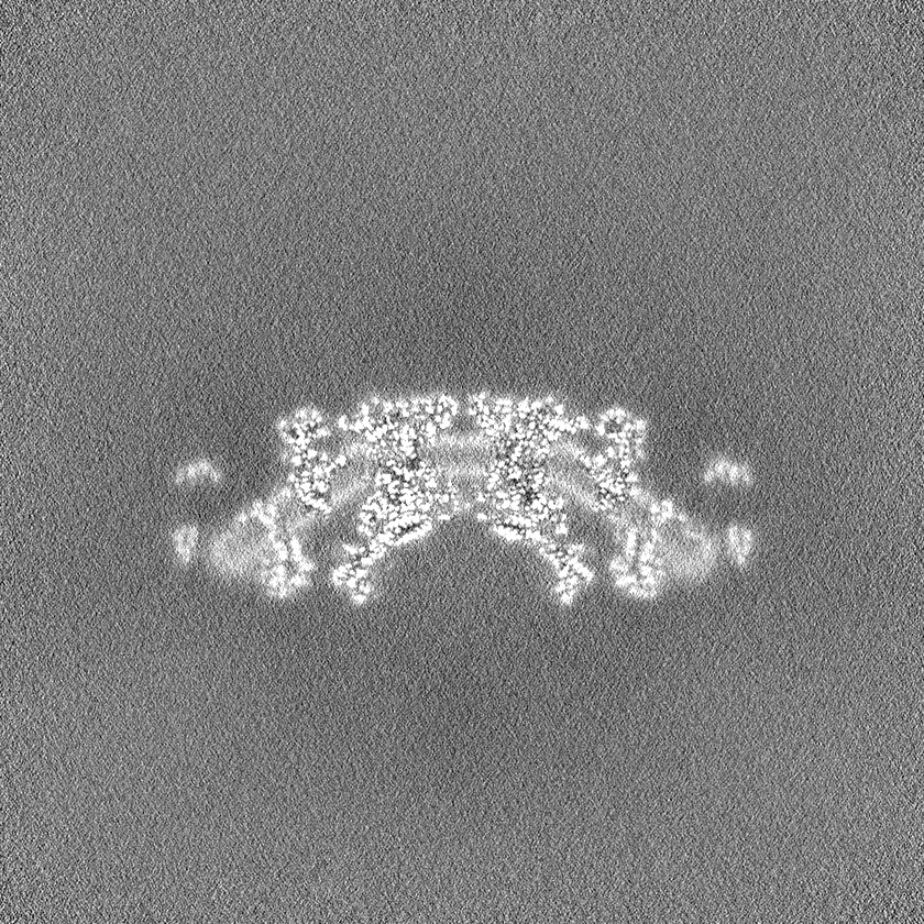
- EMDB-36375: Cryo-EM map of Tetrahymena thermophila megacomplex IV2+(I+III2+II)2 -
+ Open data

- Basic information
| Entry |  | |||||||||
|---|---|---|---|---|---|---|---|---|---|---|
| Title | Cryo-EM map of Tetrahymena thermophila megacomplex IV2+(I+III2+II)2 | |||||||||
 Map data Map data | ||||||||||
 Sample Sample |
| |||||||||
 Keywords Keywords | Electron transport chain / megacomplex / membrane protein / Tetrahymena thermophila / ELECTRON TRANSPORT | |||||||||
| Biological species |   Tetrahymena thermophila (eukaryote) Tetrahymena thermophila (eukaryote) | |||||||||
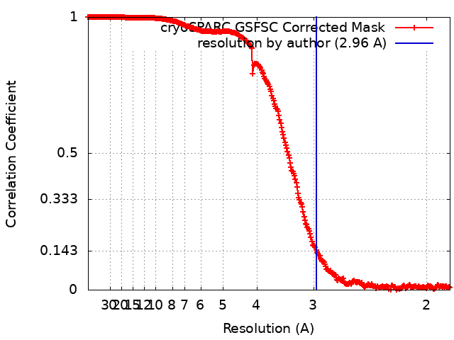
| Method | single particle reconstruction / cryo EM / Resolution: 2.96 Å | |||||||||
 Authors Authors | Wu MC / Hu YQ / Han FZ / He ZX / Tian HT / Zhou L | |||||||||
| Funding support |  China, 1 items China, 1 items
| |||||||||
 Citation Citation | Journal: Nat Commun / Year: 2023 Title: Structures of Tetrahymena thermophila respiratory megacomplexes on the tubular mitochondrial cristae. Authors: Fangzhu Han / Yiqi Hu / Mengchen Wu / Zhaoxiang He / Hongtao Tian / Long Zhou / Abstract: Tetrahymena thermophila, a classic ciliate model organism, has been shown to possess tubular mitochondrial cristae and highly divergent electron transport chain involving four transmembrane protein ...Tetrahymena thermophila, a classic ciliate model organism, has been shown to possess tubular mitochondrial cristae and highly divergent electron transport chain involving four transmembrane protein complexes (I-IV). Here we report cryo-EM structures of its ~8 MDa megacomplex IV+ (I + III+ II), as well as a ~ 10.6 MDa megacomplex (IV + I + III+ II) at lower resolution. In megacomplex IV+ (I + III+ II), each CIV protomer associates one copy of supercomplex I + III and one copy of CII, forming a half ring-shaped architecture that adapts to the membrane curvature of mitochondrial cristae. Megacomplex (IV+ I + III+ II) defines the relative position between neighbouring half rings and maintains the proximity between CIV and CIII cytochrome c binding sites. Our findings expand the current understanding of divergence in eukaryotic electron transport chain organization and how it is related to mitochondrial morphology. | |||||||||
| History |
|
- Structure visualization
| Supplemental images |
|---|
- Downloads & links
-EMDB archive
| Map data | emd_36375.map.gz | 2.1 GB |  EMDB map data format EMDB map data format | |
|---|---|---|---|---|
| Header (meta data) | emd-36375-v30.xmlemd-36375.xml | 17.8 KB 17.8 KB | Display Display | EMDB header |
| FSC (resolution estimation) | emd_36375_fsc.xml | 27.7 KB | Display | FSC data file |
| Images |  emd_36375.png emd_36375.png | 66.5 KB | ||
| Filedesc metadata | emd-36375.cif.gz | 4.5 KB | ||
| Others | emd_36375_half_map_1.map.gzemd_36375_half_map_2.map.gz | 2.1 GB 2.1 GB | ||
| Archive directory |  http://ftp.pdbj.org/pub/emdb/structures/EMD-36375ftp://ftp.pdbj.org/pub/emdb/structures/EMD-36375 http://ftp.pdbj.org/pub/emdb/structures/EMD-36375ftp://ftp.pdbj.org/pub/emdb/structures/EMD-36375 | HTTPS FTP |
-Validation report
| Summary document | emd_36375_validation.pdf.gz | 1.1 MB | Display | EMDB validaton report |
|---|---|---|---|---|
| Full document | emd_36375_full_validation.pdf.gz | 1.1 MB | Display | |
| Data in XML | emd_36375_validation.xml.gz | 38.2 KB | Display | |
| Data in CIF | emd_36375_validation.cif.gz | 51.1 KB | Display | |
| Arichive directory | https://ftp.pdbj.org/pub/emdb/validation_reports/EMD-36375ftp://ftp.pdbj.org/pub/emdb/validation_reports/EMD-36375 | HTTPS FTP |
-Related structure data


-Links
| EMDB pages | EMDB (EBI/PDBe) / EMDataResource |
|---|
-Map
| File | Download / File: emd_36375.map.gz / Format: CCP4 / Size: 2.2 GB / Type: IMAGE STORED AS FLOATING POINT NUMBER (4 BYTES) | ||||||||||||||||||||||||||||||||||||
|---|---|---|---|---|---|---|---|---|---|---|---|---|---|---|---|---|---|---|---|---|---|---|---|---|---|---|---|---|---|---|---|---|---|---|---|---|---|
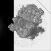
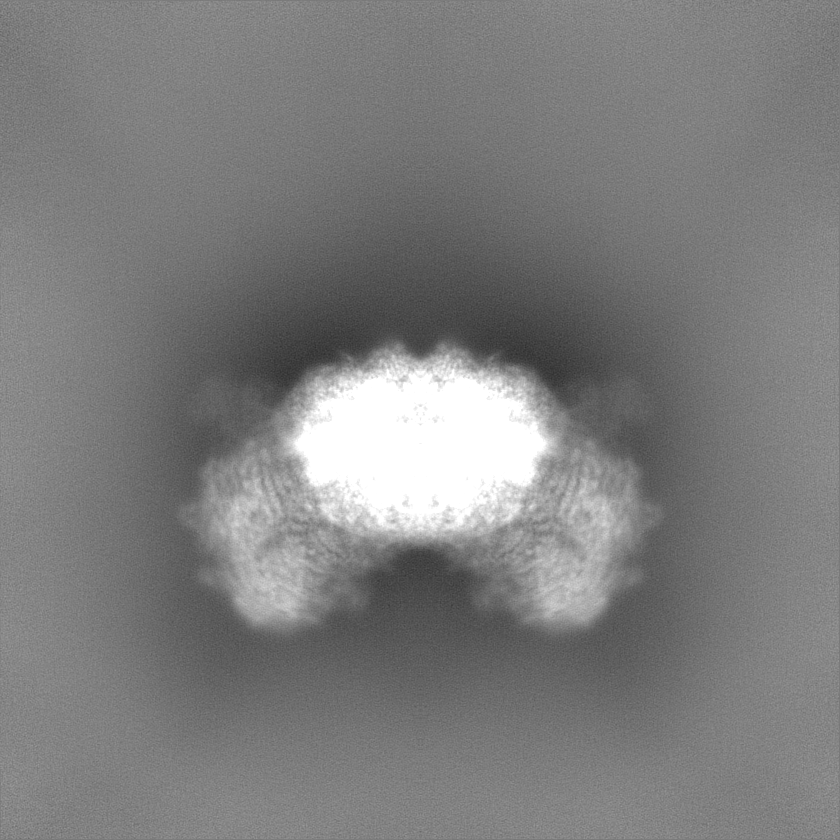
| Projections & slices | Image control
Images are generated by Spider. | ||||||||||||||||||||||||||||||||||||
| Voxel size | X=Y=Z: 0.93 Å | ||||||||||||||||||||||||||||||||||||
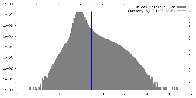
| Density |
| ||||||||||||||||||||||||||||||||||||
| Symmetry | Space group: 1 | ||||||||||||||||||||||||||||||||||||
| Details | EMDB XML:
|
 Z (Sec.)
 Y (Row.)
 X (Col.)


-Supplemental data
-Half map: #1
| File | emd_36375_half_map_1.map | ||||||||||||
|---|---|---|---|---|---|---|---|---|---|---|---|---|---|
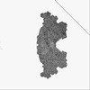
| Projections & Slices |
| ||||||||||||
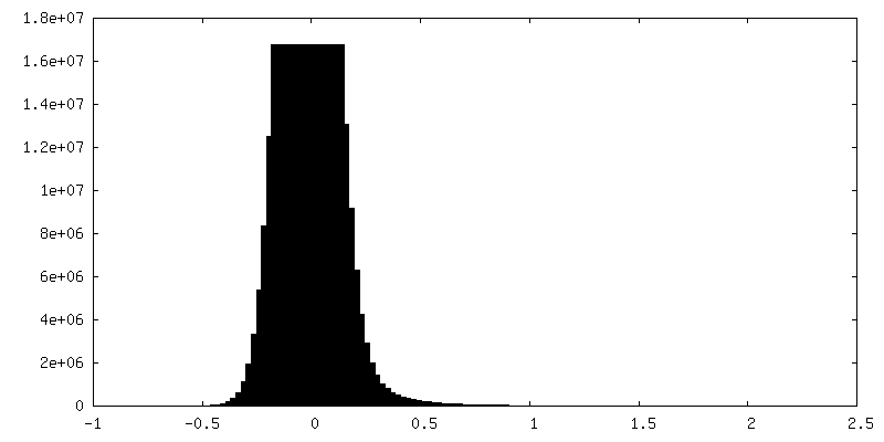
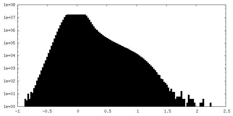
| Density Histograms |


-Half map: #2
| File | emd_36375_half_map_2.map | ||||||||||||
|---|---|---|---|---|---|---|---|---|---|---|---|---|---|
| Projections & Slices |
| ||||||||||||
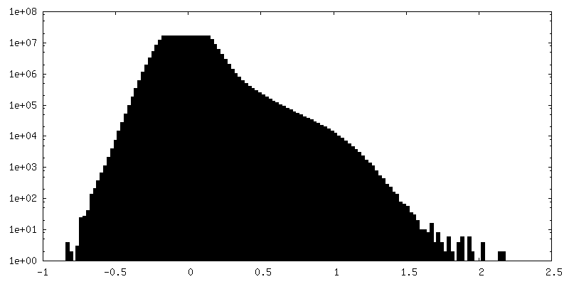
| Density Histograms |

- Sample components
-Entire : Tetrahymena thermophila megacomplex IV2+(I+III2+II)2
| Entire | Name: Tetrahymena thermophila megacomplex IV2+(I+III2+II)2 |
|---|---|
| Components |
|
-Supramolecule #1: Tetrahymena thermophila megacomplex IV2+(I+III2+II)2
| Supramolecule | Name: Tetrahymena thermophila megacomplex IV2+(I+III2+II)2 / type: complex / ID: 1 / Parent: 0 |
|---|---|
| Source (natural) | Organism: Tetrahymena thermophila (eukaryote) / Strain: SB210 |
-Experimental details
-Structure determination
| Method | cryo EM |
|---|---|
 Processing Processing | single particle reconstruction |
| Aggregation state | particle |
-Sample preparation
| Concentration | 4 mg/mL | |||||||||||||||
|---|---|---|---|---|---|---|---|---|---|---|---|---|---|---|---|---|
| Buffer | pH: 7.4 Component:
Details: SEC buffer (20 mM Tris pH 7.4, 50 mM NaCl, 0.002% PMSF, 0.005% LMNG (w/v)) | |||||||||||||||
| Grid | Model: Quantifoil R0.6/1 / Material: COPPER / Mesh: 300 / Pretreatment - Type: GLOW DISCHARGE / Pretreatment - Time: 120 sec. / Pretreatment - Atmosphere: AIR / Pretreatment - Pressure: 0.039 kPa / Details: 25mA | |||||||||||||||
| Vitrification | Cryogen name: ETHANE / Chamber humidity: 100 % / Chamber temperature: 277.15 K / Instrument: FEI VITROBOT MARK IV |
- Electron microscopy
| Microscope | FEI TITAN KRIOS |
|---|---|
| Image recording | Film or detector model: FEI FALCON IV (4k x 4k) / Digitization - Dimensions - Width: 4096 pixel / Digitization - Dimensions - Height: 4096 pixel / Number grids imaged: 2 / Number real images: 16772 / Average exposure time: 7.0 sec. / Average electron dose: 61.5 e/Å2 |
| Electron beam | Acceleration voltage: 300 kV / Electron source:  FIELD EMISSION GUN FIELD EMISSION GUN |
| Electron optics | Illumination mode: FLOOD BEAM / Imaging mode: BRIGHT FIELD / Cs: 2.7 mm / Nominal defocus max: 2.0 µm / Nominal defocus min: 0.8 µm |
| Sample stage | Specimen holder model: FEI TITAN KRIOS AUTOGRID HOLDER / Cooling holder cryogen: NITROGEN |
| Experimental equipment |  Model: Titan Krios / Image courtesy: FEI Company |
+Image processing
-Atomic model buiding 1
| Refinement | Space: REAL / Protocol: RIGID BODY FIT |
|---|
