[English] 日本語
Yorodumi
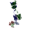
- EMDB-36176: Cryo-EM structure of Gi1-bound mGlu2-mGlu4 heterodimer -

+
Open data


ID or keywords:

Loading...

-
Basic information

Entry
Database: EMDB / ID: EMD-36176
TitleCryo-EM structure of Gi1-bound mGlu2-mGlu4 heterodimer
Map data
Sample
  • Complex: Gi1-bound mGlu2-mGlu4 heterodimer
    • Protein or peptide: x 6 types
  • Ligand: x 6 types
KeywordsComplex structure / Gi1-bound mGlu2-mGlu4 heterodimer / membrane protein
Function / homology
Function and homology information


adenylate cyclase-inhibiting G protein-coupled glutamate receptor signaling pathway / regulation of response to drug / group II metabotropic glutamate receptor activity / adenylate cyclase inhibiting G protein-coupled glutamate receptor activity / behavioral response to nicotine / G protein-coupled glutamate receptor signaling pathway / intracellular glutamate homeostasis / astrocyte projection / negative regulation of adenylate cyclase activity / neurotransmitter secretion ...adenylate cyclase-inhibiting G protein-coupled glutamate receptor signaling pathway / regulation of response to drug / group II metabotropic glutamate receptor activity / adenylate cyclase inhibiting G protein-coupled glutamate receptor activity / behavioral response to nicotine / G protein-coupled glutamate receptor signaling pathway / intracellular glutamate homeostasis / astrocyte projection / negative regulation of adenylate cyclase activity / neurotransmitter secretion / Class C/3 (Metabotropic glutamate/pheromone receptors) / glutamate secretion / glutamate receptor activity / regulation of glutamate secretion / long-term synaptic depression / regulation of dopamine secretion / calcium channel regulator activity / Adenylate cyclase inhibitory pathway / positive regulation of protein localization to cell cortex / regulation of cAMP-mediated signaling / D2 dopamine receptor binding / G protein-coupled serotonin receptor binding / regulation of neuron apoptotic process / regulation of mitotic spindle organization / cellular response to forskolin / regulation of synaptic transmission, glutamatergic / adenylate cyclase-inhibiting G protein-coupled receptor signaling pathway / presynaptic modulation of chemical synaptic transmission / response to cocaine / Regulation of insulin secretion / G protein-coupled receptor binding / G protein-coupled receptor activity / G-protein beta/gamma-subunit complex binding / Olfactory Signaling Pathway / Activation of the phototransduction cascade / G beta:gamma signalling through PLC beta / Presynaptic function of Kainate receptors / Thromboxane signalling through TP receptor / adenylate cyclase-modulating G protein-coupled receptor signaling pathway / G-protein activation / G protein-coupled acetylcholine receptor signaling pathway / Activation of G protein gated Potassium channels / Inhibition of voltage gated Ca2+ channels via Gbeta/gamma subunits / Prostacyclin signalling through prostacyclin receptor / Glucagon signaling in metabolic regulation / G beta:gamma signalling through CDC42 / ADP signalling through P2Y purinoceptor 12 / G beta:gamma signalling through BTK / Sensory perception of sweet, bitter, and umami (glutamate) taste / response to peptide hormone / Synthesis, secretion, and inactivation of Glucagon-like Peptide-1 (GLP-1) / photoreceptor disc membrane / Adrenaline,noradrenaline inhibits insulin secretion / Glucagon-type ligand receptors / Vasopressin regulates renal water homeostasis via Aquaporins / G alpha (z) signalling events / cellular response to catecholamine stimulus / Glucagon-like Peptide-1 (GLP1) regulates insulin secretion / ADORA2B mediated anti-inflammatory cytokines production / adenylate cyclase-activating dopamine receptor signaling pathway / ADP signalling through P2Y purinoceptor 1 / G beta:gamma signalling through PI3Kgamma / cellular response to prostaglandin E stimulus / Cooperation of PDCL (PhLP1) and TRiC/CCT in G-protein beta folding / sensory perception of taste / GPER1 signaling / GDP binding / G-protein beta-subunit binding / heterotrimeric G-protein complex / Inactivation, recovery and regulation of the phototransduction cascade / extracellular vesicle / G alpha (12/13) signalling events / signaling receptor complex adaptor activity / Thrombin signalling through proteinase activated receptors (PARs) / presynapse / retina development in camera-type eye / GTPase binding / presynaptic membrane / Ca2+ pathway / gene expression / phospholipase C-activating G protein-coupled receptor signaling pathway / cell cortex / midbody / G alpha (i) signalling events / fibroblast proliferation / chemical synaptic transmission / cytoplasmic vesicle / G alpha (s) signalling events / scaffold protein binding / G alpha (q) signalling events / Ras protein signal transduction / cell population proliferation / Extra-nuclear estrogen signaling / positive regulation of MAPK cascade / positive regulation of phosphatidylinositol 3-kinase/protein kinase B signal transduction / cell cycle / G protein-coupled receptor signaling pathway / cell division / lysosomal membrane / axon
Similarity search - Function
GPCR, family 3, metabotropic glutamate receptor 4 / GPCR, family 3, metabotropic glutamate receptor 2 / GPCR, family 3, metabotropic glutamate receptor / G-protein coupled receptors family 3 signature 1. / G-protein coupled receptors family 3 signature 2. / GPCR, family 3, nine cysteines domain / GPCR, family 3, nine cysteines domain superfamily / Nine Cysteines Domain of family 3 GPCR / GPCR, family 3, conserved site / G-protein coupled receptors family 3 signature 3. ...GPCR, family 3, metabotropic glutamate receptor 4 / GPCR, family 3, metabotropic glutamate receptor 2 / GPCR, family 3, metabotropic glutamate receptor / G-protein coupled receptors family 3 signature 1. / G-protein coupled receptors family 3 signature 2. / GPCR, family 3, nine cysteines domain / GPCR, family 3, nine cysteines domain superfamily / Nine Cysteines Domain of family 3 GPCR / GPCR, family 3, conserved site / G-protein coupled receptors family 3 signature 3. / GPCR, family 3 / GPCR family 3, C-terminal / 7 transmembrane sweet-taste receptor of 3 GCPR / G-protein coupled receptors family 3 profile. / G-protein alpha subunit, group I / Receptor, ligand binding region / Receptor family ligand binding region / G-alpha domain profile. / Guanine nucleotide binding protein (G-protein), alpha subunit / G protein alpha subunit, helical insertion / G-protein alpha subunit / G protein alpha subunit / Periplasmic binding protein-like I / G-protein, gamma subunit / G-protein gamma subunit domain profile. / GGL domain / G-protein gamma-like domain superfamily / G-protein gamma-like domain / GGL domain / G protein gamma subunit-like motifs / Guanine nucleotide-binding protein, beta subunit / G-protein, beta subunit / G-protein beta WD-40 repeat / WD40 repeat, conserved site / Trp-Asp (WD) repeats signature. / WD domain, G-beta repeat / WD40 repeats / WD40 repeat / Trp-Asp (WD) repeats profile. / Trp-Asp (WD) repeats circular profile. / WD40-repeat-containing domain superfamily / WD40/YVTN repeat-like-containing domain superfamily / P-loop containing nucleoside triphosphate hydrolase
Similarity search - Domain/homology
Guanine nucleotide-binding protein G(I)/G(S)/G(O) subunit gamma-2 / Guanine nucleotide-binding protein G(I)/G(S)/G(T) subunit beta-1 / Guanine nucleotide-binding protein G(i) subunit alpha-1 / Metabotropic glutamate receptor 2 / Metabotropic glutamate receptor 4
Similarity search - Component
Biological speciesHomo sapiens (human) / Escherichia coli (E. coli)
Methodsingle particle reconstruction / cryo EM / Resolution: 3.6 Å
AuthorsWang X / Wang M / Xu T / Feng Y / Han S / Zhao Q / Wu B
Funding support China, 2 items
OrganizationGrant numberCountry
National Science Foundation (NSF, China)31825010 China
National Science Foundation (NSF, China)82121005 China
CitationJournal: Cell Res / Year: 2023
Title: Structural insights into dimerization and activation of the mGlu2-mGlu3 and mGlu2-mGlu4 heterodimers.
Authors: Xinwei Wang / Mu Wang / Tuo Xu / Ye Feng / Qiang Shao / Shuo Han / Xiaojing Chu / Yechun Xu / Shuling Lin / Qiang Zhao / Beili Wu /
Abstract: Heterodimerization of the metabotropic glutamate receptors (mGlus) has shown importance in the functional modulation of the receptors and offers potential drug targets for treating central nervous ...Heterodimerization of the metabotropic glutamate receptors (mGlus) has shown importance in the functional modulation of the receptors and offers potential drug targets for treating central nervous system diseases. However, due to a lack of molecular details of the mGlu heterodimers, understanding of the mechanisms underlying mGlu heterodimerization and activation is limited. Here we report twelve cryo-electron microscopy (cryo-EM) structures of the mGlu2-mGlu3 and mGlu2-mGlu4 heterodimers in different conformational states, including inactive, intermediate inactive, intermediate active and fully active conformations. These structures provide a full picture of conformational rearrangement of mGlu2-mGlu3 upon activation. The Venus flytrap domains undergo a sequential conformational change, while the transmembrane domains exhibit a substantial rearrangement from an inactive, symmetric dimer with diverse dimerization patterns to an active, asymmetric dimer in a conserved dimerization mode. Combined with functional data, these structures reveal that stability of the inactive conformations of the subunits and the subunit-G protein interaction pattern are determinants of asymmetric signal transduction of the heterodimers. Furthermore, a novel binding site for two mGlu4 positive allosteric modulators was observed in the asymmetric dimer interfaces of the mGlu2-mGlu4 heterodimer and mGlu4 homodimer, and may serve as a drug recognition site. These findings greatly extend our knowledge about signal transduction of the mGlus.
History
DepositionMay 12, 2023-
Header (metadata) releaseJun 21, 2023-
Map releaseJun 21, 2023-
UpdateOct 18, 2023-
Current statusOct 18, 2023Processing site: PDBc / Status: Released

-
Structure visualization

Supplemental images

Downloads & links

-
Map

FileDownload / File: emd_36176.map.gz / Format: CCP4 / Size: 178 MB / Type: IMAGE STORED AS FLOATING POINT NUMBER (4 BYTES)
Voxel sizeX=Y=Z: 1.071 Å
Density
Contour LevelBy AUTHOR: 5.5
Minimum - Maximum-60.57694 - 79.046930000000003
Average (Standard dev.)0.0009591965 (±0.93268466)
SymmetrySpace group: 1
Details

EMDB XML:

Map geometry
Axis orderXYZ
Origin000
Dimensions360360360
Spacing360360360
CellA=B=C: 385.56 Å
α=β=γ: 90.0 °

-
Supplemental data

-
Sample components

+
Entire : Gi1-bound mGlu2-mGlu4 heterodimer

EntireName: Gi1-bound mGlu2-mGlu4 heterodimer
Components
  • Complex: Gi1-bound mGlu2-mGlu4 heterodimer
    • Protein or peptide: Metabotropic glutamate receptor 2
    • Protein or peptide: Metabotropic glutamate receptor 4
    • Protein or peptide: Guanine nucleotide-binding protein G(i) subunit alpha-1
    • Protein or peptide: Guanine nucleotide-binding protein G(I)/G(S)/G(T) subunit beta-1
    • Protein or peptide: Guanine nucleotide-binding protein G(I)/G(S)/G(O) subunit gamma-2
    • Protein or peptide: scFvSingle-chain variable fragment
  • Ligand: 1-butyl-3-chloranyl-4-(4-phenylpiperidin-1-yl)pyridin-2-one
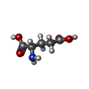
  • Ligand: GLUTAMIC ACID
  • Ligand: CHOLESTEROL
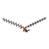
  • Ligand: DI-PALMITOYL-3-SN-PHOSPHATIDYLETHANOLAMINE
  • Ligand: 2-acetamido-2-deoxy-beta-D-glucopyranose
  • Ligand: 5-methyl-N-(4-methylpyrimidin-2-yl)-4-(1H-pyrazol-4-yl)-1,3-thiazol-2-amine

+
Supramolecule #1: Gi1-bound mGlu2-mGlu4 heterodimer

SupramoleculeName: Gi1-bound mGlu2-mGlu4 heterodimer / type: complex / ID: 1 / Parent: 0 / Macromolecule list: #1-#6
Source (natural)Organism: Homo sapiens (human)

+
Macromolecule #1: Metabotropic glutamate receptor 2

MacromoleculeName: Metabotropic glutamate receptor 2 / type: protein_or_peptide / ID: 1 / Number of copies: 1 / Enantiomer: LEVO
Source (natural)Organism: Homo sapiens (human)
Molecular weightTheoretical: 95.684273 KDa
Recombinant expressionOrganism: mammal environmental sample (environmental samples)
SequenceString: DYKDDDDGAP EGPAKKVLTL EGDLVLGGLF PVHQKGGPAE DCGPVNEHRG IQRLEAMLFA LDRINRDPHL LPGVRLGAHI LDSCSKDTH ALEQALDFVR ASLSRGADGS RHICPDGSYA THGDAPTAIT GVIGGSYSDV SIQVANLLRL FQIPQISYAS T SAKLSDKS ...String:
DYKDDDDGAP EGPAKKVLTL EGDLVLGGLF PVHQKGGPAE DCGPVNEHRG IQRLEAMLFA LDRINRDPHL LPGVRLGAHI LDSCSKDTH ALEQALDFVR ASLSRGADGS RHICPDGSYA THGDAPTAIT GVIGGSYSDV SIQVANLLRL FQIPQISYAS T SAKLSDKS RYDYFARTVP PDFFQAKAMA EILRFFNWTY VSTVASEGDY GETGIEAFEL EARARNICVA TSEKVGRAMS RA AFEGVVR ALLQKPSARV AVLFTRSEDA RELLAASQRL NASFTWVASD GWGALESVVA GSEGAAEGAI TIELASYPIS DFA SYFQSL DPWNNSRNPW FREFWEQRFR CSFRQRDCAA HSLRAVPFEQ ESKIMFVVNA VYAMAHALHN MHRALCPNTT RLCD AMRPV NGRRLYKDFV LNVKFDAPFR PADTHNEVRF DRFGDGIGRY NIFTYLRAGS GRYRYQKVGY WAEGLTLDTS LIPWA SPSA GPLPASRCSE PCLQNEVKSV QPGEVCCWLC IPCQPYEYRL DEFTCADCGL GYWPNASLTG CFELPQEYIR WGDAWA VGP VTIACLGALA TLFVLGVFVR HNATPVVKAS GRELCYILLG GVFLCYCMTF IFIAKPSTAV CTLRRLGLGT AFSVCYS AL LTKTNRIARI FGGAREGAQR PRFISPASQV AICLALISGQ LLIVVAWLVV EAPGTGKETA PERREVVTLR CNHRDASM L GSLAYNVLLI ALCTLYAFKT RKCPENFNEA KFIGFTMYTT CIIWLAFLPI FYVTSSDYRV QTTTMCVSVS LSGSVVLGC LFAPKLHIIL FQPQKNVVSH RAPTSRFGSA AARASSSLGQ GSGSQFVPTV CNGREVVDST TSSLLEVLFQ

UniProtKB: Metabotropic glutamate receptor 2

+
Macromolecule #2: Metabotropic glutamate receptor 4

MacromoleculeName: Metabotropic glutamate receptor 4 / type: protein_or_peptide / ID: 2 / Number of copies: 1 / Enantiomer: LEVO
Source (natural)Organism: Homo sapiens (human)
Molecular weightTheoretical: 102.560789 KDa
Recombinant expressionOrganism: mammal environmental sample (environmental samples)
SequenceString: DYKDDDDGAP WSHPQFEKGS GSWSHPQFEK KPKGHPHMNS IRIDGDITLG GLFPVHGRGS EGKPCGELKK EKGIHRLEAM LFALDRINN DPDLLPNITL GARILDTCSR DTHALEQSLT FVQALIEKDG TEVRCGSGGP PIITKPERVV GVIGASGSSV S IMVANILR ...String:
DYKDDDDGAP WSHPQFEKGS GSWSHPQFEK KPKGHPHMNS IRIDGDITLG GLFPVHGRGS EGKPCGELKK EKGIHRLEAM LFALDRINN DPDLLPNITL GARILDTCSR DTHALEQSLT FVQALIEKDG TEVRCGSGGP PIITKPERVV GVIGASGSSV S IMVANILR LFKIPQISYA STAPDLSDNS RYDFFSRVVP SDTYQAQAMV DIVRALKWNY VSTVASEGSY GESGVEAFIQ KS REDGGVC IAQSVKIPRE PKAGEFDKII RRLLETSNAR AVIIFANEDD IRRVLEAARR ANQTGHFFWM GSDSWGSKIA PVL HLEEVA EGAVTILPKR MSVRGFDRYF SSRTLDNNRR NIWFAEFWED NFHCKLSRHA LKKGSHVKKC TNRERIGQDS AYEQ EGKVQ FVIDAVYAMG HALHAMHRDL CPGRVGLCPR MDPVDGTQLL KYIRNVNFSG IAGNPVTFNE NGDAPGRYDI YQYQL RNDS AEYKVIGSWT DHLHLRIERM HWPGSGQQLP RSICSLPCQP GERKKTVKGM PCCWHCEPCT GYQYQVDRYT CKTCPY DMR PTENRTGCRP IPIIKLEWGS PWAVLPLFLA VVGIAATLFV VITFVRYNDT PIVKASGREL SYVLLAGIFL CYATTFL MI AEPDLGTCSL RRIFLGLGMS ISYAALLTKT NRIYRIFEQG KRSVSAPRFI SPASQLAITF SLISLQLLGI CVWFVVDP S HSVVDFQDQR TLDPRFARGV LKCDISDLSL ICLLGYSMLL MVTCTVYAIK TRGVPETFNE AKPIGFTMYT TCIVWLAFI PIFFGTSQSA DKLYIQTTTL TVSVSLSASV SLGMLYMPKV YIILFHPEQN VPKRKRSLKA VVTAATMSNK FTQKGNFRPN GEAKSELCE NLEAPALATK QTYVTYTNHA ILEVLFQ

UniProtKB: Metabotropic glutamate receptor 4

+
Macromolecule #3: Guanine nucleotide-binding protein G(i) subunit alpha-1

MacromoleculeName: Guanine nucleotide-binding protein G(i) subunit alpha-1
type: protein_or_peptide / ID: 3 / Number of copies: 1 / Enantiomer: LEVO
Source (natural)Organism: Homo sapiens (human)
Molecular weightTheoretical: 40.414047 KDa
Recombinant expressionOrganism: Baculovirus expression vector pFastBac1-HM
SequenceString: MGCTLSAEDK AAVERSKMID RNLREDGEKA AREVKLLLLG AGESGKNTIV KQMKIIHEAG YSEEECKQYK AVVYSNTIQS IIAIIRAMG RLKIDFGDSA RADDARQLFV LAGAAEEGFM TAELAGVIKR LWKDSGVQAC FNRSREYQLN DSAAYYLNDL D RIAQPNYI ...String:
MGCTLSAEDK AAVERSKMID RNLREDGEKA AREVKLLLLG AGESGKNTIV KQMKIIHEAG YSEEECKQYK AVVYSNTIQS IIAIIRAMG RLKIDFGDSA RADDARQLFV LAGAAEEGFM TAELAGVIKR LWKDSGVQAC FNRSREYQLN DSAAYYLNDL D RIAQPNYI PTQQDVLRTR VKTTGIVETH FTFKDLHFKM FDVGAQRSER KKWIHCFEGV TAIIFCVALS DYDLVLAEDE EM NRMHASM KLFDSICNNK WFTDTSIILF LNKKDLFEEK IKKSPLTICY PEYAGSNTYE EAAAYIQCQF EDLNKRKDTK EIY THFTCS TDTKNVQFVF DAVTDVIIKN NLKDCGLF

UniProtKB: Guanine nucleotide-binding protein G(i) subunit alpha-1

+
Macromolecule #4: Guanine nucleotide-binding protein G(I)/G(S)/G(T) subunit beta-1

MacromoleculeName: Guanine nucleotide-binding protein G(I)/G(S)/G(T) subunit beta-1
type: protein_or_peptide / ID: 4 / Number of copies: 1 / Enantiomer: LEVO
Source (natural)Organism: Homo sapiens (human)
Molecular weightTheoretical: 38.744371 KDa
Recombinant expressionOrganism: Baculovirus expression vector pFastBac1-HM
SequenceString: MHHHHHHGSL LQSELDQLRQ EAEQLKNQIR DARKACADAT LSQITNNIDP VGRIQMRTRR TLRGHLAKIY AMHWGTDSRL LVSASQDGK LIIWDSYTTN KVHAIPLRSS WVMTCAYAPS GNYVACGGLD NICSIYNLKT REGNVRVSRE LAGHTGYLSC C RFLDDNQI ...String:
MHHHHHHGSL LQSELDQLRQ EAEQLKNQIR DARKACADAT LSQITNNIDP VGRIQMRTRR TLRGHLAKIY AMHWGTDSRL LVSASQDGK LIIWDSYTTN KVHAIPLRSS WVMTCAYAPS GNYVACGGLD NICSIYNLKT REGNVRVSRE LAGHTGYLSC C RFLDDNQI VTSSGDTTCA LWDIETGQQT TTFTGHTGDV MSLSLAPDTR LFVSGACDAS AKLWDVREGM CRQTFTGHES DI NAICFFP NGNAFATGSD DATCRLFDLR ADQELMTYSH DNIICGITSV SFSKSGRLLL AGYDDFNCNV WDALKADRAG VLA GHDNRV SCLGVTDDGM AVATGSWDSF LKIWN

UniProtKB: Guanine nucleotide-binding protein G(I)/G(S)/G(T) subunit beta-1

+
Macromolecule #5: Guanine nucleotide-binding protein G(I)/G(S)/G(O) subunit gamma-2

MacromoleculeName: Guanine nucleotide-binding protein G(I)/G(S)/G(O) subunit gamma-2
type: protein_or_peptide / ID: 5 / Number of copies: 1 / Enantiomer: LEVO
Source (natural)Organism: Homo sapiens (human)
Molecular weightTheoretical: 7.861143 KDa
Recombinant expressionOrganism: Baculovirus expression vector pFastBac1-HM
SequenceString:
MASNNTASIA QARKLVEQLK MEANIDRIKV SKAAADLMAY CEAHAKEDPL LTPVPASENP FREKKFFCAI L

UniProtKB: Guanine nucleotide-binding protein G(I)/G(S)/G(O) subunit gamma-2

+
Macromolecule #6: scFv

MacromoleculeName: scFv / type: protein_or_peptide / ID: 6 / Number of copies: 1 / Enantiomer: LEVO
Source (natural)Organism: Escherichia coli (E. coli)
Molecular weightTheoretical: 27.409588 KDa
Recombinant expressionOrganism: Baculovirus expression vector pFastBac1-HM
SequenceString: DVQLVESGGG LVQPGGSRKL SCSASGFAFS SFGMHWVRQA PEKGLEWVAY ISSGSGTIYY ADTVKGRFTI SRDDPKNTLF LQMTSLRSE DTAMYYCVRS IYYYGSSPFD FWGQGTTLTV SSGGGGSGGG GSGGGGSDIV MTQATSSVPV TPGESVSISC R SSKSLLHS ...String:
DVQLVESGGG LVQPGGSRKL SCSASGFAFS SFGMHWVRQA PEKGLEWVAY ISSGSGTIYY ADTVKGRFTI SRDDPKNTLF LQMTSLRSE DTAMYYCVRS IYYYGSSPFD FWGQGTTLTV SSGGGGSGGG GSGGGGSDIV MTQATSSVPV TPGESVSISC R SSKSLLHS NGNTYLYWFL QRPGQSPQLL IYRMSNLASG VPDRFSGSGS GTAFTLTISR LEAEDVGVYY CMQHLEYPLT FG AGTKLEL KAAALEVLFQ

+
Macromolecule #7: 1-butyl-3-chloranyl-4-(4-phenylpiperidin-1-yl)pyridin-2-one

MacromoleculeName: 1-butyl-3-chloranyl-4-(4-phenylpiperidin-1-yl)pyridin-2-one
type: ligand / ID: 7 / Number of copies: 1 / Formula: HZR
Molecular weightTheoretical: 344.878 Da
Chemical component information

ChemComp-HZR:
1-butyl-3-chloranyl-4-(4-phenylpiperidin-1-yl)pyridin-2-one / ADX-71149

+
Macromolecule #8: GLUTAMIC ACID

MacromoleculeName: GLUTAMIC ACID / type: ligand / ID: 8 / Number of copies: 2 / Formula: GLU
Molecular weightTheoretical: 147.129 Da
Chemical component information

ChemComp-GLU:
GLUTAMIC ACID / Glutamic acid

+
Macromolecule #9: CHOLESTEROL

MacromoleculeName: CHOLESTEROL / type: ligand / ID: 9 / Number of copies: 5 / Formula: CLR
Molecular weightTheoretical: 386.654 Da
Chemical component information

ChemComp-CLR:
CHOLESTEROL / Cholesterol

+
Macromolecule #10: DI-PALMITOYL-3-SN-PHOSPHATIDYLETHANOLAMINE

MacromoleculeName: DI-PALMITOYL-3-SN-PHOSPHATIDYLETHANOLAMINE / type: ligand / ID: 10 / Number of copies: 1 / Formula: PEF
Molecular weightTheoretical: 691.959 Da
Chemical component information

ChemComp-PEF:
DI-PALMITOYL-3-SN-PHOSPHATIDYLETHANOLAMINE / phospholipid*YM / Phosphatidylethanolamine

+
Macromolecule #11: 2-acetamido-2-deoxy-beta-D-glucopyranose

MacromoleculeName: 2-acetamido-2-deoxy-beta-D-glucopyranose / type: ligand / ID: 11 / Number of copies: 2 / Formula: NAG
Molecular weightTheoretical: 221.208 Da
Chemical component information

ChemComp-NAG:
2-acetamido-2-deoxy-beta-D-glucopyranose / N-Acetylglucosamine

+
Macromolecule #12: 5-methyl-N-(4-methylpyrimidin-2-yl)-4-(1H-pyrazol-4-yl)-1,3-thiaz...

MacromoleculeName: 5-methyl-N-(4-methylpyrimidin-2-yl)-4-(1H-pyrazol-4-yl)-1,3-thiazol-2-amine
type: ligand / ID: 12 / Number of copies: 1 / Formula: BQI
Molecular weightTheoretical: 272.329 Da
Chemical component information

ChemComp-BQI:
5-methyl-N-(4-methylpyrimidin-2-yl)-4-(1H-pyrazol-4-yl)-1,3-thiazol-2-amine

-
Experimental details

-
Structure determination

Methodcryo EM
Processingsingle particle reconstruction
Aggregation stateparticle

-
Sample preparation

BufferpH: 7.5
VitrificationCryogen name: ETHANE

-
Electron microscopy

MicroscopeFEI TITAN KRIOS
Electron beamAcceleration voltage: 300 kV / Electron source: FIELD EMISSION GUN
Electron opticsIllumination mode: SPOT SCAN / Imaging mode: BRIGHT FIELDBright-field microscopy / Nominal defocus max: 1.5 µm / Nominal defocus min: 0.8 µm
Image recordingFilm or detector model: GATAN K3 BIOQUANTUM (6k x 4k) / Average electron dose: 70.0 e/Å2
Experimental equipment
Model: Titan Krios / Image courtesy: FEI Company

-
Image processing

Startup modelType of model: INSILICO MODEL
Initial angle assignmentType: MAXIMUM LIKELIHOOD
Final angle assignmentType: MAXIMUM LIKELIHOOD
Final reconstructionResolution.type: BY AUTHOR / Resolution: 3.6 Å / Resolution method: FSC 0.143 CUT-OFF / Number images used: 939819

+
About Yorodumi

-
News

-
Feb 9, 2022. New format data for meta-information of EMDB entries

New format data for meta-information of EMDB entries

  • Version 3 of the EMDB header file is now the official format.
  • The previous official version 1.9 will be removed from the archive.

Related info.:EMDB header

External links:wwPDB to switch to version 3 of the EMDB data model

-
Aug 12, 2020. Covid-19 info

Covid-19 info

URL: https://pdbj.org/emnavi/covid19.php

New page: Covid-19 featured information page in EM Navigator.

Related info.:Covid-19 info / Mar 5, 2020. Novel coronavirus structure data

+
Mar 5, 2020. Novel coronavirus structure data

Novel coronavirus structure data

Related info.:Yorodumi Speices / Aug 12, 2020. Covid-19 info

External links:COVID-19 featured content - PDBj / Molecule of the Month (242):Coronavirus Proteases

+
Jan 31, 2019. EMDB accession codes are about to change! (news from PDBe EMDB page)

EMDB accession codes are about to change! (news from PDBe EMDB page)

  • The allocation of 4 digits for EMDB accession codes will soon come to an end. Whilst these codes will remain in use, new EMDB accession codes will include an additional digit and will expand incrementally as the available range of codes is exhausted. The current 4-digit format prefixed with “EMD-” (i.e. EMD-XXXX) will advance to a 5-digit format (i.e. EMD-XXXXX), and so on. It is currently estimated that the 4-digit codes will be depleted around Spring 2019, at which point the 5-digit format will come into force.
  • The EM Navigator/Yorodumi systems omit the EMD- prefix.

Related info.:Q: What is EMD? / ID/Accession-code notation in Yorodumi/EM Navigator

External links:EMDB Accession Codes are Changing Soon! / Contact to PDBj

+
Jul 12, 2017. Major update of PDB

Major update of PDB

  • wwPDB released updated PDB data conforming to the new PDBx/mmCIF dictionary.
  • This is a major update changing the version number from 4 to 5, and with Remediation, in which all the entries are updated.
  • In this update, many items about electron microscopy experimental information are reorganized (e.g. em_software).
  • Now, EM Navigator and Yorodumi are based on the updated data.

External links:wwPDB Remediation / Enriched Model Files Conforming to OneDep Data Standards Now Available in the PDB FTP Archive

-
Yorodumi

Thousand views of thousand structures

  • Yorodumi is a browser for structure data from EMDB, PDB, SASBDB, etc.
  • This page is also the successor to EM Navigator detail page, and also detail information page/front-end page for Omokage search.
  • The word "yorodu" (or yorozu) is an old Japanese word meaning "ten thousand". "mi" (miru) is to see.

Related info.:EMDB / PDB / SASBDB / Comparison of 3 databanks / Yorodumi Search / Aug 31, 2016. New EM Navigator & Yorodumi / Yorodumi Papers / Jmol/JSmol / Function and homology information / Changes in new EM Navigator and Yorodumi

Read more