 ムービー
ムービー コントローラー


+ データを開く

- 基本情報
基本情報
| 登録情報 | データベース: EMDB / ID: EMD-3586 | |||||||||
|---|---|---|---|---|---|---|---|---|---|---|
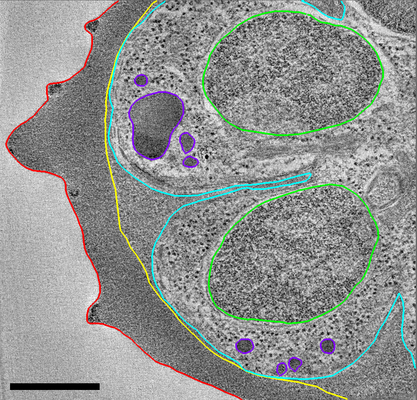
| タイトル | Malaria-infected red blood cell section showing partially segmented schizont | |||||||||
 マップデータ マップデータ | Resin section tomogram of Plasmodium falciparum infected erythrocyte | |||||||||
 試料 試料 |
| |||||||||
| 生物種 |  | |||||||||
| 手法 | 電子線トモグラフィー法 / ネガティブ染色法 | |||||||||
 データ登録者 データ登録者 | Hale VL / Saibil HR | |||||||||
 引用 引用 | ジャーナル: Proc Natl Acad Sci U S A / 年: 2017 タイトル: Parasitophorous vacuole poration precedes its rupture and rapid host erythrocyte cytoskeleton collapse in egress. 著者: Victoria L Hale / Jean M Watermeyer / Fiona Hackett / Gema Vizcay-Barrena / Christiaan van Ooij / James A Thomas / Matthew C Spink / Maria Harkiolaki / Elizabeth Duke / Roland A Fleck / ...著者: Victoria L Hale / Jean M Watermeyer / Fiona Hackett / Gema Vizcay-Barrena / Christiaan van Ooij / James A Thomas / Matthew C Spink / Maria Harkiolaki / Elizabeth Duke / Roland A Fleck / Michael J Blackman / Helen R Saibil /  要旨: In the asexual blood stages of malarial infection, merozoites invade erythrocytes and replicate within a parasitophorous vacuole to form daughter cells that eventually exit (egress) by sequential ...In the asexual blood stages of malarial infection, merozoites invade erythrocytes and replicate within a parasitophorous vacuole to form daughter cells that eventually exit (egress) by sequential rupture of the vacuole and erythrocyte membranes. The current model is that PKG, a malarial cGMP-dependent protein kinase, triggers egress, activating malarial proteases and other effectors. Using selective inhibitors of either PKG or cysteine proteases to separately inhibit the sequential steps in membrane perforation, combined with video microscopy, electron tomography, electron energy loss spectroscopy, and soft X-ray tomography of mature intracellular parasites, we resolve intermediate steps in egress. We show that the parasitophorous vacuole membrane (PVM) is permeabilized 10-30 min before its PKG-triggered breakdown into multilayered vesicles. Just before PVM breakdown, the host red cell undergoes an abrupt, dramatic shape change due to the sudden breakdown of the erythrocyte cytoskeleton, before permeabilization and eventual rupture of the erythrocyte membrane to release the parasites. In contrast to the previous view of PKG-triggered initiation of egress and a gradual dismantling of the host erythrocyte cytoskeleton over the course of schizont development, our findings identify an initial step in egress and show that host cell cytoskeleton breakdown is restricted to a narrow time window within the final stages of egress. | |||||||||
| 履歴 |
|
- 構造の表示
構造の表示
| ムービー |
 ムービービューア ムービービューア |
|---|---|
| 構造ビューア | EMマップ: SurfViewMolmilJmol/JSmol |
| 添付画像 |
- ダウンロードとリンク
-EMDBアーカイブ
| マップデータ | emd_3586.map.gz | 412.8 MB | EMDBマップデータ形式 | |
|---|---|---|---|---|
| ヘッダ (付随情報) | emd-3586-v30.xmlemd-3586.xml | 9.4 KB 9.4 KB | 表示 表示 | EMDBヘッダ |
| 画像 |  emd_3586.png emd_3586.png | 288.8 KB | ||
| アーカイブディレクトリ |  http://ftp.pdbj.org/pub/emdb/structures/EMD-3586ftp://ftp.pdbj.org/pub/emdb/structures/EMD-3586 http://ftp.pdbj.org/pub/emdb/structures/EMD-3586ftp://ftp.pdbj.org/pub/emdb/structures/EMD-3586 | HTTPS FTP |
-関連構造データ
| 関連構造データ |  3587C  3606C  3610C C: 同じ文献を引用 ( |
|---|---|
| 電子顕微鏡画像生データ | EMPIAR-10087 (タイトル: Soft X-ray tomography of Plasmodium falciparum infected human erythrocytes stalled in egress by the inhibitors Compound 2 and E64 Data size: 280.6 MB Data #1: Soft X-ray tomograms of Plasmodium falciparum infected human erythrocytes stalled in egress [Soft X-ray tomograms]) |
-リンク
| EMDBのページ | EMDB (EBI/PDBe) / EMDataResource |
|---|
-マップ
| ファイル | ダウンロード / ファイル: emd_3586.map.gz / 形式: CCP4 / 大きさ: 849.2 MB / タイプ: IMAGE STORED AS SIGNED BYTE | ||||||||||||||||||||||||||||||||||||||||||||||||||||||||||||
|---|---|---|---|---|---|---|---|---|---|---|---|---|---|---|---|---|---|---|---|---|---|---|---|---|---|---|---|---|---|---|---|---|---|---|---|---|---|---|---|---|---|---|---|---|---|---|---|---|---|---|---|---|---|---|---|---|---|---|---|---|---|
| 注釈 | Resin section tomogram of Plasmodium falciparum infected erythrocyte | ||||||||||||||||||||||||||||||||||||||||||||||||||||||||||||
| ボクセルのサイズ | X=Y=Z: 11.7 Å | ||||||||||||||||||||||||||||||||||||||||||||||||||||||||||||
| 密度 |
| ||||||||||||||||||||||||||||||||||||||||||||||||||||||||||||
| 対称性 | 空間群: 1 | ||||||||||||||||||||||||||||||||||||||||||||||||||||||||||||
| 詳細 | EMDB XML:
CCP4マップ ヘッダ情報:
| ||||||||||||||||||||||||||||||||||||||||||||||||||||||||||||
-添付データ
- 試料の構成要素
-全体 : Plasmodium falciparum infected human erythrocyte treated with the...
| 全体 | 名称: Plasmodium falciparum infected human erythrocyte treated with the egress inhibitor Compound 1 |
|---|---|
| 要素 |
|
-超分子 #1: Plasmodium falciparum infected human erythrocyte treated with the...
| 超分子 | 名称: Plasmodium falciparum infected human erythrocyte treated with the egress inhibitor Compound 1 タイプ: cell / ID: 1 / 親要素: 0 詳細: High pressure frozen and freeze-substituted cell section |
|---|---|
| 由来(天然) | 生物種: |
-実験情報
-構造解析
| 手法 | ネガティブ染色法 |
|---|---|
 解析 解析 | 電子線トモグラフィー法 |
| 試料の集合状態 | cell |
-試料調製
| 緩衝液 | pH: 7 / 詳細: RPMI medium |
|---|---|
| 染色 | タイプ: NEGATIVE / 材質: Uranyl Acetate |
| 糖包埋 | 材質: HM20 / 詳細: Freeze substitution |
| グリッド | 材質: COPPER / 支持フィルム - 材質: CARBON / 支持フィルム - トポロジー: CONTINUOUS / 前処理 - タイプ: GLOW DISCHARGE |
| 加圧凍結法 | 装置: OTHER 詳細: The value given for _emd_high_pressure_freezing.instrument is Baltech HPM010. This is not in a list of allowed values set(['LEICA EM PACT2', 'LEICA EM PACT', 'EMS-002 RAPID IMMERSION ...詳細: The value given for _emd_high_pressure_freezing.instrument is Baltech HPM010. This is not in a list of allowed values set(['LEICA EM PACT2', 'LEICA EM PACT', 'EMS-002 RAPID IMMERSION FREEZER', 'OTHER', 'LEICA EM HPM100', 'BAL-TEC HPM 010']) so OTHER is written into the XML file. |
| 切片作成 | ウルトラミクロトーム - 装置: Leica EM UC7 / ウルトラミクロトーム - 温度: 293 K / ウルトラミクロトーム - 最終 厚さ: 250 |
| 位置合わせマーカー | 直径: 10 nm |
- 電子顕微鏡法
| 顕微鏡 | FEI TECNAI F20 |
|---|---|
| 撮影 | フィルム・検出器のモデル: GATAN ULTRASCAN 4000 (4k x 4k) 平均電子線量: 100.0 e/Å2 |
| 電子線 | 加速電圧: 200 kV / 電子線源:  FIELD EMISSION GUN FIELD EMISSION GUN |
| 電子光学系 | 照射モード: FLOOD BEAM / 撮影モード: BRIGHT FIELD |
| 試料ステージ | 試料ホルダーモデル: OTHER |
| 実験機器 |  モデル: Tecnai F20 / 画像提供: FEI Company |
-画像解析
| 最終 再構成 | アルゴリズム: BACK PROJECTION / ソフトウェア - 名称: IMOD / 使用した粒子像数: 133 |
|---|
