[English] 日本語
 Yorodumi
- EMDB-35452: Cryo-EM structure of ochratoxin A-detoxifying amidohydrolase ADH3 -
+ Open data
- Basic information
| Entry |  | |||||||||
|---|---|---|---|---|---|---|---|---|---|---|
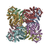
| Title | Cryo-EM structure of ochratoxin A-detoxifying amidohydrolase ADH3 | |||||||||
 Map data Map data | ||||||||||
 Sample Sample |
| |||||||||
 Keywords Keywords | amidohydrolase / octamer / ochratoxin A degradtion / cryo-EM structure / HYDROLASE | |||||||||
| Function / homology | hydrolase activity, acting on carbon-nitrogen (but not peptide) bonds / Amidohydrolase family / Metal-dependent hydrolase, composite domain superfamily / Amidohydrolase-related / Metal-dependent hydrolase / Amidohydrolase family protein Function and homology information Function and homology information | |||||||||
| Biological species |  Stenotrophomonas acidaminiphila (bacteria) Stenotrophomonas acidaminiphila (bacteria) | |||||||||
| Method | single particle reconstruction / cryo EM / Resolution: 2.71 Å | |||||||||
 Authors Authors | Dai LH / Niu D / Huang J-W / Li X / Shen PP / Li H / Hu YM / Yang Y / Chen C-C / Guo R-T | |||||||||
| Funding support |  China, 1 items China, 1 items
| |||||||||
 Citation Citation |  Journal: J Hazard Mater / Year: 2023 Journal: J Hazard Mater / Year: 2023Title: Cryo-EM structure and rational engineering of a superefficient ochratoxin A-detoxifying amidohydrolase. Authors: Longhai Dai / Du Niu / Jian-Wen Huang / Xian Li / Panpan Shen / Hao Li / Zhenzhen Xie / Jian Min / Yumei Hu / Yu Yang / Rey-Ting Guo / Chun-Chi Chen /  Abstract: Ochratoxin A (OTA) is among the most prevalent mycotoxins detected in agroproducts, posing serious threats to human and livestock health. Using enzymes to conduct OTA detoxification is an appealing ...Ochratoxin A (OTA) is among the most prevalent mycotoxins detected in agroproducts, posing serious threats to human and livestock health. Using enzymes to conduct OTA detoxification is an appealing potential strategy. The recently identified amidohydrolase from Stenotrophomonas acidaminiphila, termed ADH3, is the most efficient OTA-detoxifying enzyme reported thus far and can hydrolyze OTA to nontoxic ochratoxin α (OTα) and L-β-phenylalanine (Phe). To elucidate the catalytic mechanism of ADH3, we solved the single-particle cryo-electron microscopy (cryo-EM) structures of apo-form, Phe- and OTA-bound ADH3 to an overall resolution of 2.5-2.7 Å. The role of OTA-binding residues was investigated by structural, mutagenesis and biochemical analyses. We also rationally engineered ADH3 and obtained variant S88E, whose catalytic activity was elevated by 3.7-fold. Structural analysis of variant S88E indicates that the E88 side chain provides additional hydrogen bond interactions to the OTα moiety. Furthermore, the OTA-hydrolytic activity of variant S88E expressed in Pichia pastoris is comparable to that of Escherichia coli-expressed enzyme, revealing the feasibility of employing the industrial yeast strain to produce ADH3 and its variants for further applications. These results unveil a wealth of information about the catalytic mechanism of ADH3-mediated OTA degradation and provide a blueprint for rational engineering of high-efficiency OTA-detoxifying machineries. | |||||||||
| History |
|
- Structure visualization
| Supplemental images |
|---|
- Downloads & links
-EMDB archive
| Map data |  emd_35452.map.gz emd_35452.map.gz | 168 MB |  EMDB map data format EMDB map data format | |
|---|---|---|---|---|
| Header (meta data) |  emd-35452-v30.xml emd-35452-v30.xml emd-35452.xml emd-35452.xml | 16.9 KB 16.9 KB | Display Display |  EMDB header EMDB header |
| FSC (resolution estimation) |  emd_35452_fsc.xml emd_35452_fsc.xml | 11.9 KB | Display |  FSC data file FSC data file |
| Images |  emd_35452.png emd_35452.png | 93.2 KB | ||
| Filedesc metadata |  emd-35452.cif.gz emd-35452.cif.gz | 6.1 KB | ||
| Others |  emd_35452_half_map_1.map.gz emd_35452_half_map_1.map.gz emd_35452_half_map_2.map.gz emd_35452_half_map_2.map.gz | 164.9 MB 164.9 MB | ||
| Archive directory |  http://ftp.pdbj.org/pub/emdb/structures/EMD-35452 http://ftp.pdbj.org/pub/emdb/structures/EMD-35452 ftp://ftp.pdbj.org/pub/emdb/structures/EMD-35452 ftp://ftp.pdbj.org/pub/emdb/structures/EMD-35452 | HTTPS FTP |
-Validation report
| Summary document |  emd_35452_validation.pdf.gz emd_35452_validation.pdf.gz | 1005.7 KB | Display |  EMDB validaton report EMDB validaton report |
|---|---|---|---|---|
| Full document |  emd_35452_full_validation.pdf.gz emd_35452_full_validation.pdf.gz | 1005.3 KB | Display | |
| Data in XML |  emd_35452_validation.xml.gz emd_35452_validation.xml.gz | 20.7 KB | Display | |
| Data in CIF |  emd_35452_validation.cif.gz emd_35452_validation.cif.gz | 26.9 KB | Display | |
| Arichive directory |  https://ftp.pdbj.org/pub/emdb/validation_reports/EMD-35452 https://ftp.pdbj.org/pub/emdb/validation_reports/EMD-35452 ftp://ftp.pdbj.org/pub/emdb/validation_reports/EMD-35452 ftp://ftp.pdbj.org/pub/emdb/validation_reports/EMD-35452 | HTTPS FTP |
-Related structure data
| Related structure data |  8ihqMC  8ihrC  8ihsC  8j85C M: atomic model generated by this map C: citing same article ( |
|---|---|
| Similar structure data | Similarity search - Function & homology  F&H Search F&H Search |
- Links
Links
| EMDB pages |  EMDB (EBI/PDBe) / EMDB (EBI/PDBe) /  EMDataResource EMDataResource |
|---|---|
| Related items in Molecule of the Month |
- Map
Map
| File |  Download / File: emd_35452.map.gz / Format: CCP4 / Size: 178 MB / Type: IMAGE STORED AS FLOATING POINT NUMBER (4 BYTES) Download / File: emd_35452.map.gz / Format: CCP4 / Size: 178 MB / Type: IMAGE STORED AS FLOATING POINT NUMBER (4 BYTES) | ||||||||||||||||||||||||||||||||||||
|---|---|---|---|---|---|---|---|---|---|---|---|---|---|---|---|---|---|---|---|---|---|---|---|---|---|---|---|---|---|---|---|---|---|---|---|---|---|
| Projections & slices | Image control
Images are generated by Spider. | ||||||||||||||||||||||||||||||||||||
| Voxel size | X=Y=Z: 0.85 Å | ||||||||||||||||||||||||||||||||||||
| Density |
| ||||||||||||||||||||||||||||||||||||
| Symmetry | Space group: 1 | ||||||||||||||||||||||||||||||||||||
| Details | EMDB XML:
|
-Supplemental data
-Half map: #1
| File | emd_35452_half_map_1.map | ||||||||||||
|---|---|---|---|---|---|---|---|---|---|---|---|---|---|
| Projections & Slices |
| ||||||||||||
| Density Histograms |
-Half map: #2
| File | emd_35452_half_map_2.map | ||||||||||||
|---|---|---|---|---|---|---|---|---|---|---|---|---|---|
| Projections & Slices |
| ||||||||||||
| Density Histograms |
- Sample components
-Entire : ADH3
| Entire | Name: ADH3 |
|---|---|
| Components |
|
-Supramolecule #1: ADH3
| Supramolecule | Name: ADH3 / type: complex / ID: 1 / Parent: 0 / Macromolecule list: #1 |
|---|---|
| Source (natural) | Organism:  Stenotrophomonas acidaminiphila (bacteria) Stenotrophomonas acidaminiphila (bacteria) |
-Macromolecule #1: Amidohydrolase family protein
| Macromolecule | Name: Amidohydrolase family protein / type: protein_or_peptide / ID: 1 / Number of copies: 8 / Enantiomer: LEVO |
|---|---|
| Source (natural) | Organism:  Stenotrophomonas acidaminiphila (bacteria) Stenotrophomonas acidaminiphila (bacteria) |
| Molecular weight | Theoretical: 45.717617 KDa |
| Recombinant expression | Organism:  |
| Sequence | String: MPIRRRFASL LLLACAPAWA EPVAVQCGRL FDARSGQLKG PHTLLVADGR IRQVLPGTGA DAAGARVVDL GDKVCLPGWT DLHVHLGSQ SSPQSYSEDF RLDPVDHAFR AVGYAEKTLM AGFTSVRDLG GEVSPHLRDA INQGLVRGPR IFAAGKSIAT T GGHADPTN ...String: MPIRRRFASL LLLACAPAWA EPVAVQCGRL FDARSGQLKG PHTLLVADGR IRQVLPGTGA DAAGARVVDL GDKVCLPGWT DLHVHLGSQ SSPQSYSEDF RLDPVDHAFR AVGYAEKTLM AGFTSVRDLG GEVSPHLRDA INQGLVRGPR IFAAGKSIAT T GGHADPTN GWNERLAHLV GAPGPAEGVV NSVDEARQAV RQRYKEGSDL I(KCX)ITATGGVL SYARSGDAPQ FTVDEIKA V VDTARDYGFR VAAHAHGTEG MKRAVQAGVT SIEHGTYMDD EVMRLMKQHG TWYVPTFYAG RFVTEKAAID GYFPEVVRP KAARIGALIS QTAAKAYRNG VRIAFGTDQG VGPHGDNARE FVYMVEAGIP AAYALQAATV HAAQVLGVDD QGVLEPGKRA DVIALAGNP LEDINAVLDV RFVMKDGVIY KQ UniProtKB: Amidohydrolase family protein |
-Macromolecule #2: ZINC ION
| Macromolecule | Name: ZINC ION / type: ligand / ID: 2 / Number of copies: 16 / Formula: ZN |
|---|---|
| Molecular weight | Theoretical: 65.409 Da |
-Experimental details
-Structure determination
| Method | cryo EM |
|---|---|
 Processing Processing | single particle reconstruction |
| Aggregation state | particle |
- Sample preparation
| Concentration | 0.6 mg/mL |
|---|---|
| Buffer | pH: 7.5 / Details: 20 mM Tris-HCL,pH 7.5 |
| Vitrification | Cryogen name: ETHANE |
- Electron microscopy
| Microscope | FEI TITAN KRIOS |
|---|---|
| Image recording | Film or detector model: GATAN K3 (6k x 4k) / Average electron dose: 52.0 e/Å2 |
| Electron beam | Acceleration voltage: 300 kV / Electron source:  FIELD EMISSION GUN FIELD EMISSION GUN |
| Electron optics | Illumination mode: FLOOD BEAM / Imaging mode: BRIGHT FIELD / Nominal defocus max: 2.4 µm / Nominal defocus min: 1.0 µm |
| Experimental equipment |  Model: Titan Krios / Image courtesy: FEI Company |
 Movie
Movie Controller


 Z (Sec.)
 Y (Row.)
 X (Col.)


