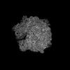
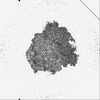
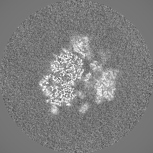
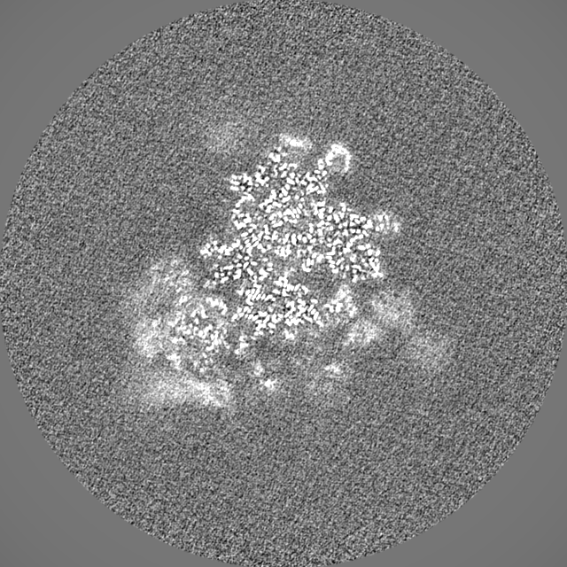
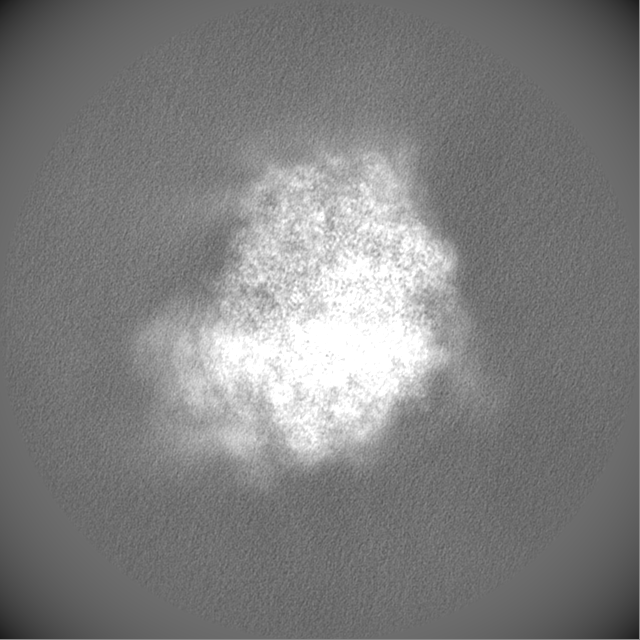
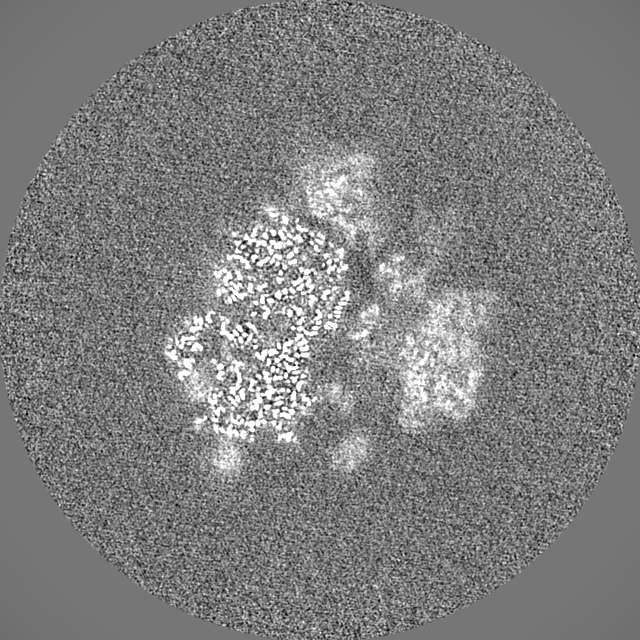
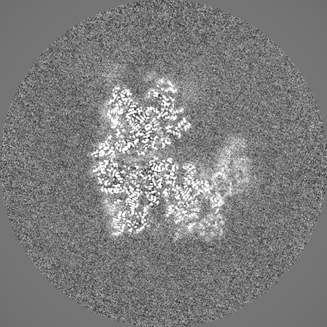
- EMDB-35413: Dibekacin-added human 80S ribosome -
+
データを開く
IDまたはキーワード:
読み込み中...
-
基本情報
登録情報
データベース: EMDB / ID: EMD-35413
タイトル
Dibekacin-added human 80S ribosome
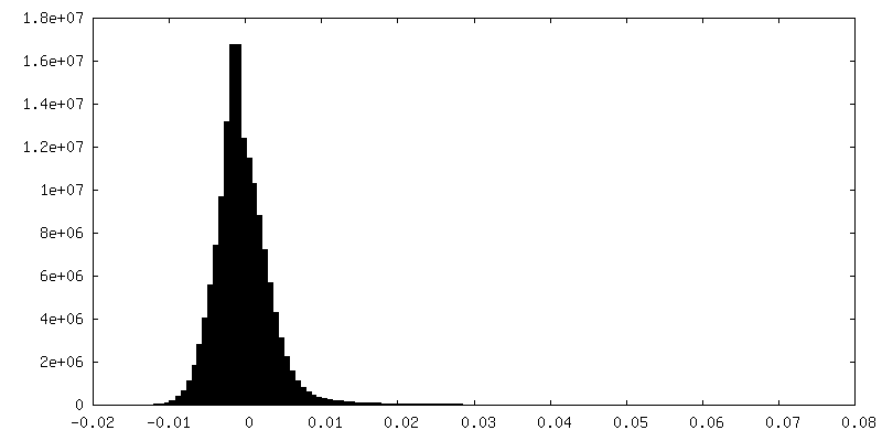
マップデータ
試料
複合体: DBK-added human 80S ribosome
RNA: x 4種
タンパク質・ペプチド: x 76種
リガンド: x 3種
キーワード
Ribosome
機能・相同性
機能・相同性情報
translation at presynapse / exit from mitosis / eukaryotic 80S initiation complex / negative regulation of protein neddylation / response to insecticide / optic nerve development / negative regulation of endoplasmic reticulum unfolded protein response / regulation of G1 to G0 transition / axial mesoderm development / oxidized pyrimidine DNA binding ...translation at presynapse / exit from mitosis / eukaryotic 80S initiation complex / negative regulation of protein neddylation / response to insecticide / optic nerve development / negative regulation of endoplasmic reticulum unfolded protein response / regulation of G1 to G0 transition / axial mesoderm development / oxidized pyrimidine DNA binding / response to TNF agonist / negative regulation of formation of translation preinitiation complex / positive regulation of base-excision repair / regulation of translation involved in cellular response to UV / ribosomal protein import into nucleus / positive regulation of respiratory burst involved in inflammatory response / positive regulation of intrinsic apoptotic signaling pathway in response to DNA damage / protein-DNA complex disassembly / positive regulation of gastrulation / regulation of adenylate cyclase-activating G protein-coupled receptor signaling pathway / 90S preribosome assembly / protein tyrosine kinase inhibitor activity / IRE1-RACK1-PP2A complex / positive regulation of endodeoxyribonuclease activity / nucleolus organization / positive regulation of Golgi to plasma membrane protein transport / positive regulation of intrinsic apoptotic signaling pathway in response to DNA damage by p53 class mediator / retinal ganglion cell axon guidance / TNFR1-mediated ceramide production / negative regulation of RNA splicing / negative regulation of DNA repair / GAIT complex / positive regulation of DNA damage response, signal transduction by p53 class mediator / TORC2 complex binding / alpha-beta T cell differentiation / G1 to G0 transition / supercoiled DNA binding / neural crest cell differentiation / positive regulation of ubiquitin-protein transferase activity / NF-kappaB complex / cysteine-type endopeptidase activator activity involved in apoptotic process / oxidized purine DNA binding / negative regulation of intrinsic apoptotic signaling pathway in response to hydrogen peroxide / negative regulation of bicellular tight junction assembly / regulation of establishment of cell polarity / ubiquitin-like protein conjugating enzyme binding / middle ear morphogenesis / negative regulation of phagocytosis / rRNA modification in the nucleus and cytosol / Formation of the ternary complex, and subsequently, the 43S complex / erythrocyte homeostasis / cytoplasmic side of rough endoplasmic reticulum membrane / laminin receptor activity / negative regulation of ubiquitin protein ligase activity / ion channel inhibitor activity / protein kinase A binding / pigmentation / Ribosomal scanning and start codon recognition / homeostatic process / Translation initiation complex formation / positive regulation of mitochondrial depolarization / macrophage chemotaxis / positive regulation of T cell receptor signaling pathway / fibroblast growth factor binding / negative regulation of Wnt signaling pathway / lung morphogenesis / male meiosis I / monocyte chemotaxis / positive regulation of activated T cell proliferation / positive regulation of natural killer cell proliferation / negative regulation of translational frameshifting / Protein hydroxylation / TOR signaling / BH3 domain binding / regulation of cell division / SARS-CoV-1 modulates host translation machinery / mTORC1-mediated signalling / cellular response to ethanol / iron-sulfur cluster binding / Peptide chain elongation / Selenocysteine synthesis / Formation of a pool of free 40S subunits / positive regulation of intrinsic apoptotic signaling pathway by p53 class mediator / endonucleolytic cleavage to generate mature 3'-end of SSU-rRNA from (SSU-rRNA, 5.8S rRNA, LSU-rRNA) / Eukaryotic Translation Termination / ubiquitin ligase inhibitor activity / blastocyst development / cellular response to actinomycin D / Response of EIF2AK4 (GCN2) to amino acid deficiency / positive regulation of signal transduction by p53 class mediator / negative regulation of ubiquitin-dependent protein catabolic process / SRP-dependent cotranslational protein targeting to membrane / protein serine/threonine kinase inhibitor activity / Viral mRNA Translation / negative regulation of respiratory burst involved in inflammatory response / Nonsense Mediated Decay (NMD) independent of the Exon Junction Complex (EJC) / protein localization to nucleus / GTP hydrolysis and joining of the 60S ribosomal subunit / L13a-mediated translational silencing of Ceruloplasmin expression / Major pathway of rRNA processing in the nucleolus and cytosol 類似検索 - 分子機能
40S ribosomal protein SA / 40S ribosomal protein SA, C-terminal domain / 40S ribosomal protein SA C-terminus / Ribosomal protein L6, N-terminal / Ribosomal protein L6, N-terminal domain / Ubiquitin-like protein FUBI / Ribosomal protein L30e / Ribosomal protein L28e / Ribosomal L15/L27a, N-terminal / Ribosomal protein L2, archaeal-type ...40S ribosomal protein SA / 40S ribosomal protein SA, C-terminal domain / 40S ribosomal protein SA C-terminus / Ribosomal protein L6, N-terminal / Ribosomal protein L6, N-terminal domain / Ubiquitin-like protein FUBI / Ribosomal protein L30e / Ribosomal protein L28e / Ribosomal L15/L27a, N-terminal / Ribosomal protein L2, archaeal-type / Ribosomal protein L23 / Ribosomal L28e/Mak16 / Ribosomal L28e protein family / Ribosomal protein L19e, C-terminal domain / metallochaperone-like domain / TRASH domain / : / Ribosomal protein S26e signature. / Ribosomal protein L41 / Ribosomal protein L41 / Ribosomal protein S21e, conserved site / Ribosomal protein S21e signature. / Ribosomal protein L1, conserved site / Ribosomal protein L1 signature. / Ribosomal protein S26e / Ribosomal protein S26e superfamily / Ribosomal protein S26e / : / Ribosomal protein S12e signature. / Ribosomal protein L1 / Ribosomal protein S12e / Ribosomal protein L29e / Ribosomal L29e protein family / Ribosomal protein S19e, conserved site / Ribosomal protein S19e signature. / Ribosomal protein L13e, conserved site / Ribosomal protein L13e signature. / Ribosomal protein S5, eukaryotic/archaeal / Small (40S) ribosomal subunit Asc1/RACK1 / Ribosomal protein S21e / Ribosomal protein S21e superfamily / Ribosomal protein S21e / Ribosomal protein S2, eukaryotic / Ribosomal protein L22e / Ribosomal protein L22e superfamily / Ribosomal L22e protein family / Ribosomal protein L27e, conserved site / S27a-like superfamily / Ribosomal protein L27e signature. / Ribosomal protein L10e, conserved site / Ribosomal protein L10e signature. / Ribosomal protein L38e / Ribosomal protein L38e superfamily / Ribosomal L38e protein family / Ribosomal protein L10e / 40S Ribosomal protein S10 / Ribosomal protein L1, 3-layer alpha/beta-sandwich / : / Ribosomal protein L44e signature. / Ribosomal protein S7e signature. / Ribosomal protein L24e, conserved site / Ribosomal protein L24e signature. / Ribosomal protein L13e / Ribosomal protein L13e / Ribosomal protein L19/L19e conserved site / Ribosomal protein L19, eukaryotic / Ribosomal protein L19e signature. / Plectin/S10, N-terminal / Plectin/S10 domain / : / 60S ribosomal protein L18a/ L20, eukaryotes / Ribosomal protein L6e signature. / Ribosomal protein S10, eukaryotic/archaeal / Ribosomal protein S8e subdomain, eukaryotes / : / Ribosomal protein S17e, conserved site / Ribosomal protein S17e signature. / Ribosomal protein S25 / S25 ribosomal protein / Ribosomal protein L44e / Ribosomal protein S3Ae, conserved site / Ribosomal protein L44 / Ribosomal protein S3Ae signature. / Ribosomal protein S27a / Ribosomal protein S27a / Ribosomal protein S27a / Ribosomal protein S30 / Ribosomal protein S30 / Ribosomal protein L34e, conserved site / Ribosomal protein L34e signature. / Ribosomal protein S2, eukaryotic/archaeal / Ribosomal protein L5 eukaryotic, C-terminal / 50S ribosomal protein L18Ae/60S ribosomal protein L20 and L18a / Ribosomal L40e family / Ribosomal L18 C-terminal region / Ribosomal protein 50S-L18Ae/60S-L20/60S-L18A / Ribosomal proteins 50S-L18Ae/60S-L20/60S-L18A / Ribosomal protein 60S L18 and 50S L18e / Ribosomal protein L30e signature 1. / Ribosomal_L40e 類似検索 - ドメイン・相同性
Small ribosomal subunit protein eS17 / Small ribosomal subunit protein uS2 / Small ribosomal subunit protein uS5 / Large ribosomal subunit protein eL33 / Large ribosomal subunit protein uL30 / Large ribosomal subunit protein uL22 / Small ribosomal subunit protein uS3 / Small ribosomal subunit protein eS12 / Large ribosomal subunit protein eL13 / Large ribosomal subunit protein uL6 ...Small ribosomal subunit protein eS17 / Small ribosomal subunit protein uS2 / Small ribosomal subunit protein uS5 / Large ribosomal subunit protein eL33 / Large ribosomal subunit protein uL30 / Large ribosomal subunit protein uL22 / Small ribosomal subunit protein uS3 / Small ribosomal subunit protein eS12 / Large ribosomal subunit protein eL13 / Large ribosomal subunit protein uL6 / Large ribosomal subunit protein eL22 / Large ribosomal subunit protein uL4 / Small ribosomal subunit protein eS19 / Large ribosomal subunit protein uL3 / Large ribosomal subunit protein uL13 / Small ribosomal subunit protein eS27 / Large ribosomal subunit protein uL29 / Large ribosomal subunit protein uL15 / Large ribosomal subunit protein uL18 / Large ribosomal subunit protein eL21 / Large ribosomal subunit protein eL28 / Small ribosomal subunit protein uS4 / Small ribosomal subunit protein uS7 / Small ribosomal subunit protein eS10 / Large ribosomal subunit protein eL29 / Large ribosomal subunit protein eL34 / Large ribosomal subunit protein eL14 / Small ribosomal subunit protein uS10 / Small ribosomal subunit protein eS1 / Large ribosomal subunit protein uL24 / Large ribosomal subunit protein eL15 / Large ribosomal subunit protein eL27 / Large ribosomal subunit protein eL43 / Large ribosomal subunit protein eL37 / Small ribosomal subunit protein eS7 / Small ribosomal subunit protein eS8 / Small ribosomal subunit protein uS8 / Small ribosomal subunit protein uS9 / Small ribosomal subunit protein uS11 / Small ribosomal subunit protein uS12 / Small ribosomal subunit protein uS13 / Small ribosomal subunit protein uS14 / Small ribosomal subunit protein uS15 / Small ribosomal subunit protein uS17 / Large ribosomal subunit protein eL8 / Small ribosomal subunit protein eS4, X isoform / Large ribosomal subunit protein uL23 / Small ribosomal subunit protein eS6 / Large ribosomal subunit protein uL14 / Small ribosomal subunit protein uS19 / Small ribosomal subunit protein eS24 / Small ribosomal subunit protein eS25 / Small ribosomal subunit protein eS26 / Small ribosomal subunit protein eS28 / Ubiquitin-like FUBI-ribosomal protein eS30 fusion protein / Large ribosomal subunit protein eL30 / Large ribosomal subunit protein eL39 / Large ribosomal subunit protein eL31 / Large ribosomal subunit protein uL1 / Large ribosomal subunit protein eL32 / Large ribosomal subunit protein uL5 / Large ribosomal subunit protein uL2 / Small ribosomal subunit protein eS32 / Ubiquitin-ribosomal protein eS31 fusion protein / Ubiquitin-ribosomal protein eL40 fusion protein / Large ribosomal subunit protein eL38 / Small ribosomal subunit protein eS21 / Small ribosomal subunit protein RACK1 / Large ribosomal subunit protein eL24 / Large ribosomal subunit protein eL42 / Large ribosomal subunit protein eL19 / Large ribosomal subunit protein eL20 / Large ribosomal subunit protein eL6 / Large ribosomal subunit protein eL18 / Ribosomal protein uL16-like / Large ribosomal subunit protein eL36 類似検索 - 構成要素
Japan Agency for Medical Research and Development (AMED)
21fk0108093j0003
日本
Japan Science and Technology
JPMJPR20EG
日本
引用
ジャーナル: J Biochem / 年: 2024 タイトル: Direct visualization of ribosomes in the cell-free system revealed the functional evolution of aminoglycoside. 著者: Junta Tomono / Kosuke Asano / Takuma Chiashi / Masato Suzuki / Masayuki Igarashi / Yoshiaki Takahashi / Yoshikazu Tanaka / Takeshi Yokoyama / 要旨: The rapid emergence of multi-drug-resistant bacteria has raised a serious public health concern. Therefore, new antibiotic developments have been highly desired. Here, we propose a new method to ...The rapid emergence of multi-drug-resistant bacteria has raised a serious public health concern. Therefore, new antibiotic developments have been highly desired. Here, we propose a new method to visualize antibiotic actions on translating ribosomes in the cell-free system under macromolecular crowding conditions by cryo-electron microscopy, designated as the DARC method: the Direct visualization of Antibiotic binding on Ribosomes in the Cell-free translation system. This new method allows for acquiring a more comprehensive understanding of the mode of action of antibiotics on the translation inhibition without ribosome purification. Furthermore, with the direct link to biochemical analysis at the same condition as cryo-EM observation, we revealed the evolution of 2-DOS aminoglycosides from dibekacin (DBK) to arbekacin (ABK) by acquiring the synthetic tailored anchoring motif to lead to stronger binding affinity to ribosomes. Our cryo-EM structures of DBK and ABK bound ribosomes in the cell-free environment clearly depicted a synthetic tailored γ-amino-α-hydroxybutyryl (HABA) motif formed additional interactions with the ribosome enhancing antibiotic bindings. This new approach would be valuable for understanding the function of antibiotics for more efficient drug development.
 ムービー
ムービー コントローラー


 データを開く
 基本情報
基本情報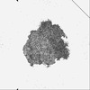
 マップデータ
 試料
試料 キーワード
キーワード 機能・相同性情報
 Homo sapiens (ヒト)
 データ登録者
 日本, 2件
  引用
引用 構造の表示
構造の表示
 ダウンロードとリンク
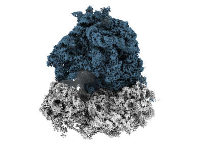 emd_35413.png
 http://ftp.pdbj.org/pub/emdb/structures/EMD-35413


 Z (Sec.)
 Y (Row.)
 X (Col.)


 試料の構成要素
 解析
解析 電子顕微鏡法
 FIELD EMISSION GUN
