+ Open data
- Basic information
| Entry |  | |||||||||||||||
|---|---|---|---|---|---|---|---|---|---|---|---|---|---|---|---|---|
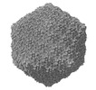
| Title | Capsid of DT57C bacteriophage in the full state | |||||||||||||||
 Map data Map data | Reconstruction of the capsid of full DT57C bacteriophage | |||||||||||||||
 Sample Sample |
| |||||||||||||||
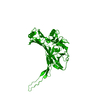
 Keywords Keywords | Capsid / T5 / MHP / VIRUS | |||||||||||||||
| Function / homology | : / Phage capsid / Phage capsid family / virion component / Major head protein Function and homology information Function and homology information | |||||||||||||||
| Biological species |  Escherichia phage DT57C (virus) Escherichia phage DT57C (virus) | |||||||||||||||
| Method | single particle reconstruction / cryo EM / Resolution: 2.9 Å | |||||||||||||||
 Authors Authors | Ayala R / Moiseenko AV / Chen TH / Kulikov EE / Golomidova AK / Orekhov PS / Street MA / Sokolova OS / Letarov AV / Wolf M | |||||||||||||||
| Funding support |  Russian Federation, Russian Federation,  Japan, 4 items Japan, 4 items
| |||||||||||||||
 Citation Citation |  Journal: Nat Commun / Year: 2023 Journal: Nat Commun / Year: 2023Title: Nearly complete structure of bacteriophage DT57C reveals architecture of head-to-tail interface and lateral tail fibers. Authors: Rafael Ayala / Andrey V Moiseenko / Ting-Hua Chen / Eugene E Kulikov / Alla K Golomidova / Philipp S Orekhov / Maya A Street / Olga S Sokolova / Andrey V Letarov / Matthias Wolf /     Abstract: The T5 family of viruses are tailed bacteriophages characterized by a long non-contractile tail. The bacteriophage DT57C is closely related to the paradigmal T5 phage, though it recognizes a ...The T5 family of viruses are tailed bacteriophages characterized by a long non-contractile tail. The bacteriophage DT57C is closely related to the paradigmal T5 phage, though it recognizes a different receptor (BtuB) and features highly divergent lateral tail fibers (LTF). Considerable portions of T5-like phages remain structurally uncharacterized. Here, we present the structure of DT57C determined by cryo-EM, and an atomic model of the virus, which was further explored using all-atom molecular dynamics simulations. The structure revealed a unique way of LTF attachment assisted by a dodecameric collar protein LtfC, and an unusual composition of the phage neck constructed of three protein rings. The tape measure protein (TMP) is organized within the tail tube in a three-stranded parallel α-helical coiled coil which makes direct contact with the genomic DNA. The presence of the C-terminal fragment of the TMP that remains within the tail tip suggests that the tail tip complex returns to its original state after DNA ejection. Our results provide a complete atomic structure of a T5-like phage, provide insights into the process of DNA ejection as well as a structural basis for the design of engineered phages and future mechanistic studies. | |||||||||||||||
| History |
|
- Structure visualization
| Supplemental images |
|---|
- Downloads & links
-EMDB archive
| Map data |  emd_34920.map.gz emd_34920.map.gz | 2.6 GB |  EMDB map data format EMDB map data format | |
|---|---|---|---|---|
| Header (meta data) |  emd-34920-v30.xml emd-34920-v30.xml emd-34920.xml emd-34920.xml | 17.9 KB 17.9 KB | Display Display |  EMDB header EMDB header |
| FSC (resolution estimation) |  emd_34920_fsc.xml emd_34920_fsc.xml | 29.5 KB | Display |  FSC data file FSC data file |
| Images |  emd_34920.png emd_34920.png | 153.6 KB | ||
| Filedesc metadata |  emd-34920.cif.gz emd-34920.cif.gz | 6 KB | ||
| Others |  emd_34920_half_map_1.map.gz emd_34920_half_map_1.map.gz emd_34920_half_map_2.map.gz emd_34920_half_map_2.map.gz | 2.5 GB 2.5 GB | ||
| Archive directory |  http://ftp.pdbj.org/pub/emdb/structures/EMD-34920 http://ftp.pdbj.org/pub/emdb/structures/EMD-34920 ftp://ftp.pdbj.org/pub/emdb/structures/EMD-34920 ftp://ftp.pdbj.org/pub/emdb/structures/EMD-34920 | HTTPS FTP |
-Validation report
| Summary document |  emd_34920_validation.pdf.gz emd_34920_validation.pdf.gz | 1008 KB | Display |  EMDB validaton report EMDB validaton report |
|---|---|---|---|---|
| Full document |  emd_34920_full_validation.pdf.gz emd_34920_full_validation.pdf.gz | 1007.6 KB | Display | |
| Data in XML |  emd_34920_validation.xml.gz emd_34920_validation.xml.gz | 38.2 KB | Display | |
| Data in CIF |  emd_34920_validation.cif.gz emd_34920_validation.cif.gz | 52.5 KB | Display | |
| Arichive directory |  https://ftp.pdbj.org/pub/emdb/validation_reports/EMD-34920 https://ftp.pdbj.org/pub/emdb/validation_reports/EMD-34920 ftp://ftp.pdbj.org/pub/emdb/validation_reports/EMD-34920 ftp://ftp.pdbj.org/pub/emdb/validation_reports/EMD-34920 | HTTPS FTP |
-Related structure data
| Related structure data |  8ho3MC  8hqkC  8hqoC  8hqzC  8hreC  8hrgC M: atomic model generated by this map C: citing same article ( |
|---|---|
| Similar structure data | Similarity search - Function & homology  F&H Search F&H Search |
- Links
Links
| EMDB pages |  EMDB (EBI/PDBe) / EMDB (EBI/PDBe) /  EMDataResource EMDataResource |
|---|---|
| Related items in Molecule of the Month |
- Map
Map
| File |  Download / File: emd_34920.map.gz / Format: CCP4 / Size: 2.7 GB / Type: IMAGE STORED AS FLOATING POINT NUMBER (4 BYTES) Download / File: emd_34920.map.gz / Format: CCP4 / Size: 2.7 GB / Type: IMAGE STORED AS FLOATING POINT NUMBER (4 BYTES) | ||||||||||||||||||||||||||||||||||||
|---|---|---|---|---|---|---|---|---|---|---|---|---|---|---|---|---|---|---|---|---|---|---|---|---|---|---|---|---|---|---|---|---|---|---|---|---|---|
| Annotation | Reconstruction of the capsid of full DT57C bacteriophage | ||||||||||||||||||||||||||||||||||||
| Projections & slices | Image control
Images are generated by Spider. | ||||||||||||||||||||||||||||||||||||
| Voxel size | X=Y=Z: 1.387 Å | ||||||||||||||||||||||||||||||||||||
| Density |
| ||||||||||||||||||||||||||||||||||||
| Symmetry | Space group: 1 | ||||||||||||||||||||||||||||||||||||
| Details | EMDB XML:
|
-Supplemental data
-Half map: Half map B
| File | emd_34920_half_map_1.map | ||||||||||||
|---|---|---|---|---|---|---|---|---|---|---|---|---|---|
| Annotation | Half map B | ||||||||||||
| Projections & Slices |
| ||||||||||||
| Density Histograms |
-Half map: Half map A
| File | emd_34920_half_map_2.map | ||||||||||||
|---|---|---|---|---|---|---|---|---|---|---|---|---|---|
| Annotation | Half map A | ||||||||||||
| Projections & Slices |
| ||||||||||||
| Density Histograms |
- Sample components
-Entire : Escherichia phage DT57C
| Entire | Name:  Escherichia phage DT57C (virus) Escherichia phage DT57C (virus) |
|---|---|
| Components |
|
-Supramolecule #1: Escherichia phage DT57C
| Supramolecule | Name: Escherichia phage DT57C / type: virus / ID: 1 / Parent: 0 / Macromolecule list: all / NCBI-ID: 2681606 / Sci species name: Escherichia phage DT57C / Virus type: VIRION / Virus isolate: STRAIN / Virus enveloped: No / Virus empty: No |
|---|
-Macromolecule #1: Major head protein
| Macromolecule | Name: Major head protein / type: protein_or_peptide / ID: 1 / Number of copies: 13 / Enantiomer: LEVO |
|---|---|
| Source (natural) | Organism:  Escherichia phage DT57C (virus) Escherichia phage DT57C (virus) |
| Molecular weight | Theoretical: 50.691398 KDa |
| Sequence | String: MTIDINKLKE ELGLGDLAKS LEGLTAAQKA QEAERMRKEQ EEKELARMNA LVSKAVGEDR QKLEQALELV KSLDEKSKKS AELFAQTVE KQQETIVGLQ DEIKSLLTAR EGRSFVGDSV AKALYGTQET FEDEVEKLVL LSYVMEKGVF ETEHGQKHLK A VNQSSSVE ...String: MTIDINKLKE ELGLGDLAKS LEGLTAAQKA QEAERMRKEQ EEKELARMNA LVSKAVGEDR QKLEQALELV KSLDEKSKKS AELFAQTVE KQQETIVGLQ DEIKSLLTAR EGRSFVGDSV AKALYGTQET FEDEVEKLVL LSYVMEKGVF ETEHGQKHLK A VNQSSSVE VSSESYETIF SQRIIRDLQK ELVVGALFEE LPMSSKILTM LVEPDAGRAT WVAASAYGSD NTTGSEVTGA LT EIHFSTY KLAAKSFITD ETEEDAIFSL LPLLRKRLIE AHAVSIEEAF MTGDGSGKPK GLLTLASEDS AKVTTEAKAD GSV LVTAKT ISKLRRKLGR HGLKLSKLVL IVSMDAYYDL LEDEEWQDVA QVGNDAVKLQ GQVGRIYGLP VVVSEYFPAK AAGK EFAVI VYKDNFVMPR QRAVTVERER QAGKQRDAYY VTQRVNLQRY FENGVVSGAY AAS UniProtKB: Major head protein |
-Experimental details
-Structure determination
| Method | cryo EM |
|---|---|
 Processing Processing | single particle reconstruction |
| Aggregation state | particle |
- Sample preparation
| Buffer | pH: 7.5 |
|---|---|
| Vitrification | Cryogen name: ETHANE-PROPANE |
- Electron microscopy
| Microscope | FEI TITAN KRIOS |
|---|---|
| Image recording | Film or detector model: FEI FALCON III (4k x 4k) / Average electron dose: 67.0 e/Å2 |
| Electron beam | Acceleration voltage: 300 kV / Electron source:  FIELD EMISSION GUN FIELD EMISSION GUN |
| Electron optics | Illumination mode: FLOOD BEAM / Imaging mode: BRIGHT FIELD / Nominal defocus max: 4.0 µm / Nominal defocus min: 1.0 µm |
| Experimental equipment |  Model: Titan Krios / Image courtesy: FEI Company |
 Movie
Movie Controller


 Z (Sec.)
 Y (Row.)
 X (Col.)


