Movie
Movie Controller


+ Open data

- Basic information
| Entry | 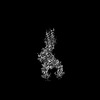 | |||||||||
|---|---|---|---|---|---|---|---|---|---|---|
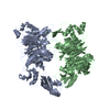
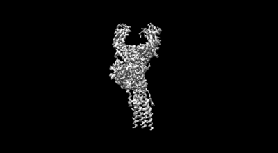
| Title | Structure of a membrane protein G | |||||||||
 Map data Map data | ||||||||||
 Sample Sample |
| |||||||||
 Keywords Keywords | membrane / IMMUNE SYSTEM | |||||||||
| Function / homology |  Function and homology information Function and homology informationIgM B cell receptor complex / B cell receptor complex / Fc-gamma receptor I complex binding / CD22 mediated BCR regulation / complement-dependent cytotoxicity / IgG immunoglobulin complex / antibody-dependent cellular cytotoxicity / immunoglobulin receptor binding / immunoglobulin complex, circulating / Classical antibody-mediated complement activation ...IgM B cell receptor complex / B cell receptor complex / Fc-gamma receptor I complex binding / CD22 mediated BCR regulation / complement-dependent cytotoxicity / IgG immunoglobulin complex / antibody-dependent cellular cytotoxicity / immunoglobulin receptor binding / immunoglobulin complex, circulating / Classical antibody-mediated complement activation / Initial triggering of complement / B cell activation / B cell proliferation / FCGR activation / complement activation, classical pathway / Role of phospholipids in phagocytosis / antigen binding / multivesicular body / FCGR3A-mediated IL10 synthesis / Antigen activates B Cell Receptor (BCR) leading to generation of second messengers / Regulation of Complement cascade / B cell differentiation / B cell receptor signaling pathway / FCGR3A-mediated phagocytosis / response to bacterium / Regulation of actin dynamics for phagocytic cup formation / antibacterial humoral response / transmembrane signaling receptor activity / Interleukin-4 and Interleukin-13 signaling / blood microparticle / Potential therapeutics for SARS / adaptive immune response / immune response / membrane raft / external side of plasma membrane / signal transduction / extracellular space / extracellular exosome / extracellular region / identical protein binding / plasma membrane Similarity search - Function | |||||||||
| Biological species |  Homo sapiens (human) Homo sapiens (human) | |||||||||
| Method | single particle reconstruction / cryo EM / Resolution: 3.03 Å | |||||||||
 Authors Authors | Ma X / Zhu Y | |||||||||
| Funding support |  China, 1 items China, 1 items
| |||||||||
 Citation Citation | Journal: Science / Year: 2022 Title: Cryo-EM structures of two human B cell receptor isotypes. Authors: Xinyu Ma / Yuwei Zhu / De Dong / Yan Chen / Shubo Wang / Dehui Yang / Zhuo Ma / Anqi Zhang / Fan Zhang / Changyou Guo / Zhiwei Huang / Abstract: The B cell receptor (BCR) complex plays a critical role in B cell development and immune responses. The assembly mechanisms underlying the BCR complex remain unknown. We determined the cryo-electron ...The B cell receptor (BCR) complex plays a critical role in B cell development and immune responses. The assembly mechanisms underlying the BCR complex remain unknown. We determined the cryo-electron microscopy (cryo-EM) structures of human IgG-BCR and IgM-BCR, which consist of membrane-bound immunoglobulin molecules (mIg) and Igα/β subunits at a 1:1 stoichiometry. Assembly of both BCR complexes involves their extracellular domains, membrane-proximal connection peptides, and transmembrane (TM) helices. The TM helices of mIgG and mIgM share a conserved set of hydrophobic and polar interactions with Igα/β TM helices. By contrast, the IgG-Cγ3 and IgM-Cμ4 domains interact with extracellular Ig-like domains of Igα/β through head-to-tail and side-by-side modes, respectively. This work reveals the structural basis for BCR assembly and provides insights into BCR triggering. | |||||||||
| History |
|
- Structure visualization
| Supplemental images |
|---|
- Downloads & links
-EMDB archive
| Map data | emd_32762.map.gz | 59.8 MB | EMDB map data format | |
|---|---|---|---|---|
| Header (meta data) | emd-32762-v30.xmlemd-32762.xml | 11.7 KB 11.7 KB | Display Display | EMDB header |
| Images |  emd_32762.png emd_32762.png | 19.3 KB | ||
| Filedesc metadata | emd-32762.cif.gz | 5.2 KB | ||
| Archive directory |  http://ftp.pdbj.org/pub/emdb/structures/EMD-32762ftp://ftp.pdbj.org/pub/emdb/structures/EMD-32762 http://ftp.pdbj.org/pub/emdb/structures/EMD-32762ftp://ftp.pdbj.org/pub/emdb/structures/EMD-32762 | HTTPS FTP |
-Related structure data
| Related structure data | 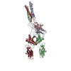 7wsoMC  7xt6C M: atomic model generated by this map C: citing same article ( |
|---|---|
| Similar structure data |
-Links
| EMDB pages | EMDB (EBI/PDBe) / EMDataResource |
|---|---|
| Related items in Molecule of the Month |


-Map
| File | Download / File: emd_32762.map.gz / Format: CCP4 / Size: 64 MB / Type: IMAGE STORED AS FLOATING POINT NUMBER (4 BYTES) | ||||||||||||||||||||||||||||||||||||
|---|---|---|---|---|---|---|---|---|---|---|---|---|---|---|---|---|---|---|---|---|---|---|---|---|---|---|---|---|---|---|---|---|---|---|---|---|---|
| Projections & slices | Image control
Images are generated by Spider. | ||||||||||||||||||||||||||||||||||||
| Voxel size | X=Y=Z: 1.1 Å | ||||||||||||||||||||||||||||||||||||
| Density |
| ||||||||||||||||||||||||||||||||||||
| Symmetry | Space group: 1 | ||||||||||||||||||||||||||||||||||||
| Details | EMDB XML:
|
 Z (Sec.)
 Y (Row.)
 X (Col.)


-Supplemental data
- Sample components
-Entire : complex
| Entire | Name: complex |
|---|---|
| Components |
|
-Supramolecule #1: complex
| Supramolecule | Name: complex / type: complex / ID: 1 / Parent: 0 / Macromolecule list: all |
|---|---|
| Source (natural) | Organism: Homo sapiens (human) |
-Macromolecule #1: Immunoglobulin heavy constant gamma 1
| Macromolecule | Name: Immunoglobulin heavy constant gamma 1 / type: protein_or_peptide / ID: 1 / Number of copies: 2 / Enantiomer: LEVO |
|---|---|
| Source (natural) | Organism: Homo sapiens (human) |
| Molecular weight | Theoretical: 28.533205 KDa |
| Recombinant expression | Organism: Homo sapiens (human) |
| Sequence | String: GPSVFLFPPK PKDTLMISRT PEVTCVVVDV SHEDPEVKFN WYVDGVEVHN AKTKPREEQY NSTYRVVSVL TVLHQDWLNG KEYKCKVSN KALPAPIEKT ISKAKGQPRE PQVYTLPPSR DELTKNQVSL TCLVKGFYPS DIAVEWESNG QPENNYKTTP P VLDSDGSF ...String: GPSVFLFPPK PKDTLMISRT PEVTCVVVDV SHEDPEVKFN WYVDGVEVHN AKTKPREEQY NSTYRVVSVL TVLHQDWLNG KEYKCKVSN KALPAPIEKT ISKAKGQPRE PQVYTLPPSR DELTKNQVSL TCLVKGFYPS DIAVEWESNG QPENNYKTTP P VLDSDGSF FLYSKLTVDK SRWQQGNVFS CSVMHEALHN HYTQKSLSLS PELQLEESCA EAQDGELDGL WTTITIFITL FL LSVCYSA TVTFF UniProtKB: Immunoglobulin heavy constant gamma 1 |
-Macromolecule #2: B-cell antigen receptor complex-associated protein alpha chain
| Macromolecule | Name: B-cell antigen receptor complex-associated protein alpha chain type: protein_or_peptide / ID: 2 / Number of copies: 1 / Enantiomer: LEVO |
|---|---|
| Source (natural) | Organism: Homo sapiens (human) |
| Molecular weight | Theoretical: 15.504718 KDa |
| Recombinant expression | Organism: Homo sapiens (human) |
| Sequence | String: LWMHKVPASL MVSLGEDAHF QCPHNSSNNA NVTWWRVLHG NYTWPPEFLG PGEDPNGTLI IQNVNKSHGG IYVCRVQEGN ESYQQSCGT YLRVRQPPPR PFLDMGEGTK NRIITAEGII LLFCAVVPGT LLLFRKRW UniProtKB: B-cell antigen receptor complex-associated protein alpha chain |
-Macromolecule #3: B-cell antigen receptor complex-associated protein beta chain
| Macromolecule | Name: B-cell antigen receptor complex-associated protein beta chain type: protein_or_peptide / ID: 3 / Number of copies: 1 / Enantiomer: LEVO |
|---|---|
| Source (natural) | Organism: Homo sapiens (human) |
| Molecular weight | Theoretical: 16.143744 KDa |
| Recombinant expression | Organism: Homo sapiens (human) |
| Sequence | String: SRIWQSPRFI ARKRGFTVKM HCYMNSASGN VSWLWKQEMD ENPQQLKLEK GRMEESQNES LATLTIQGIR FEDNGIYFCQ QKCNNTSEV YQGCGTELRV MGFSTLAQLK QRNTLKDGII MIQTLLIILF IIVPIFLLLD UniProtKB: B-cell antigen receptor complex-associated protein beta chain |
-Experimental details
-Structure determination
| Method | cryo EM |
|---|---|
 Processing Processing | single particle reconstruction |
| Aggregation state | particle |
-Sample preparation
| Buffer | pH: 7 |
|---|---|
| Vitrification | Cryogen name: NITROGEN |
- Electron microscopy
| Microscope | FEI TITAN KRIOS |
|---|---|
| Image recording | Film or detector model: GATAN K2 QUANTUM (4k x 4k) / Average electron dose: 1.5 e/Å2 |
| Electron beam | Acceleration voltage: 300 kV / Electron source:  FIELD EMISSION GUN FIELD EMISSION GUN |
| Electron optics | Illumination mode: FLOOD BEAM / Imaging mode: BRIGHT FIELD / Nominal defocus max: 2.5 µm / Nominal defocus min: 1.2 µm |
| Experimental equipment |  Model: Titan Krios / Image courtesy: FEI Company |
-Image processing
| Startup model | Type of model: PDB ENTRY PDB model - PDB ID: |
|---|---|
| Final reconstruction | Resolution.type: BY AUTHOR / Resolution: 3.03 Å / Resolution method: FSC 0.143 CUT-OFF / Number images used: 213086 |
| Initial angle assignment | Type: MAXIMUM LIKELIHOOD |
| Final angle assignment | Type: MAXIMUM LIKELIHOOD |