+ Open data
- Basic information
| Entry |  | |||||||||
|---|---|---|---|---|---|---|---|---|---|---|
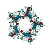
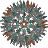
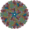
| Title | Cryo-EM structure of alphavirus, Getah virus | |||||||||
 Map data Map data | Cryo-EM structure of Getah capsid protein, Block-based reconstruction (Block 1). | |||||||||
 Sample Sample |
| |||||||||
 Keywords Keywords | alphavirus / Getah virus / Togaviridae / structural genomics / VIRAL PROTEIN | |||||||||
| Function / homology |  Function and homology information Function and homology informationtogavirin / T=4 icosahedral viral capsid / symbiont-mediated suppression of host toll-like receptor signaling pathway / host cell cytoplasm / symbiont entry into host cell / serine-type endopeptidase activity / fusion of virus membrane with host endosome membrane / host cell nucleus / virion attachment to host cell / host cell plasma membrane ...togavirin / T=4 icosahedral viral capsid / symbiont-mediated suppression of host toll-like receptor signaling pathway / host cell cytoplasm / symbiont entry into host cell / serine-type endopeptidase activity / fusion of virus membrane with host endosome membrane / host cell nucleus / virion attachment to host cell / host cell plasma membrane / structural molecule activity / virion membrane / proteolysis / RNA binding / membrane Similarity search - Function | |||||||||
| Biological species |  Getah virus Getah virus | |||||||||
| Method | single particle reconstruction / cryo EM / Resolution: 3.8 Å | |||||||||
 Authors Authors | Wang M / Sun ZZ | |||||||||
| Funding support | 1 items
| |||||||||
 Citation Citation |  Journal: To Be Published Journal: To Be PublishedTitle: Implications for the pathogenicity and antigenicity of alpha viruses revealed by a 3.5 angstrom Cryo-EM structure of Getah virus Authors: Wang M / Sun ZZ / Wang JF | |||||||||
| History |
|
- Structure visualization
| Supplemental images |
|---|
- Downloads & links
-EMDB archive
| Map data |  emd_32426.map.gz emd_32426.map.gz | 117 MB |  EMDB map data format EMDB map data format | |
|---|---|---|---|---|
| Header (meta data) |  emd-32426-v30.xml emd-32426-v30.xml emd-32426.xml emd-32426.xml | 10.9 KB 10.9 KB | Display Display |  EMDB header EMDB header |
| Images |  emd_32426.png emd_32426.png | 44.6 KB | ||
| Filedesc metadata |  emd-32426.cif.gz emd-32426.cif.gz | 5.4 KB | ||
| Archive directory |  http://ftp.pdbj.org/pub/emdb/structures/EMD-32426 http://ftp.pdbj.org/pub/emdb/structures/EMD-32426 ftp://ftp.pdbj.org/pub/emdb/structures/EMD-32426 ftp://ftp.pdbj.org/pub/emdb/structures/EMD-32426 | HTTPS FTP |
-Validation report
| Summary document |  emd_32426_validation.pdf.gz emd_32426_validation.pdf.gz | 648.9 KB | Display |  EMDB validaton report EMDB validaton report |
|---|---|---|---|---|
| Full document |  emd_32426_full_validation.pdf.gz emd_32426_full_validation.pdf.gz | 648.5 KB | Display | |
| Data in XML |  emd_32426_validation.xml.gz emd_32426_validation.xml.gz | 6.5 KB | Display | |
| Data in CIF |  emd_32426_validation.cif.gz emd_32426_validation.cif.gz | 7.4 KB | Display | |
| Arichive directory |  https://ftp.pdbj.org/pub/emdb/validation_reports/EMD-32426 https://ftp.pdbj.org/pub/emdb/validation_reports/EMD-32426 ftp://ftp.pdbj.org/pub/emdb/validation_reports/EMD-32426 ftp://ftp.pdbj.org/pub/emdb/validation_reports/EMD-32426 | HTTPS FTP |
-Related structure data
| Related structure data |  7wcoMC  7vgaC  7wc2C M: atomic model generated by this map C: citing same article ( |
|---|---|
| Similar structure data | Similarity search - Function & homology  F&H Search F&H Search |
- Links
Links
| EMDB pages |  EMDB (EBI/PDBe) / EMDB (EBI/PDBe) /  EMDataResource EMDataResource |
|---|---|
| Related items in Molecule of the Month |
- Map
Map
| File |  Download / File: emd_32426.map.gz / Format: CCP4 / Size: 125 MB / Type: IMAGE STORED AS FLOATING POINT NUMBER (4 BYTES) Download / File: emd_32426.map.gz / Format: CCP4 / Size: 125 MB / Type: IMAGE STORED AS FLOATING POINT NUMBER (4 BYTES) | ||||||||||||||||||||||||||||||||||||
|---|---|---|---|---|---|---|---|---|---|---|---|---|---|---|---|---|---|---|---|---|---|---|---|---|---|---|---|---|---|---|---|---|---|---|---|---|---|
| Annotation | Cryo-EM structure of Getah capsid protein, Block-based reconstruction (Block 1). | ||||||||||||||||||||||||||||||||||||
| Projections & slices | Image control
Images are generated by Spider. | ||||||||||||||||||||||||||||||||||||
| Voxel size | X=Y=Z: 1.36 Å | ||||||||||||||||||||||||||||||||||||
| Density |
| ||||||||||||||||||||||||||||||||||||
| Symmetry | Space group: 1 | ||||||||||||||||||||||||||||||||||||
| Details | EMDB XML:
|
-Supplemental data
- Sample components
-Entire : Getah virus
| Entire | Name:  Getah virus Getah virus |
|---|---|
| Components |
|
-Supramolecule #1: Getah virus
| Supramolecule | Name: Getah virus / type: virus / ID: 1 / Parent: 0 / Macromolecule list: all / NCBI-ID: 59300 / Sci species name: Getah virus / Virus type: VIRION / Virus isolate: SUBSPECIES / Virus enveloped: Yes / Virus empty: No |
|---|
-Macromolecule #1: Spike glycoprotein E1
| Macromolecule | Name: Spike glycoprotein E1 / type: protein_or_peptide / ID: 1 / Number of copies: 1 / Enantiomer: LEVO |
|---|---|
| Source (natural) | Organism:  Getah virus Getah virus |
| Molecular weight | Theoretical: 47.62077 KDa |
| Sequence | String: YEHTATIPNV VGFPYKAHIE RNGFSPMTLQ LEVLGTSLEP TLNLEYITCE YKTVVPSPYI KCCGTSECRS MERPDYQCQV YTGVYPFMW GGAYCFCDTE NTQLSEAYVD RSDVCKHDHA AAYKAHTAAM KATIRISYGN LNQTTTAFVN GEHTVTVGGS R FTFGPIST ...String: YEHTATIPNV VGFPYKAHIE RNGFSPMTLQ LEVLGTSLEP TLNLEYITCE YKTVVPSPYI KCCGTSECRS MERPDYQCQV YTGVYPFMW GGAYCFCDTE NTQLSEAYVD RSDVCKHDHA AAYKAHTAAM KATIRISYGN LNQTTTAFVN GEHTVTVGGS R FTFGPIST AWTPFDNKIV VYKNDVYNQD FPPYGSGQPG RFGDIQSRTV ESKDLYANTA LKLSRPSSGT VHVPYTQTPS SF KYWIKER GTSLNDKAPF GCVIKTNPVR AENCAVGNIP VSMDIPDTAF TRVIDAPAVT NLECQVAVCT HSSDFGGIAT LTF KTDKPG KCAVHSHSNV ATIQEAAVDI KTDGKITLHF STASASPAFK VSVCSAKTTC MAACEPPKDH IVPYGASHNN QVFP DMSGT AMTWVQRVAG GLGGLTLAAV AVLILVTCVT MRR UniProtKB: Structural polyprotein |
-Macromolecule #2: Spike glycoprotein E2
| Macromolecule | Name: Spike glycoprotein E2 / type: protein_or_peptide / ID: 2 / Number of copies: 1 / Enantiomer: LEVO |
|---|---|
| Source (natural) | Organism:  Getah virus Getah virus |
| Molecular weight | Theoretical: 46.416793 KDa |
| Sequence | String: SVTEHFNVYK ATKPYLAYCA DCGDGQFCYS PVAIEKIRDE ASDGMIKIQV AAQIGINKGG THEHNKIRYI AGHDMKEANR DSLQVHTSG VCAIRGTMGH FIVAYCPPGD ELKVQFQDAE SHTQACKVQY KHAPAPVGRE KFTVRPHFGI EVPCTTYQLT T APTEEEID ...String: SVTEHFNVYK ATKPYLAYCA DCGDGQFCYS PVAIEKIRDE ASDGMIKIQV AAQIGINKGG THEHNKIRYI AGHDMKEANR DSLQVHTSG VCAIRGTMGH FIVAYCPPGD ELKVQFQDAE SHTQACKVQY KHAPAPVGRE KFTVRPHFGI EVPCTTYQLT T APTEEEID MHTPPDIPDI TLLSQQSGNV KITAGGKTIR YNCTCGSGNV GTTSSDKTIN SCKIAQCHAA VTNHDKWQYT SS FVPRADQ LSRKGKVHVP FPLTNSTCRV PVARAPGVTY GKRELTVKLH PDHPTLLTYR SLGADPRPYE EWIDRYVERT IPV TEEGIE YRWGNNPPVR LWAQLTTEGK PHGWPHEIIL YYYGLYPAAT IAAVSAAGLA VVLSLLASCY MFATARRKCL TPYA LTPGA VVPVTLGVLC CAPRAHA UniProtKB: Structural polyprotein |
-Macromolecule #3: Capsid protein
| Macromolecule | Name: Capsid protein / type: protein_or_peptide / ID: 3 / Number of copies: 1 / Enantiomer: LEVO / EC number: togavirin |
|---|---|
| Source (natural) | Organism:  Getah virus Getah virus |
| Molecular weight | Theoretical: 30.200117 KDa |
| Sequence | String: MNYIPTQTFY GRRWRPRPAY RPWRVPMQPA PPMVIPELQT PIVQAQQMQQ LISAVSALTT KQNGKAPKKP KKKPQKAKAK KNEQQKKNE NKKPPPKQKN PAKKKKPGKR ERMCMKIEND CIFEVKLDGK VTGYACLVGD KVMKPAHVKG VIDNPDLAKL T YKKSSKYD ...String: MNYIPTQTFY GRRWRPRPAY RPWRVPMQPA PPMVIPELQT PIVQAQQMQQ LISAVSALTT KQNGKAPKKP KKKPQKAKAK KNEQQKKNE NKKPPPKQKN PAKKKKPGKR ERMCMKIEND CIFEVKLDGK VTGYACLVGD KVMKPAHVKG VIDNPDLAKL T YKKSSKYD LECAQIPVHM KSDASKYTHE KPEGHYNWHH GAVQYSGGRF TIPTGAGKPG DSGRPIFDNK GRVVAIVLGG AN EGARTAL SVVTWTKDMV TRYTPEGTEE W UniProtKB: Structural polyprotein |
-Experimental details
-Structure determination
| Method | cryo EM |
|---|---|
 Processing Processing | single particle reconstruction |
| Aggregation state | particle |
- Sample preparation
| Buffer | pH: 7.4 |
|---|---|
| Vitrification | Cryogen name: NITROGEN |
- Electron microscopy
| Microscope | FEI TITAN KRIOS |
|---|---|
| Image recording | Film or detector model: GATAN K2 SUMMIT (4k x 4k) / Detector mode: COUNTING / Average electron dose: 30.0 e/Å2 |
| Electron beam | Acceleration voltage: 300 kV / Electron source:  FIELD EMISSION GUN FIELD EMISSION GUN |
| Electron optics | Illumination mode: FLOOD BEAM / Imaging mode: BRIGHT FIELD / Nominal defocus max: 2.5 µm / Nominal defocus min: 1.0 µm |
| Experimental equipment |  Model: Titan Krios / Image courtesy: FEI Company |
- Image processing
| Startup model | Type of model: NONE |
|---|---|
| Final reconstruction | Resolution.type: BY AUTHOR / Resolution: 3.8 Å / Resolution method: FSC 0.143 CUT-OFF / Number images used: 30996 |
| Initial angle assignment | Type: RANDOM ASSIGNMENT |
| Final angle assignment | Type: RANDOM ASSIGNMENT |
 Movie
Movie Controller


 Z (Sec.)
 Y (Row.)
 X (Col.)


