organelle inner membrane / plasma membrane-derived chromatophore membrane / plasma membrane light-harvesting complex / bacteriochlorophyll binding / photosynthetic electron transport in photosystem II / : / photosynthesis, light reaction / membrane => GO:0016020 / metal ion binding / plasma membrane 類似検索 - 分子機能
Intrinsic membrane protein family, PufX / Intrinsic membrane protein PufX / Antenna complex, beta subunit, conserved site / Antenna complexes beta subunits signature. / Antenna complex, alpha subunit / Antenna complex, alpha subunit conserved site / Antenna complexes alpha subunits signature. / Antenna complex, alpha/beta subunit / Light-harvesting protein B beta chain / Antenna complex, beta domain superfamily ...Intrinsic membrane protein family, PufX / Intrinsic membrane protein PufX / Antenna complex, beta subunit, conserved site / Antenna complexes beta subunits signature. / Antenna complex, alpha subunit / Antenna complex, alpha subunit conserved site / Antenna complexes alpha subunits signature. / Antenna complex, alpha/beta subunit / Light-harvesting protein B beta chain / Antenna complex, beta domain superfamily / Antenna complex alpha/beta subunit / Light-harvesting complex / Photosynthetic reaction centre, H subunit / Bacterial photosynthetic reaction centre, H-chain, C-terminal / Photosynthetic reaction centre, M subunit / Photosynthetic reaction centre, H subunit, N-terminal / PRC-barrel domain / Photosynthetic reaction centre, H subunit, N-terminal domain superfamily / Photosynthetic reaction centre, H-chain N-terminal region / PRC-barrel domain / Photosynthetic reaction centre, L subunit / PRC-barrel-like superfamily / Photosynthetic reaction centre, L/M / Photosystem II protein D1/D2 superfamily / Photosynthetic reaction centre protein / Photosynthetic reaction center proteins signature. 類似検索 - ドメイン・相同性
Intrinsic membrane protein PufX / Reaction center protein L chain / Antenna pigment protein beta chain / Reaction center protein M chain / Photosynthetic reaction center subunit H / Antenna pigment protein alpha chain 類似検索 - 構成要素
生物種
Rhodobacter sphaeroides f. sp. denitrificans (バクテリア)
Japan Agency for Medical Research and Development (AMED)
JP21am0101118
日本
Japan Agency for Medical Research and Development (AMED)
JP21am0101116
日本
Japan Society for the Promotion of Science (JSPS)
JP16H04174
日本
Japan Society for the Promotion of Science (JSPS)
JP18H05153
日本
Japan Society for the Promotion of Science (JSPS)
20H05086
日本
Japan Society for the Promotion of Science (JSPS)
20H02856
日本
引用
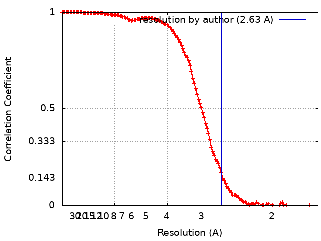
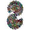
ジャーナル: Nat Commun / 年: 2022 タイトル: Asymmetric structure of the native Rhodobacter sphaeroides dimeric LH1-RC complex. 著者: Kazutoshi Tani / Ryo Kanno / Riku Kikuchi / Saki Kawamura / Kenji V P Nagashima / Malgorzata Hall / Ai Takahashi / Long-Jiang Yu / Yukihiro Kimura / Michael T Madigan / Akira Mizoguchi / ...著者: Kazutoshi Tani / Ryo Kanno / Riku Kikuchi / Saki Kawamura / Kenji V P Nagashima / Malgorzata Hall / Ai Takahashi / Long-Jiang Yu / Yukihiro Kimura / Michael T Madigan / Akira Mizoguchi / Bruno M Humbel / Zheng-Yu Wang-Otomo / 要旨: Rhodobacter sphaeroides is a model organism in bacterial photosynthesis, and its light-harvesting-reaction center (LH1-RC) complex contains both dimeric and monomeric forms. Here we present cryo-EM ...Rhodobacter sphaeroides is a model organism in bacterial photosynthesis, and its light-harvesting-reaction center (LH1-RC) complex contains both dimeric and monomeric forms. Here we present cryo-EM structures of the native LH1-RC dimer and an LH1-RC monomer lacking protein-U (ΔU). The native dimer reveals several asymmetric features including the arrangement of its two monomeric components, the structural integrity of protein-U, the overall organization of LH1, and rigidities of the proteins and pigments. PufX plays a critical role in connecting the two monomers in a dimer, with one PufX interacting at its N-terminus with another PufX and an LH1 β-polypeptide in the other monomer. One protein-U was only partially resolved in the dimeric structure, signaling different degrees of disorder in the two monomers. The ΔU LH1-RC monomer was half-moon-shaped and contained 11 α- and 10 β-polypeptides, indicating a critical role for protein-U in controlling the number of αβ-subunits required for dimer assembly and stabilization. These features are discussed in relation to membrane topology and an assembly model proposed for the native dimeric complex.
 ムービー
ムービー コントローラー


 データを開く
 基本情報
基本情報
 マップデータ
 試料
試料 キーワード
キーワード 機能・相同性情報
 Rhodobacter sphaeroides f. sp. denitrificans (バクテリア)
 データ登録者
 日本, 6件
  引用
引用

 構造の表示
構造の表示
 ダウンロードとリンク
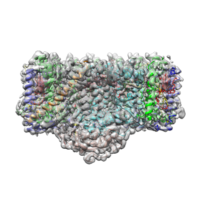 emd_32193.png
 http://ftp.pdbj.org/pub/emdb/structures/EMD-32193


 Z (Sec.)
 Y (Row.)
 X (Col.)


 試料の構成要素


 解析
解析 電子顕微鏡法
 FIELD EMISSION GUN