 Movie
Movie Controller

[English] 日本語
 Yorodumi
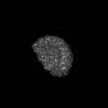
- PDB-7vy2: STRUCTURE OF PHOTOSYNTHETIC LH1-RC SUPER-COMPLEX OF RHODOBACTER S... -
+ Open data

- Basic information
| Entry | Database: PDB / ID: 7vy2 | |||||||||||||||||||||
|---|---|---|---|---|---|---|---|---|---|---|---|---|---|---|---|---|---|---|---|---|---|---|
| Title | STRUCTURE OF PHOTOSYNTHETIC LH1-RC SUPER-COMPLEX OF RHODOBACTER SPHAEROIDES DIMER | |||||||||||||||||||||
 Components Components |
| |||||||||||||||||||||
 Keywords Keywords | PHOTOSYNTHESIS / LH1-RC COMPLEX / PURPLE BACTERIA | |||||||||||||||||||||
| Function / homology |  Function and homology information Function and homology information: / organelle inner membrane / plasma membrane-derived chromatophore membrane / plasma membrane light-harvesting complex / bacteriochlorophyll binding / photosynthetic electron transport in photosystem II / photosynthesis, light reaction / membrane => GO:0016020 / metal ion binding / plasma membrane Similarity search - Function | |||||||||||||||||||||
| Biological species |  Rhodobacter sphaeroides f. sp. denitrificans (bacteria) Rhodobacter sphaeroides f. sp. denitrificans (bacteria) | |||||||||||||||||||||
| Method | ELECTRON MICROSCOPY / single particle reconstruction / cryo EM / Resolution: 2.75 Å | |||||||||||||||||||||
 Authors Authors | Tani, K. / Kanno, R. / Kawamura, S. / Kikuchi, R. / Nagashima, K.V.P. / Hall, M. / Takahashi, A. / Yu, L.-J. / Kimura, Y. / Madigan, M.T. ...Tani, K. / Kanno, R. / Kawamura, S. / Kikuchi, R. / Nagashima, K.V.P. / Hall, M. / Takahashi, A. / Yu, L.-J. / Kimura, Y. / Madigan, M.T. / Mizoguchi, A. / Humbel, B.M. / Wang-Otomo, Z.-Y. | |||||||||||||||||||||
| Funding support |  Japan, 6items Japan, 6items
| |||||||||||||||||||||
 Citation Citation | Journal: Nat Commun / Year: 2022 Title: Asymmetric structure of the native Rhodobacter sphaeroides dimeric LH1-RC complex. Authors: Kazutoshi Tani / Ryo Kanno / Riku Kikuchi / Saki Kawamura / Kenji V P Nagashima / Malgorzata Hall / Ai Takahashi / Long-Jiang Yu / Yukihiro Kimura / Michael T Madigan / Akira Mizoguchi / ...Authors: Kazutoshi Tani / Ryo Kanno / Riku Kikuchi / Saki Kawamura / Kenji V P Nagashima / Malgorzata Hall / Ai Takahashi / Long-Jiang Yu / Yukihiro Kimura / Michael T Madigan / Akira Mizoguchi / Bruno M Humbel / Zheng-Yu Wang-Otomo /   Abstract: Rhodobacter sphaeroides is a model organism in bacterial photosynthesis, and its light-harvesting-reaction center (LH1-RC) complex contains both dimeric and monomeric forms. Here we present cryo-EM ...Rhodobacter sphaeroides is a model organism in bacterial photosynthesis, and its light-harvesting-reaction center (LH1-RC) complex contains both dimeric and monomeric forms. Here we present cryo-EM structures of the native LH1-RC dimer and an LH1-RC monomer lacking protein-U (ΔU). The native dimer reveals several asymmetric features including the arrangement of its two monomeric components, the structural integrity of protein-U, the overall organization of LH1, and rigidities of the proteins and pigments. PufX plays a critical role in connecting the two monomers in a dimer, with one PufX interacting at its N-terminus with another PufX and an LH1 β-polypeptide in the other monomer. One protein-U was only partially resolved in the dimeric structure, signaling different degrees of disorder in the two monomers. The ΔU LH1-RC monomer was half-moon-shaped and contained 11 α- and 10 β-polypeptides, indicating a critical role for protein-U in controlling the number of αβ-subunits required for dimer assembly and stabilization. These features are discussed in relation to membrane topology and an assembly model proposed for the native dimeric complex. | |||||||||||||||||||||
| History |
|
- Structure visualization
| Structure viewer | Molecule: MolmilJmol/JSmol |
|---|
- Downloads & links
-Download
| PDBx/mmCIF format | 7vy2.cif.gz | 1003.2 KB | Display | PDBx/mmCIF format |
|---|---|---|---|---|
| PDB format | pdb7vy2.ent.gz | Display | PDB format | |
| PDBx/mmJSON format | 7vy2.json.gz | Tree view | PDBx/mmJSON format | |
| Others |  Other downloads Other downloads |
-Validation report
| Arichive directory | https://data.pdbj.org/pub/pdb/validation_reports/vy/7vy2ftp://data.pdbj.org/pub/pdb/validation_reports/vy/7vy2 | HTTPS FTP |
|---|
-Related structure data
| Related structure data | 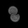 32192MC  7vy3C M: map data used to model this data C: citing same article ( |
|---|---|
| Similar structure data |
-Links
 PDBj
PDBj


- Assembly
| Deposited unit | 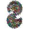
|
|---|---|
| 1 |
|
-Components
-Photosynthetic reaction center ... , 2 types, 4 molecules LlHh
| #1: Protein | Mass: 31360.416 Da / Num. of mol.: 2 / Source method: isolated from a natural source Source: (natural) Rhodobacter sphaeroides f. sp. denitrificans (bacteria)References: UniProt: A0A7Z6QV46 #3: Protein | Mass: 28091.350 Da / Num. of mol.: 2 / Source method: isolated from a natural source Source: (natural) Rhodobacter sphaeroides f. sp. denitrificans (bacteria)References: UniProt: A0A7Z6QV87 |
|---|
-Protein , 3 types, 6 molecules MmXxUu
| #2: Protein | Mass: 34412.566 Da / Num. of mol.: 2 / Source method: isolated from a natural source Source: (natural) Rhodobacter sphaeroides f. sp. denitrificans (bacteria)References: UniProt: A0A7Z6QV86 #6: Protein | Mass: 8886.395 Da / Num. of mol.: 2 / Source method: isolated from a natural source Source: (natural) Rhodobacter sphaeroides f. sp. denitrificans (bacteria)References: UniProt: A0A7Z6QV32 #7: Protein | Mass: 5555.558 Da / Num. of mol.: 2 / Source method: isolated from a natural source Source: (natural) Rhodobacter sphaeroides f. sp. denitrificans (bacteria)References: UniProt: A0A7Z6QU05 |
|---|
-Antenna pigment protein ... , 2 types, 56 molecules ADFIKOQSVY1357adfikoqsvy01030507BE...
| #4: Protein | Mass: 6473.780 Da / Num. of mol.: 28 / Source method: isolated from a natural source Source: (natural) Rhodobacter sphaeroides f. sp. denitrificans (bacteria)References: UniProt: A0A7Z6W8S0 #5: Protein/peptide | Mass: 5461.166 Da / Num. of mol.: 28 / Source method: isolated from a natural source Source: (natural) Rhodobacter sphaeroides f. sp. denitrificans (bacteria)References: UniProt: A0A7Z6QV72 |
|---|
-Sugars , 1 types, 27 molecules 
| #11: Sugar | ChemComp-LMT /  Type: D-saccharide / Mass: 510.615 Da / Num. of mol.: 27 / Source method: obtained synthetically / Formula: C24H46O11 / Comment: detergent*YM Type: D-saccharide / Mass: 510.615 Da / Num. of mol.: 27 / Source method: obtained synthetically / Formula: C24H46O11 / Comment: detergent*YM |
|---|
-Non-polymers , 9 types, 174 molecules 


| #8: Chemical | ChemComp-BCL /  Mass: 911.504 Da / Num. of mol.: 64 / Source method: obtained synthetically / Formula: C55H74MgN4O6 / Feature type: SUBJECT OF INVESTIGATION Mass: 911.504 Da / Num. of mol.: 64 / Source method: obtained synthetically / Formula: C55H74MgN4O6 / Feature type: SUBJECT OF INVESTIGATION#9: Chemical | ChemComp-BPH /  Mass: 889.215 Da / Num. of mol.: 4 / Source method: obtained synthetically / Formula: C55H76N4O6 Mass: 889.215 Da / Num. of mol.: 4 / Source method: obtained synthetically / Formula: C55H76N4O6#10: Chemical | ChemComp-U10 /  Mass: 863.343 Da / Num. of mol.: 8 / Source method: obtained synthetically / Formula: C59H90O4 Mass: 863.343 Da / Num. of mol.: 8 / Source method: obtained synthetically / Formula: C59H90O4#12: Chemical | ChemComp-PGV / (  Mass: 749.007 Da / Num. of mol.: 26 / Source method: obtained synthetically / Formula: C40H77O10P / Comment: phospholipid*YM Mass: 749.007 Da / Num. of mol.: 26 / Source method: obtained synthetically / Formula: C40H77O10P / Comment: phospholipid*YM#13: Chemical |  Mass: 55.845 Da / Num. of mol.: 2 / Source method: obtained synthetically / Formula: Fe Mass: 55.845 Da / Num. of mol.: 2 / Source method: obtained synthetically / Formula: Fe#14: Chemical | ChemComp-SPO /  Mass: 568.914 Da / Num. of mol.: 54 / Source method: obtained synthetically / Formula: C41H60O Mass: 568.914 Da / Num. of mol.: 54 / Source method: obtained synthetically / Formula: C41H60O#15: Chemical | ChemComp-CDL /  Mass: 1464.043 Da / Num. of mol.: 11 / Source method: obtained synthetically / Formula: C81H156O17P2 / Comment: phospholipid*YM Mass: 1464.043 Da / Num. of mol.: 11 / Source method: obtained synthetically / Formula: C81H156O17P2 / Comment: phospholipid*YM#16: Chemical | ChemComp-PTY /  Mass: 734.039 Da / Num. of mol.: 4 / Source method: obtained synthetically / Formula: C40H80NO8P / Comment: phospholipid*YM Mass: 734.039 Da / Num. of mol.: 4 / Source method: obtained synthetically / Formula: C40H80NO8P / Comment: phospholipid*YM#17: Chemical | ChemComp-LDA / |  Mass: 229.402 Da / Num. of mol.: 1 / Source method: obtained synthetically / Formula: C14H31NO / Comment: LDAO, detergent*YM Mass: 229.402 Da / Num. of mol.: 1 / Source method: obtained synthetically / Formula: C14H31NO / Comment: LDAO, detergent*YM |
|---|
-Details
| Has ligand of interest | Y |
|---|---|
| Has protein modification | Y |
-Experimental details
-Experiment
| Experiment | Method: ELECTRON MICROSCOPY |
|---|---|
| EM experiment | Aggregation state: PARTICLE / 3D reconstruction method: single particle reconstruction |
- Sample preparation
| Component |
| ||||||||||||||||||
|---|---|---|---|---|---|---|---|---|---|---|---|---|---|---|---|---|---|---|---|
| Molecular weight |
| ||||||||||||||||||
| Source (natural) |
| ||||||||||||||||||
| Buffer solution | pH: 8 | ||||||||||||||||||
| Specimen | Conc.: 3 mg/ml / Embedding applied: NO / Shadowing applied: NO / Staining applied: NO / Vitrification applied: YES / Details: This sample was monodisperse. | ||||||||||||||||||
| Vitrification | Instrument: LEICA EM GP / Cryogen name: ETHANE / Humidity: 80 % / Chamber temperature: 277 K |
- Electron microscopy imaging
| Experimental equipment |  Model: Titan Krios / Image courtesy: FEI Company |
|---|---|
| Microscopy | Model: FEI TITAN KRIOS |
| Electron gun | Electron source:  FIELD EMISSION GUN / Accelerating voltage: 300 kV / Illumination mode: FLOOD BEAM FIELD EMISSION GUN / Accelerating voltage: 300 kV / Illumination mode: FLOOD BEAM |
| Electron lens | Mode: BRIGHT FIELD / Nominal defocus max: 3000 nm / Nominal defocus min: 1000 nm / Alignment procedure: COMA FREE |
| Specimen holder | Cryogen: NITROGEN / Specimen holder model: FEI TITAN KRIOS AUTOGRID HOLDER |
| Image recording | Average exposure time: 1.275 sec. / Electron dose: 42 e/Å2 / Detector mode: COUNTING / Film or detector model: FEI FALCON III (4k x 4k) |
- Processing
| Software | Name: PHENIX / Version: 1.19.2_4158: / Classification: refinement | ||||||||||||||||||||||||||||||||
|---|---|---|---|---|---|---|---|---|---|---|---|---|---|---|---|---|---|---|---|---|---|---|---|---|---|---|---|---|---|---|---|---|---|
| EM software |
| ||||||||||||||||||||||||||||||||
| CTF correction | Type: PHASE FLIPPING ONLY | ||||||||||||||||||||||||||||||||
| Particle selection | Num. of particles selected: 164496 | ||||||||||||||||||||||||||||||||
| Symmetry | Point symmetry: C1 (asymmetric) | ||||||||||||||||||||||||||||||||
| 3D reconstruction | Resolution: 2.75 Å / Resolution method: FSC 0.143 CUT-OFF / Num. of particles: 124916 / Algorithm: FOURIER SPACE / Symmetry type: POINT | ||||||||||||||||||||||||||||||||
| Atomic model building | B value: 60 / Protocol: RIGID BODY FIT / Space: REAL / Target criteria: Correlation coefficient | ||||||||||||||||||||||||||||||||
| Atomic model building | PDB-ID: 7F0L Accession code: 7F0L / Source name: PDB / Type: experimental model | ||||||||||||||||||||||||||||||||
| Refinement | Cross valid method: NONE Stereochemistry target values: GeoStd + Monomer Library + CDL v1.2 | ||||||||||||||||||||||||||||||||
| Displacement parameters | Biso mean: 47.43 Å2 | ||||||||||||||||||||||||||||||||
| Refine LS restraints |
|

