Protein or peptide: Parathyroid hormone/parathyroid hormone-related peptide receptor
Ligand: water
Keywords
G protein-coupled receptor / membrane protein / SIGNALING PROTEIN
Function / homology
Function and homology information
parathyroid hormone receptor binding / type 1 parathyroid hormone receptor binding / negative regulation of bone mineralization involved in bone maturation / positive regulation of osteoclast proliferation / negative regulation of apoptotic process in bone marrow cell / response to parathyroid hormone / positive regulation of cell proliferation in bone marrow / macromolecule biosynthetic process / parathyroid hormone receptor activity / hormone-mediated apoptotic signaling pathway ...parathyroid hormone receptor binding / type 1 parathyroid hormone receptor binding / negative regulation of bone mineralization involved in bone maturation / positive regulation of osteoclast proliferation / negative regulation of apoptotic process in bone marrow cell / response to parathyroid hormone / positive regulation of cell proliferation in bone marrow / macromolecule biosynthetic process / parathyroid hormone receptor activity / hormone-mediated apoptotic signaling pathway / magnesium ion homeostasis / positive regulation of signal transduction / response to fibroblast growth factor / phosphate ion homeostasis / cAMP metabolic process / response to vitamin D / G-protein activation / Activation of the phototransduction cascade / Glucagon-type ligand receptors / Thromboxane signalling through TP receptor / Sensory perception of sweet, bitter, and umami (glutamate) taste / G beta:gamma signalling through PI3Kgamma / G beta:gamma signalling through CDC42 / Cooperation of PDCL (PhLP1) and TRiC/CCT in G-protein beta folding / Activation of G protein gated Potassium channels / Inhibition of voltage gated Ca2+ channels via Gbeta/gamma subunits / Ca2+ pathway / G alpha (z) signalling events / High laminar flow shear stress activates signaling by PIEZO1 and PECAM1:CDH5:KDR in endothelial cells / Glucagon-like Peptide-1 (GLP1) regulates insulin secretion / Vasopressin regulates renal water homeostasis via Aquaporins / Class B/2 (Secretin family receptors) / G protein-coupled peptide receptor activity / Adrenaline,noradrenaline inhibits insulin secretion / ADP signalling through P2Y purinoceptor 12 / G alpha (q) signalling events / negative regulation of chondrocyte differentiation / osteoblast development / G alpha (i) signalling events / Activation of G protein gated Potassium channels / G-protein activation / G beta:gamma signalling through PI3Kgamma / Prostacyclin signalling through prostacyclin receptor / G beta:gamma signalling through PLC beta / ADP signalling through P2Y purinoceptor 1 / Thromboxane signalling through TP receptor / Presynaptic function of Kainate receptors / G beta:gamma signalling through CDC42 / Inhibition of voltage gated Ca2+ channels via Gbeta/gamma subunits / Thrombin signalling through proteinase activated receptors (PARs) / G alpha (12/13) signalling events / Glucagon-type ligand receptors / G beta:gamma signalling through BTK / ADP signalling through P2Y purinoceptor 12 / Adrenaline,noradrenaline inhibits insulin secretion / Cooperation of PDCL (PhLP1) and TRiC/CCT in G-protein beta folding / Ca2+ pathway / Thrombin signalling through proteinase activated receptors (PARs) / G alpha (z) signalling events / Extra-nuclear estrogen signaling / G alpha (s) signalling events / photoreceptor outer segment membrane / G alpha (q) signalling events / peptide hormone receptor binding / positive regulation of inositol phosphate biosynthetic process / G alpha (i) signalling events / spectrin binding / Glucagon-like Peptide-1 (GLP1) regulates insulin secretion / High laminar flow shear stress activates signaling by PIEZO1 and PECAM1:CDH5:KDR in endothelial cells / Vasopressin regulates renal water homeostasis via Aquaporins / bone mineralization / alkylglycerophosphoethanolamine phosphodiesterase activity / peptide hormone binding / PKA activation in glucagon signalling / G protein-coupled receptor signaling pathway, coupled to cyclic nucleotide second messenger / hair follicle placode formation / photoreceptor outer segment / Rho protein signal transduction / developmental growth / chondrocyte differentiation / D1 dopamine receptor binding / positive regulation of glycogen biosynthetic process / bone resorption / positive regulation of bone mineralization / intracellular transport / vascular endothelial cell response to laminar fluid shear stress / renal water homeostasis / response to cadmium ion / Hedgehog 'off' state / cell maturation / adenylate cyclase-activating adrenergic receptor signaling pathway / activation of adenylate cyclase activity / homeostasis of number of cells within a tissue / photoreceptor inner segment / cardiac muscle cell apoptotic process / regulation of insulin secretion / cellular response to glucagon stimulus / adenylate cyclase activator activity / trans-Golgi network membrane / skeletal system development Similarity search - Function
Parathyroid hormone / Parathyroid hormone/parathyroid hormone-related protein / Parathyroid hormone family / Parathyroid hormone family signature. / Parathyroid hormone / GPCR, family 2, parathyroid hormone receptor / G-protein coupled receptors family 2 signature 1. / : / GPCR, family 2, extracellular hormone receptor domain / G-protein coupled receptors family 2 profile 1. ...Parathyroid hormone / Parathyroid hormone/parathyroid hormone-related protein / Parathyroid hormone family / Parathyroid hormone family signature. / Parathyroid hormone / GPCR, family 2, parathyroid hormone receptor / G-protein coupled receptors family 2 signature 1. / : / GPCR, family 2, extracellular hormone receptor domain / G-protein coupled receptors family 2 profile 1. / Domain present in hormone receptors / Hormone receptor domain / GPCR family 2, extracellular hormone receptor domain superfamily / G-protein coupled receptors family 2 signature 2. / GPCR, family 2, secretin-like, conserved site / GPCR, family 2, secretin-like / 7 transmembrane receptor (Secretin family) / GPCR, family 2-like / G-protein coupled receptors family 2 profile 2. / G-protein alpha subunit, group S / Guanine nucleotide binding protein (G-protein), alpha subunit / G protein alpha subunit, helical insertion / G-protein alpha subunit / G-alpha domain profile. / G protein alpha subunit / G-protein, gamma subunit / G-protein gamma subunit domain profile. / G-protein gamma-like domain / G-protein gamma-like domain superfamily / GGL domain / G protein gamma subunit-like motifs / GGL domain / Guanine nucleotide-binding protein, beta subunit / G-protein, beta subunit / G-protein beta WD-40 repeat / WD40 repeat, conserved site / Trp-Asp (WD) repeats signature. / WD domain, G-beta repeat / Trp-Asp (WD) repeats profile. / Trp-Asp (WD) repeats circular profile. / WD40 repeats / WD40 repeat / WD40-repeat-containing domain superfamily / WD40/YVTN repeat-like-containing domain superfamily / P-loop containing nucleoside triphosphate hydrolase Similarity search - Domain/homology
Parathyroid hormone / Guanine nucleotide-binding protein G(I)/G(S)/G(T) subunit beta-1 / Guanine nucleotide-binding protein G(s) subunit alpha isoforms short / Guanine nucleotide-binding protein G(I)/G(S)/G(O) subunit gamma-2 / Parathyroid hormone/parathyroid hormone-related peptide receptor Similarity search - Component
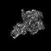
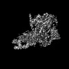
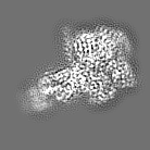
Journal: Mol Cell / Year: 2022 Title: Endogenous ligand recognition and structural transition of a human PTH receptor. Authors: Kazuhiro Kobayashi / Kouki Kawakami / Tsukasa Kusakizako / Hirotake Miyauchi / Atsuhiro Tomita / Kan Kobayashi / Wataru Shihoya / Keitaro Yamashita / Tomohiro Nishizawa / Hideaki E Kato / ...Authors: Kazuhiro Kobayashi / Kouki Kawakami / Tsukasa Kusakizako / Hirotake Miyauchi / Atsuhiro Tomita / Kan Kobayashi / Wataru Shihoya / Keitaro Yamashita / Tomohiro Nishizawa / Hideaki E Kato / Asuka Inoue / Osamu Nureki / Abstract: Endogenous parathyroid hormone (PTH) and PTH-related peptide (PTHrP) bind to the parathyroid hormone receptor 1 (PTH1R) and activate the stimulatory G-protein (Gs) signaling pathway. Intriguingly, ...Endogenous parathyroid hormone (PTH) and PTH-related peptide (PTHrP) bind to the parathyroid hormone receptor 1 (PTH1R) and activate the stimulatory G-protein (Gs) signaling pathway. Intriguingly, the two ligands have distinct signaling and physiological properties: PTH evokes prolonged Gs activation, whereas PTHrP evokes transient Gs activation with reduced bone-resorption effects. The distinct molecular actions are ascribed to the differences in ligand recognition and dissociation kinetics. Here, we report cryoelectron microscopic structures of six forms of the human PTH1R-Gs complex in the presence of PTH or PTHrP at resolutions of 2.8 -4.1 Å. A comparison of the PTH-bound and PTHrP-bound structures reveals distinct ligand-receptor interactions underlying the ligand affinity and selectivity. Furthermore, five distinct PTH-bound structures, combined with computational analyses, provide insights into the unique and complex process of ligand dissociation from the receptor and shed light on the distinct durations of signaling induced by PTH and PTHrP.
In the structure databanks used in Yorodumi, some data are registered as the other names, "COVID-19 virus" and "2019-nCoV". Here are the details of the virus and the list of structure data.
Jan 31, 2019. EMDB accession codes are about to change! (news from PDBe EMDB page)
EMDB accession codes are about to change! (news from PDBe EMDB page)
The allocation of 4 digits for EMDB accession codes will soon come to an end. Whilst these codes will remain in use, new EMDB accession codes will include an additional digit and will expand incrementally as the available range of codes is exhausted. The current 4-digit format prefixed with “EMD-” (i.e. EMD-XXXX) will advance to a 5-digit format (i.e. EMD-XXXXX), and so on. It is currently estimated that the 4-digit codes will be depleted around Spring 2019, at which point the 5-digit format will come into force.
The EM Navigator/Yorodumi systems omit the EMD- prefix.
Related info.:Q: What is EMD? / ID/Accession-code notation in Yorodumi/EM Navigator
Yorodumi is a browser for structure data from EMDB, PDB, SASBDB, etc.
This page is also the successor to EM Navigator detail page, and also detail information page/front-end page for Omokage search.
The word "yorodu" (or yorozu) is an old Japanese word meaning "ten thousand". "mi" (miru) is to see.
Related info.:EMDB / PDB / SASBDB / Comparison of 3 databanks / Yorodumi Search / Aug 31, 2016. New EM Navigator & Yorodumi / Yorodumi Papers / Jmol/JSmol / Function and homology information / Changes in new EM Navigator and Yorodumi
 Movie
Movie Controller


 Open data
 Basic information
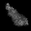
 Map data
 Sample
 Keywords
 Function and homology information
 Homo sapiens (human) /
 Authors
 Japan, 1 items
  Citation
 Structure visualization
 Downloads & links
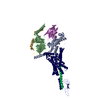
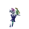
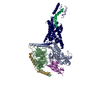
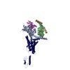
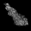
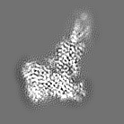
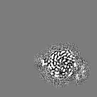
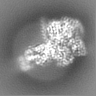
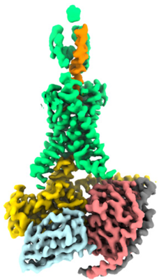 emd_32143.png
 http://ftp.pdbj.org/pub/emdb/structures/EMD-32143


 Z (Sec.)
 Y (Row.)
 X (Col.)


 Sample components

 Spodoptera frugiperda (fall armyworm)
 Processing
 Electron microscopy
 FIELD EMISSION GUN

