ムービー
ムービー コントローラー


+ データを開く

- 基本情報
基本情報
| 登録情報 | データベース: EMDB / ID: EMD-3185 | |||||||||
|---|---|---|---|---|---|---|---|---|---|---|
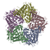
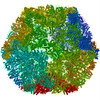
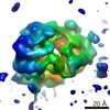
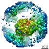
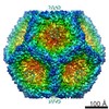
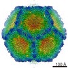
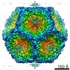
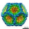
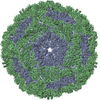
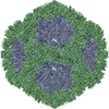
| タイトル | Structure of the in vitro assembled bacteriophage phi6 polymerase complex | |||||||||
 マップデータ マップデータ | Icosahedral reconstruction of in vitro assembled bacteriophage phi6 polymerase complex | |||||||||
 試料 試料 |
| |||||||||
 キーワード キーワード | Bacteriophage phi6 / polymerase complex | |||||||||
| 機能・相同性 |  機能・相同性情報 機能・相同性情報T=2 icosahedral viral capsid / viral procapsid / RNA uridylyltransferase activity / viral genome packaging / viral inner capsid / ribonucleoside triphosphate phosphatase activity / virion component / viral capsid / nucleoside-triphosphate phosphatase / viral nucleocapsid ...T=2 icosahedral viral capsid / viral procapsid / RNA uridylyltransferase activity / viral genome packaging / viral inner capsid / ribonucleoside triphosphate phosphatase activity / virion component / viral capsid / nucleoside-triphosphate phosphatase / viral nucleocapsid / RNA-directed RNA polymerase / viral RNA genome replication / nucleotide binding / RNA-directed RNA polymerase activity / DNA-templated transcription / RNA binding / ATP binding / metal ion binding / identical protein binding 類似検索 - 分子機能 | |||||||||
| 生物種 |  Pseudomonas phage phi6 (ファージ) Pseudomonas phage phi6 (ファージ) | |||||||||
| 手法 | 単粒子再構成法 / クライオ電子顕微鏡法 / 解像度: 4.8 Å | |||||||||
 データ登録者 データ登録者 | Ilca S / Kotecha A / Sun X / Poranen MP / Stuart DI / Huiskonen JT | |||||||||
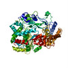
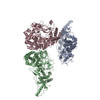
 引用 引用 | ジャーナル: Nat Commun / 年: 2015 タイトル: Localized reconstruction of subunits from electron cryomicroscopy images of macromolecular complexes. 著者: Serban L Ilca / Abhay Kotecha / Xiaoyu Sun / Minna M Poranen / David I Stuart / Juha T Huiskonen /   要旨: Electron cryomicroscopy can yield near-atomic resolution structures of highly ordered macromolecular complexes. Often however some subunits bind in a flexible manner, have different symmetry from the ...Electron cryomicroscopy can yield near-atomic resolution structures of highly ordered macromolecular complexes. Often however some subunits bind in a flexible manner, have different symmetry from the rest of the complex, or are present in sub-stoichiometric amounts, limiting the attainable resolution. Here we report a general method for the localized three-dimensional reconstruction of such subunits. After determining the particle orientations, local areas corresponding to the subunits can be extracted and treated as single particles. We demonstrate the method using three examples including a flexible assembly and complexes harbouring subunits with either partial occupancy or mismatched symmetry. Most notably, the method allows accurate fitting of the monomeric RNA-dependent RNA polymerase bound at the threefold axis of symmetry inside a viral capsid, revealing for the first time its exact orientation and interactions with the capsid proteins. Localized reconstruction is expected to provide novel biological insights in a range of challenging biological systems. | |||||||||
| 履歴 |
|
- 構造の表示
構造の表示
| ムービー |
ムービービューア |
|---|---|
| 構造ビューア | EMマップ: SurfViewMolmilJmol/JSmol |
| 添付画像 |


- ダウンロードとリンク
-EMDBアーカイブ
| マップデータ | emd_3185.map.gz | 385.2 MB | EMDBマップデータ形式 | |
|---|---|---|---|---|
| ヘッダ (付随情報) | emd-3185-v30.xmlemd-3185.xml | 14.8 KB 14.8 KB | 表示 表示 | EMDBヘッダ |
| FSC (解像度算出) | emd_3185_fsc.xml | 16.5 KB | 表示 | FSCデータファイル |
| 画像 | EMD-3185.tiffemd_3185.tiff | 1.1 MB 1.1 MB | ||
| マスクデータ | emd_3185_msk_1.map | 421.9 MB | マスクマップ | |
| アーカイブディレクトリ |  http://ftp.pdbj.org/pub/emdb/structures/EMD-3185ftp://ftp.pdbj.org/pub/emdb/structures/EMD-3185 http://ftp.pdbj.org/pub/emdb/structures/EMD-3185ftp://ftp.pdbj.org/pub/emdb/structures/EMD-3185 | HTTPS FTP |
-検証レポート
| 文書・要旨 | emd_3185_validation.pdf.gz | 332 KB | 表示 | EMDB検証レポート |
|---|---|---|---|---|
| 文書・詳細版 | emd_3185_full_validation.pdf.gz | 331.1 KB | 表示 | |
| XML形式データ | emd_3185_validation.xml.gz | 14.6 KB | 表示 | |
| アーカイブディレクトリ | https://ftp.pdbj.org/pub/emdb/validation_reports/EMD-3185ftp://ftp.pdbj.org/pub/emdb/validation_reports/EMD-3185 | HTTPS FTP |
-関連構造データ
-リンク
| EMDBのページ | EMDB (EBI/PDBe) / EMDataResource |
|---|---|
| 「今月の分子」の関連する項目 |

-マップ
| ファイル | ダウンロード / ファイル: emd_3185.map.gz / 形式: CCP4 / 大きさ: 412 MB / タイプ: IMAGE STORED AS FLOATING POINT NUMBER (4 BYTES) | ||||||||||||||||||||||||||||||||||||||||||||||||||||||||||||
|---|---|---|---|---|---|---|---|---|---|---|---|---|---|---|---|---|---|---|---|---|---|---|---|---|---|---|---|---|---|---|---|---|---|---|---|---|---|---|---|---|---|---|---|---|---|---|---|---|---|---|---|---|---|---|---|---|---|---|---|---|---|
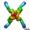
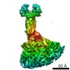
| 注釈 | Icosahedral reconstruction of in vitro assembled bacteriophage phi6 polymerase complex | ||||||||||||||||||||||||||||||||||||||||||||||||||||||||||||
| 投影像・断面図 | 画像のコントロール
画像は Spider により作成 | ||||||||||||||||||||||||||||||||||||||||||||||||||||||||||||
| ボクセルのサイズ | X=Y=Z: 1.35 Å | ||||||||||||||||||||||||||||||||||||||||||||||||||||||||||||
| 密度 |
| ||||||||||||||||||||||||||||||||||||||||||||||||||||||||||||
| 対称性 | 空間群: 1 | ||||||||||||||||||||||||||||||||||||||||||||||||||||||||||||
| 詳細 | EMDB XML:
CCP4マップ ヘッダ情報:
| ||||||||||||||||||||||||||||||||||||||||||||||||||||||||||||
 Z (Sec.)
 Y (Row.)
 X (Col.)


-添付データ
-セグメンテーションマップ: Mask used for FSC
| 注釈 | Mask used for FSC | ||||||||||||
|---|---|---|---|---|---|---|---|---|---|---|---|---|---|
| ファイル | emd_3185_msk_1.map | ||||||||||||
| 投影像・断面図 |
| ||||||||||||
| 密度ヒストグラム |


- 試料の構成要素
-全体 : Bacteriophage phi6 polymerase complex assembled in vitro from pur...
| 全体 | 名称: Bacteriophage phi6 polymerase complex assembled in vitro from purified proteins P1, P2, and P4 |
|---|---|
| 要素 |
|
-超分子 #1000: Bacteriophage phi6 polymerase complex assembled in vitro from pur...
| 超分子 | 名称: Bacteriophage phi6 polymerase complex assembled in vitro from purified proteins P1, P2, and P4 タイプ: sample / ID: 1000 / 集合状態: Icosahedral assembly with 120 copies of P1 / Number unique components: 3 |
|---|
-分子 #1: P1 protein from bacteriophage phi6
| 分子 | 名称: P1 protein from bacteriophage phi6 / タイプ: protein_or_peptide / ID: 1 / コピー数: 120 集合状態: 60 asymmetric dimers from an icosahedral shell 組換発現: Yes |
|---|---|
| 由来(天然) | 生物種: Pseudomonas phage phi6 (ファージ) |
| 分子量 | 理論値: 85 KDa |
| 組換発現 | 生物種:  Pseudomonas syringae (バクテリア) / 組換株: pathovar phaseolicola / 組換プラスミド: pLM358 Pseudomonas syringae (バクテリア) / 組換株: pathovar phaseolicola / 組換プラスミド: pLM358 |
| 配列 | UniProtKB: Major inner protein P1 |
-分子 #2: P2 protein from bacteriophage phi6
| 分子 | 名称: P2 protein from bacteriophage phi6 / タイプ: protein_or_peptide / ID: 2 / 集合状態: monomer / 組換発現: Yes |
|---|---|
| 由来(天然) | 生物種: Pseudomonas phage phi6 (ファージ) / 別称: bacteriophage phi6 |
| 分子量 | 理論値: 75 KDa |
| 組換発現 | 生物種: Pseudomonas syringae (バクテリア) / 組換株: pathovar phaseolicola / 組換プラスミド: pLM358 |
| 配列 | UniProtKB: RNA-directed RNA polymerase |
-分子 #3: P4 protein from bacteriophage phi6
| 分子 | 名称: P4 protein from bacteriophage phi6 / タイプ: protein_or_peptide / ID: 3 / 集合状態: hexamer / 組換発現: Yes |
|---|---|
| 由来(天然) | 生物種: Pseudomonas phage phi6 (ファージ) / 別称: bacteriophage phi6 |
| 分子量 | 理論値: 35 KDa |
| 組換発現 | 生物種: Pseudomonas syringae (バクテリア) / 組換株: pathovar phaseolicola / 組換プラスミド: pLM358 |
| 配列 | UniProtKB: Packaging enzyme P4 |
-実験情報
-構造解析
| 手法 | クライオ電子顕微鏡法 |
|---|---|
 解析 解析 | 単粒子再構成法 |
| 試料の集合状態 | particle |
-試料調製
| 濃度 | 2.4 mg/mL |
|---|---|
| 緩衝液 | pH: 8 / 詳細: 50 mM Tris |
| グリッド | 詳細: glow discharged Cflat grid (CF-2/1-2C-T) |
| 凍結 | 凍結剤: ETHANE / チャンバー内温度: 120 K / 装置: FEI VITROBOT MARK IV / 手法: Blot 4 seconds before plunging |
- 電子顕微鏡法
| 顕微鏡 | FEI POLARA 300 |
|---|---|
| 温度 | 最低: 81 K / 最高: 120 K / 平均: 81 K |
| 特殊光学系 | エネルギーフィルター - 名称: GIF QUANTUM LS エネルギーフィルター - エネルギー下限: 0.0 eV エネルギーフィルター - エネルギー上限: 20.0 eV |
| 詳細 | dose rate 6-8 e-/pix/s |
| 日付 | 2014年6月12日 |
| 撮影 | カテゴリ: CCD フィルム・検出器のモデル: GATAN K2 SUMMIT (4k x 4k) デジタル化 - サンプリング間隔: 5 µm / 実像数: 834 / 平均電子線量: 16 e/Å2 詳細: Every image is the average of 22 frames recorded by the direct electron detector ビット/ピクセル: 16 |
| 電子線 | 加速電圧: 300 kV / 電子線源:  FIELD EMISSION GUN FIELD EMISSION GUN |
| 電子光学系 | 倍率(補正後): 37037 / 照射モード: FLOOD BEAM / 撮影モード: BRIGHT FIELD / Cs: 2.0 mm / 最大 デフォーカス(公称値): 2.6 µm / 最小 デフォーカス(公称値): 1.1 µm / 倍率(公称値): 160000 |
| 試料ステージ | 試料ホルダーモデル: OTHER |
| 実験機器 |  モデル: Tecnai Polara / 画像提供: FEI Company |
-画像解析
| 詳細 | Particles were selected from the best classes after 2D and 3D classification |
|---|---|
| CTF補正 | 詳細: Each particle |
| 最終 再構成 | 想定した対称性 - 点群: I (正20面体型対称) / アルゴリズム: OTHER / 解像度のタイプ: BY AUTHOR / 解像度: 4.8 Å / 解像度の算出法: OTHER / ソフトウェア - 名称: Relion 詳細: Final map was postprocessed in Relion. Inverse B-factor of 200 was applied. 使用した粒子像数: 4379 |
| FSC曲線 (解像度の算出) |  |