Ministry of Education, Culture, Sports, Science and Technology (Japan)
24117002
日本
引用
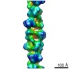
ジャーナル: mBio / 年: 2021 タイトル: Chained Structure of Dimeric F-like ATPase in Mycoplasma mobile Gliding Machinery. 著者: Takuma Toyonaga / Takayuki Kato / Akihiro Kawamoto / Noriyuki Kodera / Tasuku Hamaguchi / Yuhei O Tahara / Toshio Ando / Keiichi Namba / Makoto Miyata / 要旨: Mycoplasma mobile, a fish pathogen, exhibits gliding motility using ATP hydrolysis on solid surfaces, including animal cells. The gliding machinery can be divided into surface and internal structures. ...Mycoplasma mobile, a fish pathogen, exhibits gliding motility using ATP hydrolysis on solid surfaces, including animal cells. The gliding machinery can be divided into surface and internal structures. The internal structure of the motor is composed of 28 so-called "chains" that are each composed of 17 repeating protein units called "particles." These proteins include homologs of the catalytic α and β subunits of F-ATPase. In this study, we isolated the particles and determined their structures using negative-staining electron microscopy and high-speed atomic force microscopy. The isolated particles were composed of five proteins, MMOB1660 (α-subunit homolog), -1670 (β-subunit homolog), -1630, -1620, and -4530, and showed ATP hydrolyzing activity. The two-dimensional (2D) structure, with dimensions of 35 and 26 nm, showed a dimer of hexameric ring approximately 12 nm in diameter, resembling F-ATPase catalytic (αβ). We isolated the F-like ATPase unit, which is composed of MMOB1660, -1670, and -1630. Furthermore, we isolated the chain and analyzed the three-dimensional (3D) structure, showing that dimers of mushroom-like structures resembling F-ATPase were connected and aligned along the dimer axis at 31-nm intervals. An atomic model of F-ATPase catalytic (αβ) from PS3 was successfully fitted to each hexameric ring of the mushroom-like structure. These results suggest that the motor for gliding shares an evolutionary origin with F-ATPase. Based on the obtained structure, we propose possible force transmission processes in the gliding mechanism. FF-ATPase, a rotary ATPase, is widespread in the membranes of mitochondria, chloroplasts, and bacteria and converts ATP energy with a proton motive force across the membrane by its physical rotation. Homologous protein complexes play roles in ion and protein transport. Mycoplasma mobile, a pathogenic bacterium, was recently suggested to have a special motility system evolutionarily derived from F-ATPase. The present study isolated the protein complex from cells and supported this conclusion by clarifying the detailed structures containing common and novel features as F-ATPase relatives.
 ムービー
ムービー コントローラー


 データを開く
 基本情報
基本情報 マップデータ
 試料
試料 Mycoplasma mobile (バクテリア)
 データ登録者
 日本, 1件
  引用
引用 構造の表示
構造の表示 ムービービューア
 UCSF Chimera
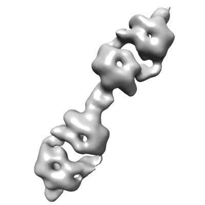
 ダウンロードとリンク
 emd_31520.png
 http://ftp.pdbj.org/pub/emdb/structures/EMD-31520
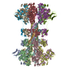
 Z (Sec.)
 Y (Row.)
 X (Col.)


 試料の構成要素
 解析
解析 電子顕微鏡法