Ministry of Education, Culture, Sports, Science and Technology (Japan)
24117002
Japan
Citation
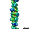
Journal: mBio / Year: 2021 Title: Chained Structure of Dimeric F-like ATPase in Mycoplasma mobile Gliding Machinery. Authors: Takuma Toyonaga / Takayuki Kato / Akihiro Kawamoto / Noriyuki Kodera / Tasuku Hamaguchi / Yuhei O Tahara / Toshio Ando / Keiichi Namba / Makoto Miyata / Abstract: Mycoplasma mobile, a fish pathogen, exhibits gliding motility using ATP hydrolysis on solid surfaces, including animal cells. The gliding machinery can be divided into surface and internal structures. ...Mycoplasma mobile, a fish pathogen, exhibits gliding motility using ATP hydrolysis on solid surfaces, including animal cells. The gliding machinery can be divided into surface and internal structures. The internal structure of the motor is composed of 28 so-called "chains" that are each composed of 17 repeating protein units called "particles." These proteins include homologs of the catalytic α and β subunits of F-ATPase. In this study, we isolated the particles and determined their structures using negative-staining electron microscopy and high-speed atomic force microscopy. The isolated particles were composed of five proteins, MMOB1660 (α-subunit homolog), -1670 (β-subunit homolog), -1630, -1620, and -4530, and showed ATP hydrolyzing activity. The two-dimensional (2D) structure, with dimensions of 35 and 26 nm, showed a dimer of hexameric ring approximately 12 nm in diameter, resembling F-ATPase catalytic (αβ). We isolated the F-like ATPase unit, which is composed of MMOB1660, -1670, and -1630. Furthermore, we isolated the chain and analyzed the three-dimensional (3D) structure, showing that dimers of mushroom-like structures resembling F-ATPase were connected and aligned along the dimer axis at 31-nm intervals. An atomic model of F-ATPase catalytic (αβ) from PS3 was successfully fitted to each hexameric ring of the mushroom-like structure. These results suggest that the motor for gliding shares an evolutionary origin with F-ATPase. Based on the obtained structure, we propose possible force transmission processes in the gliding mechanism. FF-ATPase, a rotary ATPase, is widespread in the membranes of mitochondria, chloroplasts, and bacteria and converts ATP energy with a proton motive force across the membrane by its physical rotation. Homologous protein complexes play roles in ion and protein transport. Mycoplasma mobile, a pathogenic bacterium, was recently suggested to have a special motility system evolutionarily derived from F-ATPase. The present study isolated the protein complex from cells and supported this conclusion by clarifying the detailed structures containing common and novel features as F-ATPase relatives.
In the structure databanks used in Yorodumi, some data are registered as the other names, "COVID-19 virus" and "2019-nCoV". Here are the details of the virus and the list of structure data.
Jan 31, 2019. EMDB accession codes are about to change! (news from PDBe EMDB page)
EMDB accession codes are about to change! (news from PDBe EMDB page)
The allocation of 4 digits for EMDB accession codes will soon come to an end. Whilst these codes will remain in use, new EMDB accession codes will include an additional digit and will expand incrementally as the available range of codes is exhausted. The current 4-digit format prefixed with “EMD-” (i.e. EMD-XXXX) will advance to a 5-digit format (i.e. EMD-XXXXX), and so on. It is currently estimated that the 4-digit codes will be depleted around Spring 2019, at which point the 5-digit format will come into force.
The EM Navigator/Yorodumi systems omit the EMD- prefix.
Related info.:Q: What is EMD? / ID/Accession-code notation in Yorodumi/EM Navigator
Yorodumi is a browser for structure data from EMDB, PDB, SASBDB, etc.
This page is also the successor to EM Navigator detail page, and also detail information page/front-end page for Omokage search.
The word "yorodu" (or yorozu) is an old Japanese word meaning "ten thousand". "mi" (miru) is to see.
Related info.:EMDB / PDB / SASBDB / Comparison of 3 databanks / Yorodumi Search / Aug 31, 2016. New EM Navigator & Yorodumi / Yorodumi Papers / Jmol/JSmol / Function and homology information / Changes in new EM Navigator and Yorodumi
 Movie
Movie Controller

 Yorodumi
 Open data
 Basic information
 Map data
 Sample
 Mycoplasma mobile (bacteria)
 Authors
 Japan, 1 items
  Citation
 Structure visualization
 Movie viewer
 UCSF Chimera
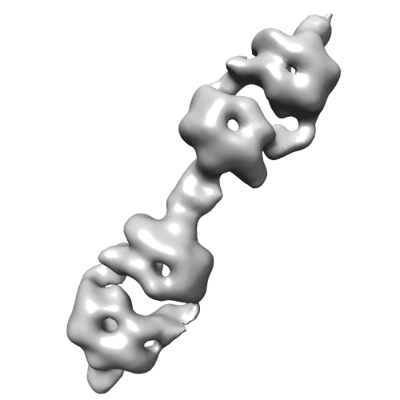
 Downloads & links
 emd_31520.png
 http://ftp.pdbj.org/pub/emdb/structures/EMD-31520
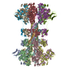
 Z (Sec.)
 Y (Row.)
 X (Col.)


 Sample components
 Processing
 Electron microscopy