 Movie
Movie Controller


+ Open data

- Basic information
| Entry |  | |||||||||
|---|---|---|---|---|---|---|---|---|---|---|
| Title | Cryo-EM structure of EBV gB in complex with nAb 3A3 | |||||||||
 Map data Map data | ||||||||||
 Sample Sample |
| |||||||||
| Biological species |   Human gammaherpesvirus 4 (Epstein-Barr virus) Human gammaherpesvirus 4 (Epstein-Barr virus) | |||||||||
| Method | single particle reconstruction / cryo EM / Resolution: 7.1 Å | |||||||||
 Authors Authors | Zheng Q / Li S / Zha Z / Chen Y / Hong J / Zhang X | |||||||||
 Citation Citation | Journal: To Be Published Title: Cryo-EM structure of EBV gB in complex with nAbs 3A3 and 3A5 Authors: Zheng Q / Li S / Zha Z / Hong J / Chen Y / Zhang X | |||||||||
| History |
|
- Structure visualization
| Supplemental images |
|---|

- Downloads & links
-EMDB archive
| Map data | emd_31516.map.gz | 59.2 MB |  EMDB map data format EMDB map data format | |
|---|---|---|---|---|
| Header (meta data) | emd-31516-v30.xmlemd-31516.xml | 8.3 KB 8.3 KB | Display Display | EMDB header |
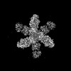
| Images |  emd_31516.png emd_31516.png | 32 KB | ||
| Archive directory |  http://ftp.pdbj.org/pub/emdb/structures/EMD-31516ftp://ftp.pdbj.org/pub/emdb/structures/EMD-31516 http://ftp.pdbj.org/pub/emdb/structures/EMD-31516ftp://ftp.pdbj.org/pub/emdb/structures/EMD-31516 | HTTPS FTP |
-Related structure data


-Links
| EMDB pages | EMDB (EBI/PDBe) / EMDataResource |
|---|
-Map
| File | Download / File: emd_31516.map.gz / Format: CCP4 / Size: 64 MB / Type: IMAGE STORED AS FLOATING POINT NUMBER (4 BYTES) | ||||||||||||||||||||||||||||||||||||
|---|---|---|---|---|---|---|---|---|---|---|---|---|---|---|---|---|---|---|---|---|---|---|---|---|---|---|---|---|---|---|---|---|---|---|---|---|---|
| Projections & slices | Image control
Images are generated by Spider. | ||||||||||||||||||||||||||||||||||||
| Voxel size | X=Y=Z: 1.12 Å | ||||||||||||||||||||||||||||||||||||
| Density |
| ||||||||||||||||||||||||||||||||||||
| Symmetry | Space group: 1 | ||||||||||||||||||||||||||||||||||||
| Details | EMDB XML:
|
 Z (Sec.)
 Y (Row.)
 X (Col.)


-Supplemental data
- Sample components
-Entire : Cryo-EM structure of EBV gB in complex with nAbs 3A3 and 3A5
| Entire | Name: Cryo-EM structure of EBV gB in complex with nAbs 3A3 and 3A5 |
|---|---|
| Components |
|
-Supramolecule #1: Cryo-EM structure of EBV gB in complex with nAbs 3A3 and 3A5
| Supramolecule | Name: Cryo-EM structure of EBV gB in complex with nAbs 3A3 and 3A5 type: complex / ID: 1 / Parent: 0 / Macromolecule list: all |
|---|---|
| Source (natural) | Organism: |
| Recombinant expression | Organism:  Homo sapiens (human) Homo sapiens (human) |
-Macromolecule #1: EBV gB
| Macromolecule | Name: EBV gB / type: protein_or_peptide / ID: 1 / Enantiomer: DEXTRO |
|---|---|
| Source (natural) | Organism: Human gammaherpesvirus 4 (Epstein-Barr virus) |
| Recombinant expression | Organism: Homo sapiens (human) |
| Sequence | String: AQTPEQPAPP ATTVQPTATR QQTSFPFRVC ELSSHGDLFR FSSDIQCPSF GTRENHTEGL LMVFKDNIIP YSFKVRSYTK IVTNILIYNG HRADSVTNRH EEKFSVESYE TDQMDTIYQC YNAVKMTKDG LTRVYVDRDG VNITVNLKPT GGLANGVRRY ASQTELYDAP ...String: AQTPEQPAPP ATTVQPTATR QQTSFPFRVC ELSSHGDLFR FSSDIQCPSF GTRENHTEGL LMVFKDNIIP YSFKVRSYTK IVTNILIYNG HRADSVTNRH EEKFSVESYE TDQMDTIYQC YNAVKMTKDG LTRVYVDRDG VNITVNLKPT GGLANGVRRY ASQTELYDAP GRVEATYRTR TTVNCLITDM MAKSNSPFDF FVTTTGQTVE MSPFYDGKNT ETFHERADSF HVRTNYKIVD YDNRGTNPQG ERRAFLDKGT YTLSWKLENR TAYCPLQHWQ TFDSTIATET GKSIHFVTDE GTSSFVTNTT VGIELPDAFK CIEEQVNKTM HEKYEAVQDR YTKGQEAITY FITSGGLLLA WLPLTPRSLA TVKNLTELTT PTSSPPSSPS PPAPPAARGS TSAAVLRRRR RNAGNATTPV PPAAPGKSLG TLNNPATVQI QFAYDSLRRQ INRMLGDLAR AWCLEQKRQN MVLRELTKIN PTTVMSSIYG KAVAAKRLGD VISVSQCVPV NQATVTLRKS MRVPGSETMC YSRPLVSFSF INDTKTYEGQ LGTDNEIFLT KKMTEVCQAT SQYYFQSGNE IHVYNDYHHF KTIELDGIAT LQTFISLNTS LIENIDFASL ELYSRDEQRA SNVFDLEGIF REYNFQAQNI A |
-Experimental details
-Structure determination
| Method | cryo EM |
|---|---|
 Processing Processing | single particle reconstruction |
| Aggregation state | particle |
-Sample preparation
| Buffer | pH: 7.4 |
|---|---|
| Vitrification | Cryogen name: ETHANE |
- Electron microscopy
| Microscope | FEI TECNAI F30 |
|---|---|
| Image recording | Film or detector model: FEI FALCON III (4k x 4k) / Average electron dose: 60.0 e/Å2 |
| Electron beam | Acceleration voltage: 300 kV / Electron source:  FIELD EMISSION GUN FIELD EMISSION GUN |
| Electron optics | Illumination mode: FLOOD BEAM / Imaging mode: BRIGHT FIELD |
| Experimental equipment |  Model: Tecnai F30 / Image courtesy: FEI Company |
-Image processing
| Final reconstruction | Resolution.type: BY AUTHOR / Resolution: 7.1 Å / Resolution method: FSC 0.143 CUT-OFF / Number images used: 9270 |
|---|---|
| Initial angle assignment | Type: RANDOM ASSIGNMENT |
| Final angle assignment | Type: MAXIMUM LIKELIHOOD |
