ムービー
ムービー コントローラー


+ データを開く

- 基本情報
基本情報
| 登録情報 | データベース: EMDB / ID: EMD-3134 | |||||||||
|---|---|---|---|---|---|---|---|---|---|---|
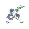
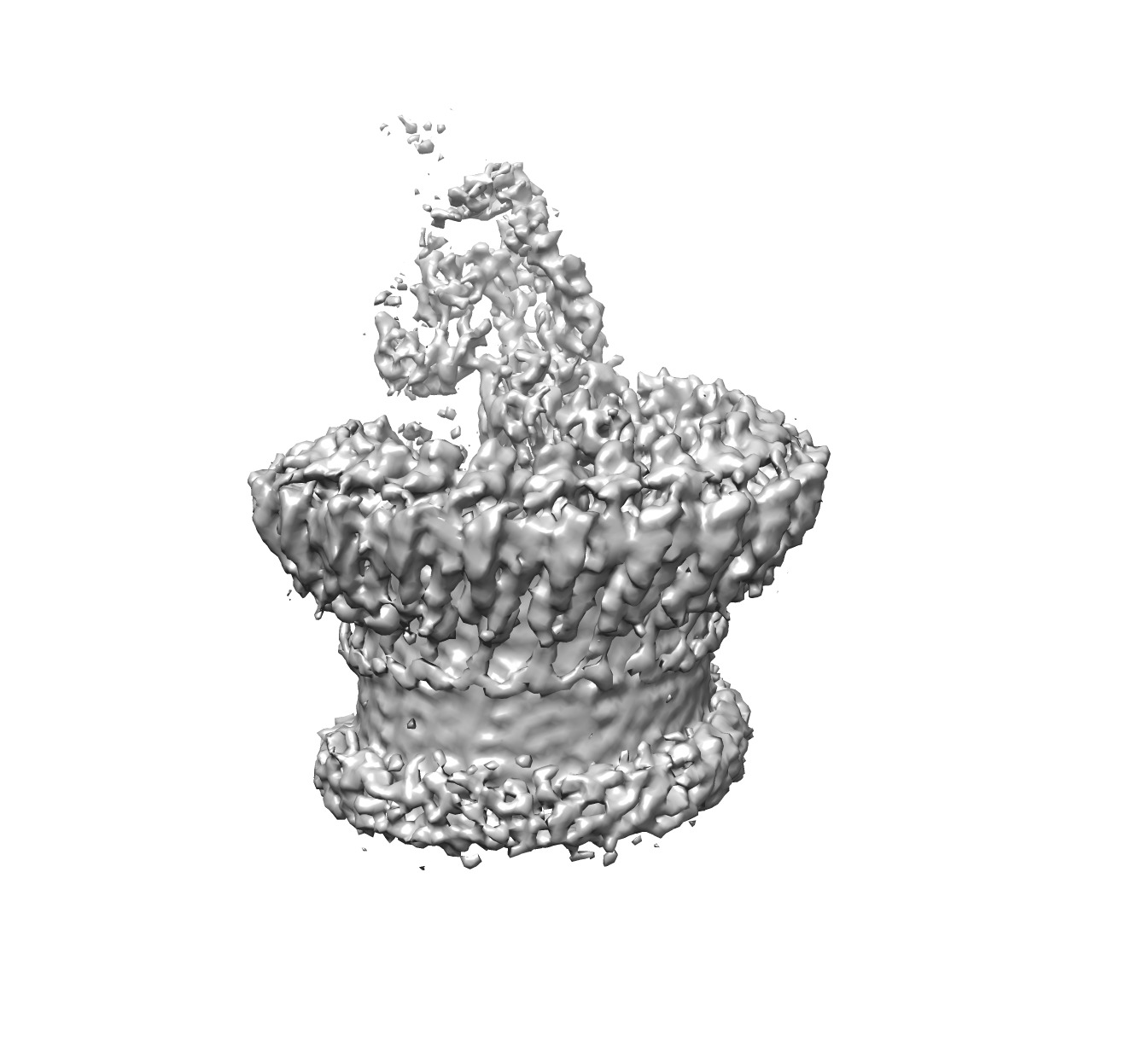
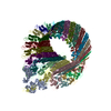
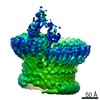
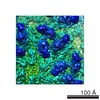
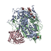
| タイトル | Electron cryo-microscopy of an immune pore | |||||||||
 マップデータ マップデータ | Reconstruction of the membrane attack complex | |||||||||
 試料 試料 |
| |||||||||
 キーワード キーワード | cryo-EM / single particles / membrane protein | |||||||||
| 生物種 |  Homo sapiens (ヒト) Homo sapiens (ヒト) | |||||||||
| 手法 | 単粒子再構成法 / クライオ電子顕微鏡法 / 解像度: 8.5 Å | |||||||||
 データ登録者 データ登録者 | Serna M / Bubeck D | |||||||||
 引用 引用 | ジャーナル: Nat Commun / 年: 2016 タイトル: Structural basis of complement membrane attack complex formation. 著者: Marina Serna / Joanna L Giles / B Paul Morgan / Doryen Bubeck /  要旨: In response to complement activation, the membrane attack complex (MAC) assembles from fluid-phase proteins to form pores in lipid bilayers. MAC directly lyses pathogens by a 'multi-hit' mechanism; ...In response to complement activation, the membrane attack complex (MAC) assembles from fluid-phase proteins to form pores in lipid bilayers. MAC directly lyses pathogens by a 'multi-hit' mechanism; however, sublytic MAC pores on host cells activate signalling pathways. Previous studies have described the structures of individual MAC components and subcomplexes; however, the molecular details of its assembly and mechanism of action remain unresolved. Here we report the electron cryo-microscopy structure of human MAC at subnanometre resolution. Structural analyses define the stoichiometry of the complete pore and identify a network of interaction interfaces that determine its assembly mechanism. MAC adopts a 'split-washer' configuration, in contrast to the predicted closed ring observed for perforin and cholesterol-dependent cytolysins. Assembly precursors partially penetrate the lipid bilayer, resulting in an irregular β-barrel pore. Our results demonstrate how differences in symmetric and asymmetric components of the MAC underpin a molecular basis for pore formation and suggest a mechanism of action that extends beyond membrane penetration. | |||||||||
| 履歴 |
|
- 構造の表示
構造の表示
| ムービー |
 ムービービューア ムービービューア |
|---|---|
| 構造ビューア | EMマップ: SurfViewMolmilJmol/JSmol |
| 添付画像 |
- ダウンロードとリンク
-EMDBアーカイブ
| マップデータ | emd_3134.map.gz | 3.5 MB | EMDBマップデータ形式 | |
|---|---|---|---|---|
| ヘッダ (付随情報) | emd-3134-v30.xmlemd-3134.xml | 13.5 KB 13.5 KB | 表示 表示 | EMDBヘッダ |
| 画像 | EMD3134_image.tif | 506.3 KB | ||
| アーカイブディレクトリ |  http://ftp.pdbj.org/pub/emdb/structures/EMD-3134ftp://ftp.pdbj.org/pub/emdb/structures/EMD-3134 http://ftp.pdbj.org/pub/emdb/structures/EMD-3134ftp://ftp.pdbj.org/pub/emdb/structures/EMD-3134 | HTTPS FTP |
-関連構造データ

-リンク
| EMDBのページ | EMDB (EBI/PDBe) / EMDataResource |
|---|
-マップ
| ファイル | ダウンロード / ファイル: emd_3134.map.gz / 形式: CCP4 / 大きさ: 34.5 MB / タイプ: IMAGE STORED AS FLOATING POINT NUMBER (4 BYTES) | ||||||||||||||||||||||||||||||||||||||||||||||||||||||||||||
|---|---|---|---|---|---|---|---|---|---|---|---|---|---|---|---|---|---|---|---|---|---|---|---|---|---|---|---|---|---|---|---|---|---|---|---|---|---|---|---|---|---|---|---|---|---|---|---|---|---|---|---|---|---|---|---|---|---|---|---|---|---|
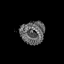
| 注釈 | Reconstruction of the membrane attack complex | ||||||||||||||||||||||||||||||||||||||||||||||||||||||||||||
| 投影像・断面図 | 画像のコントロール
画像は Spider により作成 | ||||||||||||||||||||||||||||||||||||||||||||||||||||||||||||
| ボクセルのサイズ | X=Y=Z: 2.8 Å | ||||||||||||||||||||||||||||||||||||||||||||||||||||||||||||
| 密度 |
| ||||||||||||||||||||||||||||||||||||||||||||||||||||||||||||
| 対称性 | 空間群: 1 | ||||||||||||||||||||||||||||||||||||||||||||||||||||||||||||
| 詳細 | EMDB XML:
CCP4マップ ヘッダ情報:
| ||||||||||||||||||||||||||||||||||||||||||||||||||||||||||||
 Z (Sec.)
 Y (Row.)
 X (Col.)


-添付データ
- 試料の構成要素
-全体 : Membrane attack complex
| 全体 | 名称: Membrane attack complex |
|---|---|
| 要素 |
|
-超分子 #1000: Membrane attack complex
| 超分子 | 名称: Membrane attack complex / タイプ: sample / ID: 1000 詳細: Protein complex was assembled on liposomes and detergent solubilized Number unique components: 7 |
|---|---|
| 分子量 | 理論値: 1.8 MDa |
-分子 #1: C5
| 分子 | 名称: C5 / タイプ: protein_or_peptide / ID: 1 / コピー数: 1 / 集合状態: monomer / 組換発現: No |
|---|---|
| 由来(天然) | 生物種: Homo sapiens (ヒト) / 別称: Human / 組織: Plasma |
| 分子量 | 理論値: 190 KDa |
-分子 #2: C6
| 分子 | 名称: C6 / タイプ: protein_or_peptide / ID: 2 / コピー数: 1 / 集合状態: monomer / 組換発現: No |
|---|---|
| 由来(天然) | 生物種: Homo sapiens (ヒト) / 別称: Human / 組織: Plasma |
| 分子量 | 理論値: 120 KDa |
-分子 #3: C7
| 分子 | 名称: C7 / タイプ: protein_or_peptide / ID: 3 / コピー数: 1 / 集合状態: monomer / 組換発現: No |
|---|---|
| 由来(天然) | 生物種: Homo sapiens (ヒト) / 別称: Human / 組織: Plasma |
| 分子量 | 理論値: 110 KDa |
-分子 #4: C8 alpha
| 分子 | 名称: C8 alpha / タイプ: protein_or_peptide / ID: 4 / コピー数: 1 / 集合状態: monomer / 組換発現: No |
|---|---|
| 由来(天然) | 生物種: Homo sapiens (ヒト) / 別称: Human / 組織: Plasma |
| 分子量 | 理論値: 152 KDa |
-分子 #5: C8 beta
| 分子 | 名称: C8 beta / タイプ: protein_or_peptide / ID: 5 / コピー数: 1 / 集合状態: monomer / 組換発現: No |
|---|---|
| 由来(天然) | 生物種: Homo sapiens (ヒト) / 別称: Human / 組織: Plasma |
| 分子量 | 理論値: 152 KDa |
-分子 #6: C8 gamma
| 分子 | 名称: C8 gamma / タイプ: protein_or_peptide / ID: 6 / コピー数: 1 / 集合状態: monomer / 組換発現: No |
|---|---|
| 由来(天然) | 生物種: Homo sapiens (ヒト) / 別称: Human / 組織: Plasma |
| 分子量 | 理論値: 152 KDa |
-分子 #7: C9
| 分子 | 名称: C9 / タイプ: protein_or_peptide / ID: 7 / コピー数: 18 / 集合状態: eighteen-mer / 組換発現: No |
|---|---|
| 由来(天然) | 生物種: Homo sapiens (ヒト) / 別称: Human / 組織: Plasma |
| 分子量 | 理論値: 69 KDa |
-実験情報
-構造解析
| 手法 | クライオ電子顕微鏡法 |
|---|---|
 解析 解析 | 単粒子再構成法 |
| 試料の集合状態 | particle |
-試料調製
| 緩衝液 | pH: 7.4 / 詳細: 20 mM HEPES-NaOH, 150 mM NaCl |
|---|---|
| グリッド | 詳細: 300 mesh quantifoil R1.2/1.3 grids with thin carbon support |
| 凍結 | 凍結剤: ETHANE / チャンバー内湿度: 90 % / 装置: FEI VITROBOT MARK III |
- 電子顕微鏡法
| 顕微鏡 | FEI TITAN KRIOS |
|---|---|
| 日付 | 2015年7月2日 |
| 撮影 | カテゴリ: CCD フィルム・検出器のモデル: FEI FALCON II (4k x 4k) デジタル化 - サンプリング間隔: 14.0 µm / 実像数: 622 / 平均電子線量: 45 e/Å2 |
| 電子線 | 加速電圧: 300 kV / 電子線源:  FIELD EMISSION GUN FIELD EMISSION GUN |
| 電子光学系 | 照射モード: FLOOD BEAM / 撮影モード: BRIGHT FIELD / Cs: 2.00 mm / 最大 デフォーカス(公称値): 4.0 µm / 最小 デフォーカス(公称値): 2.0 µm / 倍率(公称値): 59000 |
| 試料ステージ | 試料ホルダーモデル: FEI TITAN KRIOS AUTOGRID HOLDER |
| 実験機器 |  モデル: Titan Krios / 画像提供: FEI Company |
-画像解析
| 詳細 | Particles were manually selected. |
|---|---|
| CTF補正 | 詳細: CTFFIND3, phase flip on each particle |
| 最終 再構成 | 想定した対称性 - 点群: C1 (非対称) / アルゴリズム: OTHER / 解像度のタイプ: BY AUTHOR / 解像度: 8.5 Å / 解像度の算出法: OTHER / ソフトウェア - 名称: RELION, EMAN2 / 使用した粒子像数: 41981 |
-原子モデル構築 1
| 初期モデル | PDB ID: |
|---|---|
| ソフトウェア | 名称: Chimera |
| 精密化 | 空間: REAL / プロトコル: RIGID BODY FIT |
-原子モデル構築 2
| 初期モデル | PDB ID: |
|---|---|
| ソフトウェア | 名称: Chimera |
| 精密化 | 空間: REAL / プロトコル: RIGID BODY FIT |