 Movie
Movie Controller


+ Open data

- Basic information
| Entry | Database: EMDB / ID: EMD-3135 | |||||||||
|---|---|---|---|---|---|---|---|---|---|---|
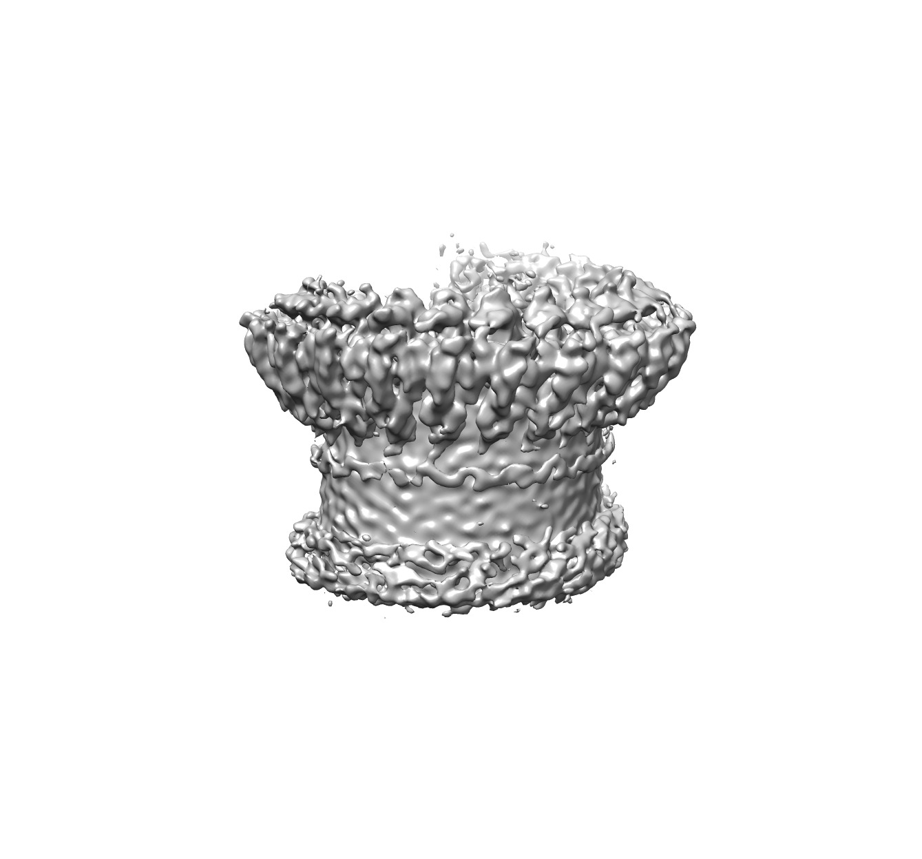
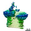
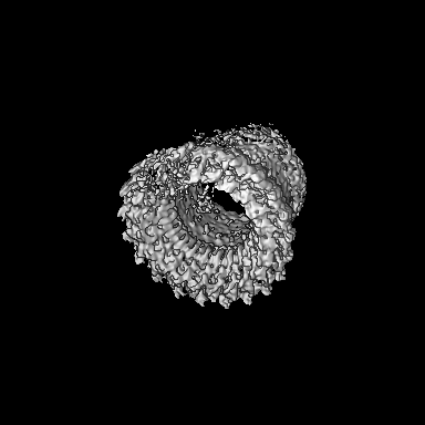
| Title | Electron cryo-microscopy of an immune pore | |||||||||
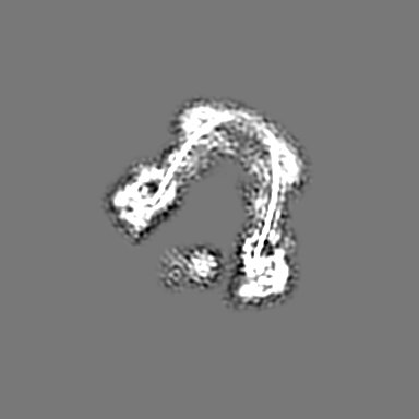
 Map data Map data | Reconstruction of the barrel of the membrane attack complex | |||||||||
 Sample Sample |
| |||||||||
 Keywords Keywords | cryo-EM / single particles / membrane protein | |||||||||
| Biological species |  Homo sapiens (human) Homo sapiens (human) | |||||||||
| Method | single particle reconstruction / cryo EM / Resolution: 7.3 Å | |||||||||
 Authors Authors | Serna M / Bubeck D | |||||||||
 Citation Citation | Journal: Nat Commun / Year: 2016 Title: Structural basis of complement membrane attack complex formation. Authors: Marina Serna / Joanna L Giles / B Paul Morgan / Doryen Bubeck /  Abstract: In response to complement activation, the membrane attack complex (MAC) assembles from fluid-phase proteins to form pores in lipid bilayers. MAC directly lyses pathogens by a 'multi-hit' mechanism; ...In response to complement activation, the membrane attack complex (MAC) assembles from fluid-phase proteins to form pores in lipid bilayers. MAC directly lyses pathogens by a 'multi-hit' mechanism; however, sublytic MAC pores on host cells activate signalling pathways. Previous studies have described the structures of individual MAC components and subcomplexes; however, the molecular details of its assembly and mechanism of action remain unresolved. Here we report the electron cryo-microscopy structure of human MAC at subnanometre resolution. Structural analyses define the stoichiometry of the complete pore and identify a network of interaction interfaces that determine its assembly mechanism. MAC adopts a 'split-washer' configuration, in contrast to the predicted closed ring observed for perforin and cholesterol-dependent cytolysins. Assembly precursors partially penetrate the lipid bilayer, resulting in an irregular β-barrel pore. Our results demonstrate how differences in symmetric and asymmetric components of the MAC underpin a molecular basis for pore formation and suggest a mechanism of action that extends beyond membrane penetration. | |||||||||
| History |
|
- Structure visualization
| Movie |
 Movie viewer Movie viewer |
|---|---|
| Structure viewer | EM map: SurfViewMolmilJmol/JSmol |
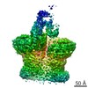
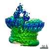
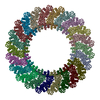
| Supplemental images |
- Downloads & links
-EMDB archive
| Map data | emd_3135.map.gz | 20.4 MB | EMDB map data format | |
|---|---|---|---|---|
| Header (meta data) | emd-3135-v30.xmlemd-3135.xml | 12.6 KB 12.6 KB | Display Display | EMDB header |
| Images | EMD3135_image.tif | 409.4 KB | ||
| Archive directory |  http://ftp.pdbj.org/pub/emdb/structures/EMD-3135ftp://ftp.pdbj.org/pub/emdb/structures/EMD-3135 http://ftp.pdbj.org/pub/emdb/structures/EMD-3135ftp://ftp.pdbj.org/pub/emdb/structures/EMD-3135 | HTTPS FTP |
-Related structure data


-Links
| EMDB pages | EMDB (EBI/PDBe) / EMDataResource |
|---|
-Map
| File | Download / File: emd_3135.map.gz / Format: CCP4 / Size: 210.9 MB / Type: IMAGE STORED AS FLOATING POINT NUMBER (4 BYTES) | ||||||||||||||||||||||||||||||||||||||||||||||||||||||||||||
|---|---|---|---|---|---|---|---|---|---|---|---|---|---|---|---|---|---|---|---|---|---|---|---|---|---|---|---|---|---|---|---|---|---|---|---|---|---|---|---|---|---|---|---|---|---|---|---|---|---|---|---|---|---|---|---|---|---|---|---|---|---|
| Annotation | Reconstruction of the barrel of the membrane attack complex | ||||||||||||||||||||||||||||||||||||||||||||||||||||||||||||
| Projections & slices | Image control
Images are generated by Spider. | ||||||||||||||||||||||||||||||||||||||||||||||||||||||||||||
| Voxel size | X=Y=Z: 1.4 Å | ||||||||||||||||||||||||||||||||||||||||||||||||||||||||||||
| Density |
| ||||||||||||||||||||||||||||||||||||||||||||||||||||||||||||
| Symmetry | Space group: 1 | ||||||||||||||||||||||||||||||||||||||||||||||||||||||||||||
| Details | EMDB XML:
CCP4 map header:
| ||||||||||||||||||||||||||||||||||||||||||||||||||||||||||||
 Z (Sec.)
 Y (Row.)
 X (Col.)


-Supplemental data
- Sample components
-Entire : Membrane attack complex
| Entire | Name: Membrane attack complex |
|---|---|
| Components |
|
-Supramolecule #1000: Membrane attack complex
| Supramolecule | Name: Membrane attack complex / type: sample / ID: 1000 Details: Protein complex was assembled on liposomes and detergent solubilized Number unique components: 7 |
|---|---|
| Molecular weight | Theoretical: 1.8 MDa |
-Macromolecule #1: C5
| Macromolecule | Name: C5 / type: protein_or_peptide / ID: 1 / Number of copies: 1 / Oligomeric state: monomer / Recombinant expression: No |
|---|---|
| Source (natural) | Organism: Homo sapiens (human) / synonym: Human / Tissue: Plasma |
| Molecular weight | Theoretical: 190 KDa |
-Macromolecule #2: C6
| Macromolecule | Name: C6 / type: protein_or_peptide / ID: 2 / Number of copies: 1 / Oligomeric state: monomer / Recombinant expression: No |
|---|---|
| Source (natural) | Organism: Homo sapiens (human) / synonym: Human / Tissue: Plasma |
| Molecular weight | Theoretical: 120 KDa |
-Macromolecule #3: C7
| Macromolecule | Name: C7 / type: protein_or_peptide / ID: 3 / Number of copies: 1 / Oligomeric state: monomer / Recombinant expression: No |
|---|---|
| Source (natural) | Organism: Homo sapiens (human) / synonym: Human / Tissue: Plasma |
| Molecular weight | Theoretical: 110 KDa |
-Macromolecule #4: C8 alpha
| Macromolecule | Name: C8 alpha / type: protein_or_peptide / ID: 4 / Number of copies: 1 / Oligomeric state: monomer / Recombinant expression: No |
|---|---|
| Source (natural) | Organism: Homo sapiens (human) / synonym: Human / Tissue: Plasma |
| Molecular weight | Theoretical: 152 KDa |
-Macromolecule #5: C8 beta
| Macromolecule | Name: C8 beta / type: protein_or_peptide / ID: 5 / Number of copies: 1 / Oligomeric state: monomer / Recombinant expression: No |
|---|---|
| Source (natural) | Organism: Homo sapiens (human) / synonym: Human / Tissue: Plasma |
| Molecular weight | Theoretical: 152 KDa |
-Macromolecule #6: C8 gamma
| Macromolecule | Name: C8 gamma / type: protein_or_peptide / ID: 6 / Number of copies: 1 / Oligomeric state: monomer / Recombinant expression: No |
|---|---|
| Source (natural) | Organism: Homo sapiens (human) / synonym: Human / Tissue: Plasma |
| Molecular weight | Theoretical: 152 KDa |
-Macromolecule #7: C9
| Macromolecule | Name: C9 / type: protein_or_peptide / ID: 7 / Number of copies: 18 / Oligomeric state: eighteen-mer / Recombinant expression: No |
|---|---|
| Source (natural) | Organism: Homo sapiens (human) / synonym: Human / Tissue: Plasma |
| Molecular weight | Theoretical: 69 KDa |
-Experimental details
-Structure determination
| Method | cryo EM |
|---|---|
 Processing Processing | single particle reconstruction |
| Aggregation state | particle |
-Sample preparation
| Buffer | pH: 7.4 / Details: 20 mM HEPES, 150 mM NaCl |
|---|---|
| Grid | Details: 300 mesh quatifol R1.2/1.3 grids with thin carbon film |
| Vitrification | Cryogen name: ETHANE / Chamber humidity: 90 % / Instrument: FEI VITROBOT MARK III |
- Electron microscopy
| Microscope | FEI TITAN KRIOS |
|---|---|
| Date | Jul 2, 2015 |
| Image recording | Category: CCD / Film or detector model: FEI FALCON II (4k x 4k) / Digitization - Sampling interval: 14.0 µm / Number real images: 622 / Average electron dose: 45 e/Å2 |
| Electron beam | Acceleration voltage: 300 kV / Electron source:  FIELD EMISSION GUN FIELD EMISSION GUN |
| Electron optics | Illumination mode: FLOOD BEAM / Imaging mode: BRIGHT FIELD / Cs: 2.00 mm / Nominal defocus max: 4.0 µm / Nominal defocus min: 2.0 µm / Nominal magnification: 59000 |
| Sample stage | Specimen holder model: FEI TITAN KRIOS AUTOGRID HOLDER |
| Experimental equipment |  Model: Titan Krios / Image courtesy: FEI Company |
-Image processing
| Details | Masked refinement |
|---|---|
| CTF correction | Details: CTFFIND3, phase flip on each particle |
| Final reconstruction | Applied symmetry - Point group: C1 (asymmetric) / Algorithm: OTHER / Resolution.type: BY AUTHOR / Resolution: 7.3 Å / Resolution method: OTHER / Software - Name: RELION, EMAN2 / Number images used: 25343 |
