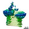
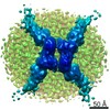
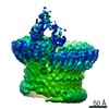
Journal: Nat Commun / Year: 2016 Title: Structure of the poly-C9 component of the complement membrane attack complex. Authors: Natalya V Dudkina / Bradley A Spicer / Cyril F Reboul / Paul J Conroy / Natalya Lukoyanova / Hans Elmlund / Ruby H P Law / Susan M Ekkel / Stephanie C Kondos / Robert J A Goode / Georg Ramm ...Authors: Natalya V Dudkina / Bradley A Spicer / Cyril F Reboul / Paul J Conroy / Natalya Lukoyanova / Hans Elmlund / Ruby H P Law / Susan M Ekkel / Stephanie C Kondos / Robert J A Goode / Georg Ramm / James C Whisstock / Helen R Saibil / Michelle A Dunstone / Abstract: The membrane attack complex (MAC)/perforin-like protein complement component 9 (C9) is the major component of the MAC, a multi-protein complex that forms pores in the membrane of target pathogens. In ...The membrane attack complex (MAC)/perforin-like protein complement component 9 (C9) is the major component of the MAC, a multi-protein complex that forms pores in the membrane of target pathogens. In contrast to homologous proteins such as perforin and the cholesterol-dependent cytolysins (CDCs), all of which require the membrane for oligomerisation, C9 assembles directly onto the nascent MAC from solution. However, the molecular mechanism of MAC assembly remains to be understood. Here we present the 8 Å cryo-EM structure of a soluble form of the poly-C9 component of the MAC. These data reveal a 22-fold symmetrical arrangement of C9 molecules that yield an 88-strand pore-forming β-barrel. The N-terminal thrombospondin-1 (TSP1) domain forms an unexpectedly extensive part of the oligomerisation interface, thus likely facilitating solution-based assembly. These TSP1 interactions may also explain how additional C9 subunits can be recruited to the growing MAC subsequent to membrane insertion.
History
Deposition
Nov 9, 2015
-
Header (metadata) release
Nov 18, 2015
-
Map release
Feb 10, 2016
-
Update
Feb 17, 2016
-
Current status
Feb 17, 2016
Processing site: PDBe / Status: Released
-
Structure visualization
Movie
Surface view with section colored by density value
Name: C9 from human plasma / type: sample / ID: 1000 / Oligomeric state: 22 / Number unique components: 1
Molecular weight
Experimental: 1.3 MDa
-
Macromolecule #1: C9
Macromolecule
Name: C9 / type: protein_or_peptide / ID: 1 / Name.synonym: complement component 9 / Number of copies: 1 / Oligomeric state: 22 / Recombinant expression: No
Source (natural)
Organism: Homo sapiens (human) / synonym: Human / Tissue: Blood
Molecular weight
Theoretical: 1.3 MDa
Sequence
UniProtKB: Complement component C9 / GO: immune system process
-
Experimental details
-
Structure determination
Method
cryo EM
Processing
single particle reconstruction
Aggregation state
particle
-
Sample preparation
Concentration
1 mg/mL
Buffer
pH: 8 / Details: 10 mM Tris-HCl, 100 mM NaCl
Vitrification
Cryogen name: ETHANE / Chamber humidity: 100 % / Chamber temperature: 91 K / Instrument: FEI VITROBOT MARK III / Method: Blot for 5s before plunging
-
Electron microscopy
Microscope
FEI POLARA 300
Temperature
Min: 80 K / Max: 90 K / Average: 85 K
Alignment procedure
Legacy - Astigmatism: Objective lens astigmatism was corrected at 100,000 times magnification
Specialist optics
Energy filter - Name: GIF Quantum / Energy filter - Lower energy threshold: 0.0 eV / Energy filter - Upper energy threshold: 20.0 eV
Date
Dec 15, 2014
Image recording
Category: CCD / Film or detector model: GATAN K2 SUMMIT (4k x 4k) / Digitization - Sampling interval: 5 µm / Number real images: 1385 / Average electron dose: 25 e/Å2 Details: IMOD was applied to 1385 frames grouped from the 70 recorded.
Electron beam
Acceleration voltage: 300 kV / Electron source: FIELD EMISSION GUN
In the structure databanks used in Yorodumi, some data are registered as the other names, "COVID-19 virus" and "2019-nCoV". Here are the details of the virus and the list of structure data.
Jan 31, 2019. EMDB accession codes are about to change! (news from PDBe EMDB page)
EMDB accession codes are about to change! (news from PDBe EMDB page)
The allocation of 4 digits for EMDB accession codes will soon come to an end. Whilst these codes will remain in use, new EMDB accession codes will include an additional digit and will expand incrementally as the available range of codes is exhausted. The current 4-digit format prefixed with “EMD-” (i.e. EMD-XXXX) will advance to a 5-digit format (i.e. EMD-XXXXX), and so on. It is currently estimated that the 4-digit codes will be depleted around Spring 2019, at which point the 5-digit format will come into force.
The EM Navigator/Yorodumi systems omit the EMD- prefix.
Related info.:Q: What is EMD? / ID/Accession-code notation in Yorodumi/EM Navigator
Yorodumi is a browser for structure data from EMDB, PDB, SASBDB, etc.
This page is also the successor to EM Navigator detail page, and also detail information page/front-end page for Omokage search.
The word "yorodu" (or yorozu) is an old Japanese word meaning "ten thousand". "mi" (miru) is to see.
Related info.:EMDB / PDB / SASBDB / Comparison of 3 databanks / Yorodumi Search / Aug 31, 2016. New EM Navigator & Yorodumi / Yorodumi Papers / Jmol/JSmol / Function and homology information / Changes in new EM Navigator and Yorodumi
 Movie
Movie Controller

 Yorodumi
 Open data
 Basic information
 Map data
 Sample
 Keywords
 Function and homology information
 Homo sapiens (human)
 Authors
 Citation

 Structure visualization
 Downloads & links
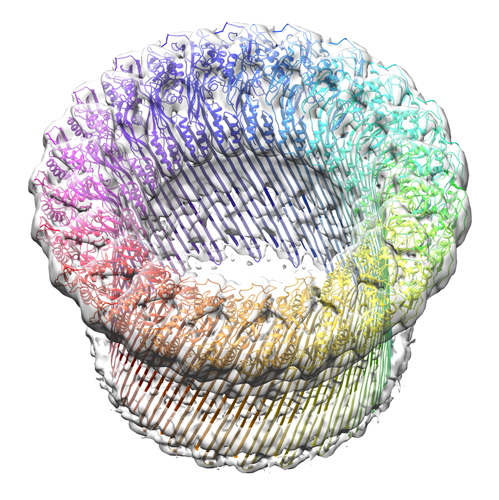 EMD-3235_image-for-emdb.png
 http://ftp.pdbj.org/pub/emdb/structures/EMD-3235


 Z (Sec.)
 Y (Row.)
 X (Col.)


 Sample components
 Processing
 Electron microscopy
 FIELD EMISSION GUN
