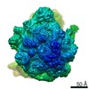
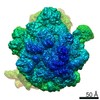
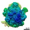
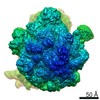
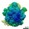
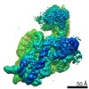
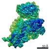
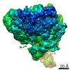
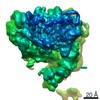
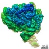
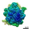
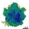
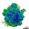
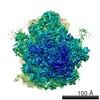
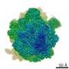
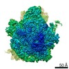
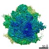
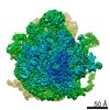
Journal: Proc Natl Acad Sci U S A / Year: 2022 Title: Annealing synchronizes the 70 ribosome into a minimum-energy conformation. Authors: Xiaofeng Chu / Xin Su / Mingdong Liu / Li Li / Tianhao Li / Yicheng Qin / Guoliang Lu / Lei Qi / Yunhui Liu / Jinzhong Lin / Qing-Tao Shen / Abstract: Researchers commonly anneal metals, alloys, and semiconductors to repair defects and improve microstructures via recrystallization. Theoretical studies indicate that simulated annealing on biological ...Researchers commonly anneal metals, alloys, and semiconductors to repair defects and improve microstructures via recrystallization. Theoretical studies indicate that simulated annealing on biological macromolecules helps predict the final structures with minimum free energy. Experimental validation of this homogenizing effect and further exploration of its applications are fascinating scientific questions that remain elusive. Here, we chose the apo-state 70 ribosome from as a model, wherein the 30 subunit undergoes a thermally driven intersubunit rotation and exhibits substantial structural flexibility as well as distinct free energy. We experimentally demonstrate that annealing at a fast cooling rate enhances the 70 ribosome homogeneity and improves local resolution on the 30 subunit. After annealing, the 70 ribosome is in a nonrotated state with respect to corresponding intermediate structures in unannealed or heated ribosomes. Manifold-based analysis further indicates that the annealed 70 ribosome takes a narrow conformational distribution and exhibits a minimum-energy state in the free-energy landscape. Our experimental results offer a facile yet robust approach to enhance protein stability, which is ideal for high-resolution cryogenic electron microscopy. Beyond structure determination, annealing shows great potential for synchronizing proteins on a single-molecule level and can be extended to study protein folding and explore conformational and energy landscapes.
History
Deposition
May 7, 2021
-
Header (metadata) release
Feb 23, 2022
-
Map release
Feb 23, 2022
-
Update
Mar 16, 2022
-
Current status
Mar 16, 2022
Processing site: PDBj / Status: Released
-
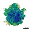
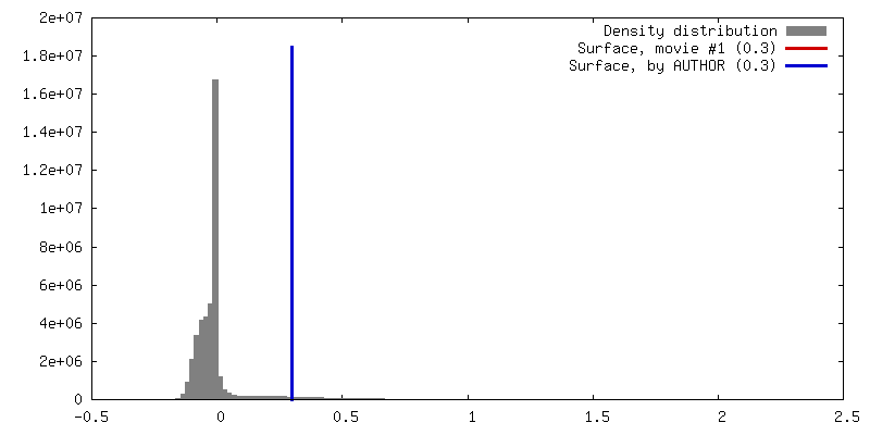
Structure visualization
Movie
Surface view with section colored by density value
In the structure databanks used in Yorodumi, some data are registered as the other names, "COVID-19 virus" and "2019-nCoV". Here are the details of the virus and the list of structure data.
Jan 31, 2019. EMDB accession codes are about to change! (news from PDBe EMDB page)
EMDB accession codes are about to change! (news from PDBe EMDB page)
The allocation of 4 digits for EMDB accession codes will soon come to an end. Whilst these codes will remain in use, new EMDB accession codes will include an additional digit and will expand incrementally as the available range of codes is exhausted. The current 4-digit format prefixed with “EMD-” (i.e. EMD-XXXX) will advance to a 5-digit format (i.e. EMD-XXXXX), and so on. It is currently estimated that the 4-digit codes will be depleted around Spring 2019, at which point the 5-digit format will come into force.
The EM Navigator/Yorodumi systems omit the EMD- prefix.
Related info.:Q: What is EMD? / ID/Accession-code notation in Yorodumi/EM Navigator
Yorodumi is a browser for structure data from EMDB, PDB, SASBDB, etc.
This page is also the successor to EM Navigator detail page, and also detail information page/front-end page for Omokage search.
The word "yorodu" (or yorozu) is an old Japanese word meaning "ten thousand". "mi" (miru) is to see.
Related info.:EMDB / PDB / SASBDB / Comparison of 3 databanks / Yorodumi Search / Aug 31, 2016. New EM Navigator & Yorodumi / Yorodumi Papers / Jmol/JSmol / Function and homology information / Changes in new EM Navigator and Yorodumi
 Movie
Movie Controller

 Yorodumi
 Open data
 Basic information
 Map data
 Sample
 Authors
 Citation
 Structure visualization
 Movie viewer
 Downloads & links
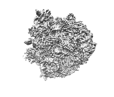 emd_31266.png
 http://ftp.pdbj.org/pub/emdb/structures/EMD-31266
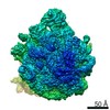


 Z (Sec.)
 Y (Row.)
 X (Col.)


 Sample components
 Processing
 Electron microscopy
 FIELD EMISSION GUN
