Japan Agency for Medical Research and Development (AMED)
19am0101117
Japan
Japan Agency for Medical Research and Development (AMED)
17pc0101020
Japan
Citation
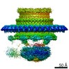
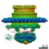
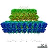
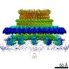
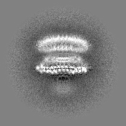
Journal: Nat Commun / Year: 2021 Title: Native flagellar MS ring is formed by 34 subunits with 23-fold and 11-fold subsymmetries. Authors: Akihiro Kawamoto / Tomoko Miyata / Fumiaki Makino / Miki Kinoshita / Tohru Minamino / Katsumi Imada / Takayuki Kato / Keiichi Namba / Abstract: The bacterial flagellar MS ring is a transmembrane complex acting as the core of the flagellar motor and template for flagellar assembly. The C ring attached to the MS ring is involved in torque ...The bacterial flagellar MS ring is a transmembrane complex acting as the core of the flagellar motor and template for flagellar assembly. The C ring attached to the MS ring is involved in torque generation and rotation switch, and a large symmetry mismatch between these two rings has been a long puzzle, especially with respect to their role in motor function. Here, using cryoEM structural analysis of the flagellar basal body and the MS ring formed by full-length FliF from Salmonella enterica, we show that the native MS ring is formed by 34 FliF subunits with no symmetry variation. Symmetry analysis of the C ring shows a variation with a peak at 34-fold, suggesting flexibility in C ring assembly. Finally, our data also indicate that FliF subunits assume two different conformations, contributing differentially to the inner and middle parts of the M ring and thus resulting in 23- and 11-fold subsymmetries in the inner and middle M ring, respectively. The internal core of the M ring, formed by 23 subunits, forms a hole of the right size to accommodate the protein export gate.
History
Deposition
Oct 7, 2020
-
Header (metadata) release
May 19, 2021
-
Map release
May 19, 2021
-
Update
Mar 27, 2024
-
Current status
Mar 27, 2024
Processing site: PDBj / Status: Released
-
Structure visualization
Movie
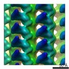
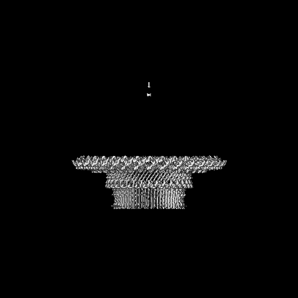
Surface view with section colored by density value
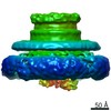
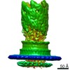
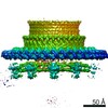
Entire : transmembrane protein complex made of FliF
Entire
Name: transmembrane protein complex made of FliF
Components
Complex: transmembrane protein complex made of FliF
Protein or peptide: Flagellar M-ring protein
-
Supramolecule #1: transmembrane protein complex made of FliF
Supramolecule
Name: transmembrane protein complex made of FliF / type: complex / ID: 1 / Parent: 0 / Macromolecule list: all Details: the MS ring formed by full-length FliF from Salmonella enterica serovar Typhimurium
In the structure databanks used in Yorodumi, some data are registered as the other names, "COVID-19 virus" and "2019-nCoV". Here are the details of the virus and the list of structure data.
Jan 31, 2019. EMDB accession codes are about to change! (news from PDBe EMDB page)
EMDB accession codes are about to change! (news from PDBe EMDB page)
The allocation of 4 digits for EMDB accession codes will soon come to an end. Whilst these codes will remain in use, new EMDB accession codes will include an additional digit and will expand incrementally as the available range of codes is exhausted. The current 4-digit format prefixed with “EMD-” (i.e. EMD-XXXX) will advance to a 5-digit format (i.e. EMD-XXXXX), and so on. It is currently estimated that the 4-digit codes will be depleted around Spring 2019, at which point the 5-digit format will come into force.
The EM Navigator/Yorodumi systems omit the EMD- prefix.
Related info.:Q: What is EMD? / ID/Accession-code notation in Yorodumi/EM Navigator
Yorodumi is a browser for structure data from EMDB, PDB, SASBDB, etc.
This page is also the successor to EM Navigator detail page, and also detail information page/front-end page for Omokage search.
The word "yorodu" (or yorozu) is an old Japanese word meaning "ten thousand". "mi" (miru) is to see.
Related info.:EMDB / PDB / SASBDB / Comparison of 3 databanks / Yorodumi Search / Aug 31, 2016. New EM Navigator & Yorodumi / Yorodumi Papers / Jmol/JSmol / Function and homology information / Changes in new EM Navigator and Yorodumi
 Movie
Movie Controller


 Open data
 Basic information
 Map data
 Sample
 Keywords
 Function and homology information
 Salmonella typhimurium (strain LT2 / SGSC1412 / ATCC 700720) (bacteria) /
  Authors
 Japan, 11 items
  Citation
 Structure visualization
 Downloads & links
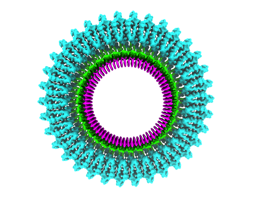 emd_30612.png
 http://ftp.pdbj.org/pub/emdb/structures/EMD-30612

 Z (Sec.)
 Y (Row.)
 X (Col.)


 Sample components
 Processing
 Electron microscopy
 FIELD EMISSION GUN
