 Movie
Movie Controller


+ Open data

- Basic information
| Entry | Database: EMDB / ID: EMD-30607 | |||||||||
|---|---|---|---|---|---|---|---|---|---|---|
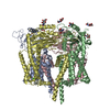
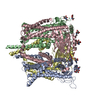
| Title | Structure of PKD1L3-CTD/PKD2L1 in calcium-bound state | |||||||||
 Map data Map data | Ca | |||||||||
 Sample Sample |
| |||||||||
 Keywords Keywords | Heterotetrameric TRP channel / Calcium / Primary cilia / PKD / TRANSPORT PROTEIN | |||||||||
| Function / homology |  Function and homology information Function and homology informationdetection of chemical stimulus involved in sensory perception of sour taste / detection of chemical stimulus involved in sensory perception of taste / pH-gated monoatomic ion channel activity / response to water / osmolarity-sensing monoatomic cation channel activity / cellular response to pH / calcium-activated potassium channel activity / muscle alpha-actinin binding / calcium-activated cation channel activity / cellular response to acidic pH ...detection of chemical stimulus involved in sensory perception of sour taste / detection of chemical stimulus involved in sensory perception of taste / pH-gated monoatomic ion channel activity / response to water / osmolarity-sensing monoatomic cation channel activity / cellular response to pH / calcium-activated potassium channel activity / muscle alpha-actinin binding / calcium-activated cation channel activity / cellular response to acidic pH / cation channel complex / non-motile cilium / : / ciliary membrane / smoothened signaling pathway / sodium channel activity / monoatomic cation transport / monoatomic cation channel activity / calcium channel complex / protein tetramerization / calcium ion transmembrane transport / calcium channel activity / actin cytoskeleton / carbohydrate binding / cytoplasmic vesicle / protein homotetramerization / transmembrane transporter binding / receptor complex / calcium ion binding / endoplasmic reticulum / identical protein binding / plasma membrane / cytosol Similarity search - Function | |||||||||
| Biological species |  | |||||||||
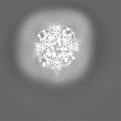
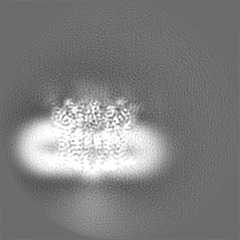
| Method | single particle reconstruction / cryo EM / Resolution: 3.1 Å | |||||||||
 Authors Authors | Su Q / Shi YG | |||||||||
| Funding support |  China, 2 items China, 2 items
| |||||||||
 Citation Citation | Journal: Nat Commun / Year: 2021 Title: Structural basis for Ca activation of the heteromeric PKD1L3/PKD2L1 channel. Authors: Qiang Su / Mengying Chen / Yan Wang / Bin Li / Dan Jing / Xiechao Zhan / Yong Yu / Yigong Shi /  Abstract: The heteromeric complex between PKD1L3, a member of the polycystic kidney disease (PKD) protein family, and PKD2L1, also known as TRPP2 or TRPP3, has been a prototype for mechanistic characterization ...The heteromeric complex between PKD1L3, a member of the polycystic kidney disease (PKD) protein family, and PKD2L1, also known as TRPP2 or TRPP3, has been a prototype for mechanistic characterization of heterotetrametric TRP-like channels. Here we show that a truncated PKD1L3/PKD2L1 complex with the C-terminal TRP-fold fragment of PKD1L3 retains both Ca and acid-induced channel activities. Cryo-EM structures of this core heterocomplex with or without supplemented Ca were determined at resolutions of 3.1 Å and 3.4 Å, respectively. The heterotetramer, with a pseudo-symmetric TRP architecture of 1:3 stoichiometry, has an asymmetric selectivity filter (SF) guarded by Lys2069 from PKD1L3 and Asp523 from the three PKD2L1 subunits. Ca-entrance to the SF vestibule is accompanied by a swing motion of Lys2069 on PKD1L3. The S6 of PKD1L3 is pushed inward by the S4-S5 linker of the nearby PKD2L1 (PKD2L1-III), resulting in an elongated intracellular gate which seals the pore domain. Comparison of the apo and Ca-loaded complexes unveils an unprecedented Ca binding site in the extracellular cleft of the voltage-sensing domain (VSD) of PKD2L1-III, but not the other three VSDs. Structure-guided mutagenic studies support this unconventional site to be responsible for Ca-induced channel activation through an allosteric mechanism. | |||||||||
| History |
|
- Structure visualization
| Movie |
Movie viewer |
|---|---|
| Structure viewer | EM map: SurfViewMolmilJmol/JSmol |
| Supplemental images |

- Downloads & links
-EMDB archive
| Map data | emd_30607.map.gz | 48.7 MB | EMDB map data format | |
|---|---|---|---|---|
| Header (meta data) | emd-30607-v30.xmlemd-30607.xml | 13.9 KB 13.9 KB | Display Display | EMDB header |
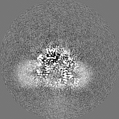
| Images |  emd_30607.png emd_30607.png | 249.1 KB | ||
| Filedesc metadata | emd-30607.cif.gz | 6.3 KB | ||
| Archive directory |  http://ftp.pdbj.org/pub/emdb/structures/EMD-30607ftp://ftp.pdbj.org/pub/emdb/structures/EMD-30607 http://ftp.pdbj.org/pub/emdb/structures/EMD-30607ftp://ftp.pdbj.org/pub/emdb/structures/EMD-30607 | HTTPS FTP |
-Validation report
| Summary document | emd_30607_validation.pdf.gz | 557 KB | Display | EMDB validaton report |
|---|---|---|---|---|
| Full document | emd_30607_full_validation.pdf.gz | 556.6 KB | Display | |
| Data in XML | emd_30607_validation.xml.gz | 5.9 KB | Display | |
| Data in CIF | emd_30607_validation.cif.gz | 6.8 KB | Display | |
| Arichive directory | https://ftp.pdbj.org/pub/emdb/validation_reports/EMD-30607ftp://ftp.pdbj.org/pub/emdb/validation_reports/EMD-30607 | HTTPS FTP |
-Related structure data
| Related structure data |  7d7fMC  7d7eC M: atomic model generated by this map C: citing same article ( |
|---|---|
| Similar structure data |


-Links
| EMDB pages | EMDB (EBI/PDBe) / EMDataResource |
|---|---|
| Related items in Molecule of the Month |


-Map
| File | Download / File: emd_30607.map.gz / Format: CCP4 / Size: 52.7 MB / Type: IMAGE STORED AS FLOATING POINT NUMBER (4 BYTES) | ||||||||||||||||||||||||||||||||||||||||||||||||||||||||||||
|---|---|---|---|---|---|---|---|---|---|---|---|---|---|---|---|---|---|---|---|---|---|---|---|---|---|---|---|---|---|---|---|---|---|---|---|---|---|---|---|---|---|---|---|---|---|---|---|---|---|---|---|---|---|---|---|---|---|---|---|---|---|
| Annotation | Ca | ||||||||||||||||||||||||||||||||||||||||||||||||||||||||||||
| Projections & slices | Image control
Images are generated by Spider. | ||||||||||||||||||||||||||||||||||||||||||||||||||||||||||||
| Voxel size | X=Y=Z: 1.087 Å | ||||||||||||||||||||||||||||||||||||||||||||||||||||||||||||
| Density |
| ||||||||||||||||||||||||||||||||||||||||||||||||||||||||||||
| Symmetry | Space group: 1 | ||||||||||||||||||||||||||||||||||||||||||||||||||||||||||||
| Details | EMDB XML:
CCP4 map header:
| ||||||||||||||||||||||||||||||||||||||||||||||||||||||||||||
 Z (Sec.)
 Y (Row.)
 X (Col.)


-Supplemental data
- Sample components
-Entire : Mouse PKD1L3 in complex with PKD2L1 in presence of 20 mM calcium
| Entire | Name: Mouse PKD1L3 in complex with PKD2L1 in presence of 20 mM calcium |
|---|---|
| Components |
|
-Supramolecule #1: Mouse PKD1L3 in complex with PKD2L1 in presence of 20 mM calcium
| Supramolecule | Name: Mouse PKD1L3 in complex with PKD2L1 in presence of 20 mM calcium type: complex / ID: 1 / Parent: 0 / Macromolecule list: #1-#2 |
|---|---|
| Source (natural) | Organism: |
-Macromolecule #1: Polycystic kidney disease 2-like 1 protein
| Macromolecule | Name: Polycystic kidney disease 2-like 1 protein / type: protein_or_peptide / ID: 1 / Number of copies: 3 / Enantiomer: LEVO |
|---|---|
| Source (natural) | Organism: |
| Molecular weight | Theoretical: 69.234734 KDa |
| Recombinant expression | Organism:  Homo sapiens (human) Homo sapiens (human) |
| Sequence | String: MGSAGWSHPQ FEKGGGSGGG SGGSAWSHPQ FEKGSAAATL VSSCCLHICR SIRGLWGTTL TENTAENREL YVKTTLRELV VYIVFLVDI CLLTYGMTSS SAYYYTKVMS ELFLHTPSDS GVSFQTISSM SDFWDFAQGP LLDSLYWTKW YNNQSLGRGS H SFIYYENL ...String: MGSAGWSHPQ FEKGGGSGGG SGGSAWSHPQ FEKGSAAATL VSSCCLHICR SIRGLWGTTL TENTAENREL YVKTTLRELV VYIVFLVDI CLLTYGMTSS SAYYYTKVMS ELFLHTPSDS GVSFQTISSM SDFWDFAQGP LLDSLYWTKW YNNQSLGRGS H SFIYYENL LLGAPRLRQL RVRNDSCVVH EDFREDILNC YDVYSPDKED QLPFGPQNGT AWTYHSQNEL GGSSHWGRLT SY SGGGYYL DLPGSRQASA EALQGLQEGL WLDRGTRVVF IDFSVYNANI NLFCILRLVV EFPATGGTIP SWQIRTVKLI RYV NNWDFF IVGCEVVFCV FIFYYVVEEI LEIHLHRLRY LSSVWNILDL VVILLSIVAV GFHIFRTLEV NRLMGKLLQQ PDTY ADFEF LAFWQTQYNN MNAVNLFFAW IKIFKYISFN KTMTQLSSTL ARCAKDILGF AIMFFIVFFA YAQLGYLLFG TQVEN FSTF VKCIFTQFRI ILGDFDYNAI DNANRILGPV YFVTYVFFVF FVLLNMFLAI INDTYSEVKE ELAGQKDQLQ LSDFLK QSY NKTLLRLRLR KERVSDVQKV LKGGEPEIQF EDFTSTLREL G UniProtKB: Polycystin-2-like protein 1 |
-Macromolecule #2: Polycystic kidney disease protein 1-like 3
| Macromolecule | Name: Polycystic kidney disease protein 1-like 3 / type: protein_or_peptide / ID: 2 / Number of copies: 1 / Enantiomer: LEVO |
|---|---|
| Source (natural) | Organism: |
| Molecular weight | Theoretical: 62.541914 KDa |
| Recombinant expression | Organism: Homo sapiens (human) |
| Sequence | String: MGSAGDYKDH DGDYKDHDID YKDDDDKGSA AAPIYTAPAM NNLAKPTRKA WKKQLSKLTG GTLVQILFLT LLMTTVYSAK DSSRFFLHR AIWKRFSHRF SEIKTVEDFY PWANGTLLPN LYGDYRGFIT DGNSFLLGNV LIRQTRIPND IFFPGSLHKQ M KSPPQHQE ...String: MGSAGDYKDH DGDYKDHDID YKDDDDKGSA AAPIYTAPAM NNLAKPTRKA WKKQLSKLTG GTLVQILFLT LLMTTVYSAK DSSRFFLHR AIWKRFSHRF SEIKTVEDFY PWANGTLLPN LYGDYRGFIT DGNSFLLGNV LIRQTRIPND IFFPGSLHKQ M KSPPQHQE DRENYGAGWV PPDTNITKVD SIWHYQNQES LGGYPIQGEL ATYSGGGYVV RLGRNHSAAT RVLQHLEQRR WL DHCTKAL FVEFTVFNAN VNLLCAVTLI LESSGVGTFL TSLQLDSLTS LQSSERGFAW IVSQVVYYLL VCYYAFIQGC RLK RQRLAF FTRKRNLLDT SIVLISFSIL GLSMQSLSLL HKKMQQYHCD RDRFISFYEA LRVNSAVTHL RGFLLLFATV RVWD LLRHH AQLQVINKTL SKAWDEVLGF ILIIVVLLSS YAMTFNLLFG WSISDYQSFF RSIVTVVGLL MGTSKHKEVI ALYPI LGSL LVLSSIILMG LVIINLFVSA ILIAFGKERK ACEKEATLTD MLLQKLSSLL GIRLHQNPSE EHADNTG UniProtKB: Polycystin-1-like protein 3 |
-Macromolecule #3: 2-acetamido-2-deoxy-beta-D-glucopyranose
| Macromolecule | Name: 2-acetamido-2-deoxy-beta-D-glucopyranose / type: ligand / ID: 3 / Number of copies: 10 / Formula: NAG |
|---|---|
| Molecular weight | Theoretical: 221.208 Da |
| Chemical component information |  ChemComp-NAG: |
-Macromolecule #4: CALCIUM ION
| Macromolecule | Name: CALCIUM ION / type: ligand / ID: 4 / Number of copies: 5 / Formula: CA |
|---|---|
| Molecular weight | Theoretical: 40.078 Da |
-Experimental details
-Structure determination
| Method | cryo EM |
|---|---|
 Processing Processing | single particle reconstruction |
| Aggregation state | particle |
-Sample preparation
| Concentration | 10 mg/mL |
|---|---|
| Buffer | pH: 7.5 |
| Vitrification | Cryogen name: ETHANE |
- Electron microscopy
| Microscope | FEI TITAN KRIOS |
|---|---|
| Image recording | Film or detector model: GATAN K3 BIOQUANTUM (6k x 4k) / Average electron dose: 50.0 e/Å2 |
| Electron beam | Acceleration voltage: 300 kV / Electron source:  FIELD EMISSION GUN FIELD EMISSION GUN |
| Electron optics | Illumination mode: FLOOD BEAM / Imaging mode: BRIGHT FIELD |
| Experimental equipment |  Model: Titan Krios / Image courtesy: FEI Company |
