 Movie
Movie Controller


+ Open data

- Basic information
| Entry | Database: EMDB / ID: EMD-30069 | |||||||||
|---|---|---|---|---|---|---|---|---|---|---|
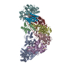
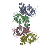
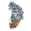
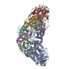
| Title | Cryo-EM structure of FMO-RC complex from green sulfur bacteria | |||||||||
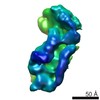
 Map data Map data | structure of FMO-RC complex from a green sulfur bacterium | |||||||||
 Sample Sample |
| |||||||||
 Keywords Keywords | Photosynthetic reaction center / Green sulfur bacteria / Energy transfer / PHOTOSYNTHESIS | |||||||||
| Function / homology |  Function and homology information Function and homology informationthylakoid / bacteriochlorophyll binding / iron-sulfur cluster binding / photosynthesis / membrane / metal ion binding Similarity search - Function | |||||||||
| Biological species |  Chlorobaculum tepidum (strain ATCC 49652 / DSM 12025 / NBRC 103806 / TLS) (bacteria) / Chlorobaculum tepidum TLS (bacteria) Chlorobaculum tepidum (strain ATCC 49652 / DSM 12025 / NBRC 103806 / TLS) (bacteria) / Chlorobaculum tepidum TLS (bacteria) | |||||||||
| Method | single particle reconstruction / cryo EM / Resolution: 2.7 Å | |||||||||
 Authors Authors | Chen JH / Zhang X | |||||||||
 Citation Citation | Journal: Science / Year: 2020 Title: Architecture of the photosynthetic complex from a green sulfur bacterium. Authors: Jing-Hua Chen / Hangjun Wu / Caihuang Xu / Xiao-Chi Liu / Zihui Huang / Shenghai Chang / Wenda Wang / Guangye Han / Tingyun Kuang / Jian-Ren Shen / Xing Zhang /   Abstract: The photosynthetic apparatus of green sulfur bacteria (GSB) contains a peripheral antenna chlorosome, light-harvesting Fenna-Matthews-Olson proteins (FMO), and a reaction center (GsbRC). We used cryo- ...The photosynthetic apparatus of green sulfur bacteria (GSB) contains a peripheral antenna chlorosome, light-harvesting Fenna-Matthews-Olson proteins (FMO), and a reaction center (GsbRC). We used cryo-electron microscopy to determine a 2.7-angstrom structure of the FMO-GsbRC supercomplex from The GsbRC binds considerably fewer (bacterio)chlorophylls [(B)Chls] than other known type I RCs do, and the organization of (B)Chls is similar to that in photosystem II. Two BChl layers in GsbRC are not connected by Chls, as seen in other RCs, but associate with two carotenoid derivatives. Relatively long distances of 22 to 33 angstroms were observed between BChls of FMO and GsbRC, consistent with the inefficient energy transfer between these entities. The structure contains common features of both type I and type II RCs and provides insight into the evolution of photosynthetic RCs. | |||||||||
| History |
|
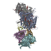
- Structure visualization
| Movie |
Movie viewer |
|---|---|
| Structure viewer | EM map: SurfViewMolmilJmol/JSmol |
| Supplemental images |

- Downloads & links
-EMDB archive
| Map data | emd_30069.map.gz | 14.5 MB | EMDB map data format | |
|---|---|---|---|---|
| Header (meta data) | emd-30069-v30.xmlemd-30069.xml | 24.2 KB 24.2 KB | Display Display | EMDB header |
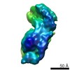
| Images | 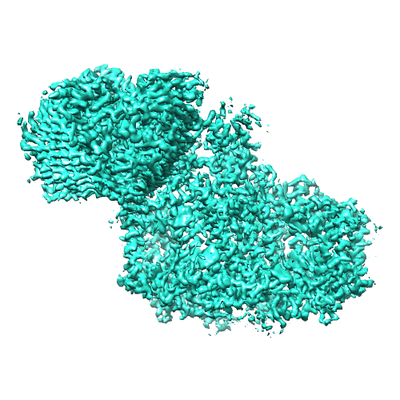 emd_30069.png emd_30069.png | 158.4 KB | ||
| Filedesc metadata | emd-30069.cif.gz | 7.4 KB | ||
| Archive directory |  http://ftp.pdbj.org/pub/emdb/structures/EMD-30069ftp://ftp.pdbj.org/pub/emdb/structures/EMD-30069 http://ftp.pdbj.org/pub/emdb/structures/EMD-30069ftp://ftp.pdbj.org/pub/emdb/structures/EMD-30069 | HTTPS FTP |
-Related structure data
| Related structure data | 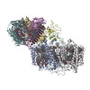 6m32MC M: atomic model generated by this map C: citing same article ( |
|---|---|
| Similar structure data |


-Links
| EMDB pages | EMDB (EBI/PDBe) / EMDataResource |
|---|---|
| Related items in Molecule of the Month |


-Map
| File | Download / File: emd_30069.map.gz / Format: CCP4 / Size: 15.6 MB / Type: IMAGE STORED AS FLOATING POINT NUMBER (4 BYTES) | ||||||||||||||||||||||||||||||||||||||||||||||||||||||||||||
|---|---|---|---|---|---|---|---|---|---|---|---|---|---|---|---|---|---|---|---|---|---|---|---|---|---|---|---|---|---|---|---|---|---|---|---|---|---|---|---|---|---|---|---|---|---|---|---|---|---|---|---|---|---|---|---|---|---|---|---|---|---|
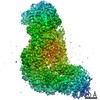
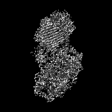
| Annotation | structure of FMO-RC complex from a green sulfur bacterium | ||||||||||||||||||||||||||||||||||||||||||||||||||||||||||||
| Projections & slices | Image control
Images are generated by Spider. | ||||||||||||||||||||||||||||||||||||||||||||||||||||||||||||
| Voxel size | X=Y=Z: 1.307 Å | ||||||||||||||||||||||||||||||||||||||||||||||||||||||||||||
| Density |
| ||||||||||||||||||||||||||||||||||||||||||||||||||||||||||||
| Symmetry | Space group: 1 | ||||||||||||||||||||||||||||||||||||||||||||||||||||||||||||
| Details | EMDB XML:
CCP4 map header:
| ||||||||||||||||||||||||||||||||||||||||||||||||||||||||||||
 Z (Sec.)
 Y (Row.)
 X (Col.)


-Supplemental data
- Sample components
+Entire : FMO-RC of green sulfur bacteria
+Supramolecule #1: FMO-RC of green sulfur bacteria
+Macromolecule #1: Bacteriochlorophyll a protein
+Macromolecule #2: P840 reaction center 17 kDa protein
+Macromolecule #3: Photosystem P840 reaction center iron-sulfur protein
+Macromolecule #4: Photosystem P840 reaction center, large subunit
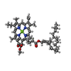
+Macromolecule #5: BACTERIOCHLOROPHYLL A

+Macromolecule #6: IRON/SULFUR CLUSTER

+Macromolecule #7: Bacteriochlorophyll A isomer
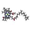
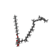
+Macromolecule #8: 2-[(1E,3E,5E,7E,9E,11E,13E,15E,17E,19E)-3,7,12,16,20,24-hexamethy...

+Macromolecule #9: [(2R,3S,4S,5R,6R)-6-[(10E,12E,14E)-2,6,10,14,19,23-hexamethyl-25-...
+Macromolecule #10: 1,2-DIPALMITOYL-PHOSPHATIDYL-GLYCEROLE

+Macromolecule #11: 1,2-DISTEAROYL-MONOGALACTOSYL-DIGLYCERIDE

+Macromolecule #12: CALCIUM ION
+Macromolecule #13: Chlorophyll A ester
-Experimental details
-Structure determination
| Method | cryo EM |
|---|---|
 Processing Processing | single particle reconstruction |
| Aggregation state | particle |
-Sample preparation
| Buffer | pH: 8 |
|---|---|
| Grid | Model: Quantifoil R1.2/1.3 / Material: COPPER / Mesh: 300 / Support film - Material: GRAPHENE OXIDE / Support film - topology: HOLEY ARRAY |
| Vitrification | Cryogen name: ETHANE / Chamber humidity: 100 % |
- Electron microscopy
| Microscope | FEI TITAN KRIOS |
|---|---|
| Image recording | Film or detector model: GATAN K2 SUMMIT (4k x 4k) / Detector mode: SUPER-RESOLUTION / Digitization - Dimensions - Width: 3710 pixel / Digitization - Dimensions - Height: 3838 pixel / Digitization - Frames/image: 1-40 / Number grids imaged: 3 / Number real images: 9156 / Average exposure time: 10.0 sec. / Average electron dose: 47.0 e/Å2 |
| Electron beam | Acceleration voltage: 300 kV / Electron source:  FIELD EMISSION GUN FIELD EMISSION GUN |
| Electron optics | C2 aperture diameter: 70.0 µm / Calibrated defocus max: 2.5 µm / Calibrated defocus min: 1.5 µm / Calibrated magnification: 38244 / Illumination mode: FLOOD BEAM / Imaging mode: BRIGHT FIELD / Cs: 2.7 mm / Nominal defocus max: 2.5 µm / Nominal defocus min: 1.5 µm / Nominal magnification: 22500 |
| Sample stage | Specimen holder model: FEI TITAN KRIOS AUTOGRID HOLDER / Cooling holder cryogen: NITROGEN |
| Experimental equipment |  Model: Titan Krios / Image courtesy: FEI Company |
