Movie
Movie Controller

[English] 日本語
 Yorodumi
- EMDB-2995: Negative stain 3D reconstruction of the yeast 26S proteasome in c... -
+ Open data

- Basic information
| Entry | Database: EMDB / ID: EMD-2995 | |||||||||
|---|---|---|---|---|---|---|---|---|---|---|
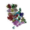
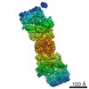
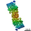
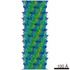
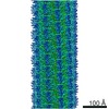
| Title | Negative stain 3D reconstruction of the yeast 26S proteasome in complex with ubiquitin-bound Ubp6 | |||||||||
 Map data Map data | Negative stain 3D reconstruction of yeast 26S holoenzyme in complex with ubiquitin-bound Ubp6 | |||||||||
 Sample Sample |
| |||||||||
 Keywords Keywords | Proteasome / UPS / Ubp6 / deubiquitinase / regulatory particle | |||||||||
| Function / homology |  Function and homology information Function and homology informationnegative regulation of ERAD pathway / negative regulation of proteasomal protein catabolic process / mitochondria-associated ubiquitin-dependent protein catabolic process / regulation of proteasomal ubiquitin-dependent protein catabolic process / proteasome regulatory particle / proteasome binding / Ub-specific processing proteases / protein deubiquitination / proteasome complex / ubiquitinyl hydrolase 1 / cysteine-type deubiquitinase activity Similarity search - Function | |||||||||
| Biological species |  | |||||||||
| Method | single particle reconstruction / negative staining / Resolution: 22.3 Å | |||||||||
 Authors Authors | Bashore C / Dambacher CM / Matyskiela M / Lander GC / Martin A | |||||||||
 Citation Citation | Journal: Nat Struct Mol Biol / Year: 2015 Title: Ubp6 deubiquitinase controls conformational dynamics and substrate degradation of the 26S proteasome. Authors: Charlene Bashore / Corey M Dambacher / Ellen A Goodall / Mary E Matyskiela / Gabriel C Lander / Andreas Martin /  Abstract: Substrates are targeted for proteasomal degradation through the attachment of ubiquitin chains that need to be removed by proteasomal deubiquitinases before substrate processing. In budding yeast, ...Substrates are targeted for proteasomal degradation through the attachment of ubiquitin chains that need to be removed by proteasomal deubiquitinases before substrate processing. In budding yeast, the deubiquitinase Ubp6 trims ubiquitin chains and affects substrate processing by the proteasome, but the underlying mechanisms and the location of Ubp6 within the holoenzyme have been elusive. Here we show that Ubp6 activity strongly responds to interactions with the base ATPase and the conformational state of the proteasome. Electron microscopy analyses reveal that ubiquitin-bound Ubp6 contacts the N ring and AAA+ ring of the ATPase hexamer and is in proximity to the deubiquitinase Rpn11. Ubiquitin-bound Ubp6 inhibits substrate deubiquitination by Rpn11, stabilizes the substrate-engaged conformation of the proteasome and allosterically interferes with the engagement of a subsequent substrate. Ubp6 may thus act as a ubiquitin-dependent 'timer' to coordinate individual processing steps at the proteasome and modulate substrate degradation. | |||||||||
| History |
|
- Structure visualization
| Movie |
Movie viewer |
|---|---|
| Structure viewer | EM map: SurfViewMolmilJmol/JSmol |
| Supplemental images |

- Downloads & links
-EMDB archive
| Map data | emd_2995.map.gz | 25.2 MB | EMDB map data format | |
|---|---|---|---|---|
| Header (meta data) | emd-2995-v30.xmlemd-2995.xml | 13.8 KB 13.8 KB | Display Display | EMDB header |
| Images | 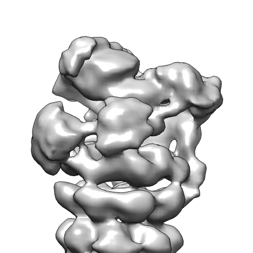 EMD-2995.png EMD-2995.png | 93.9 KB | ||
| Archive directory |  http://ftp.pdbj.org/pub/emdb/structures/EMD-2995ftp://ftp.pdbj.org/pub/emdb/structures/EMD-2995 http://ftp.pdbj.org/pub/emdb/structures/EMD-2995ftp://ftp.pdbj.org/pub/emdb/structures/EMD-2995 | HTTPS FTP |
-Related structure data


-Links
| EMDB pages | EMDB (EBI/PDBe) / EMDataResource |
|---|---|
| Related items in Molecule of the Month |


-Map
| File | Download / File: emd_2995.map.gz / Format: CCP4 / Size: 26.4 MB / Type: IMAGE STORED AS FLOATING POINT NUMBER (4 BYTES) | ||||||||||||||||||||||||||||||||||||||||||||||||||||||||||||
|---|---|---|---|---|---|---|---|---|---|---|---|---|---|---|---|---|---|---|---|---|---|---|---|---|---|---|---|---|---|---|---|---|---|---|---|---|---|---|---|---|---|---|---|---|---|---|---|---|---|---|---|---|---|---|---|---|---|---|---|---|---|
| Annotation | Negative stain 3D reconstruction of yeast 26S holoenzyme in complex with ubiquitin-bound Ubp6 | ||||||||||||||||||||||||||||||||||||||||||||||||||||||||||||
| Projections & slices | Image control
Images are generated by Spider. | ||||||||||||||||||||||||||||||||||||||||||||||||||||||||||||
| Voxel size | X=Y=Z: 4.1 Å | ||||||||||||||||||||||||||||||||||||||||||||||||||||||||||||
| Density |
| ||||||||||||||||||||||||||||||||||||||||||||||||||||||||||||
| Symmetry | Space group: 1 | ||||||||||||||||||||||||||||||||||||||||||||||||||||||||||||
| Details | EMDB XML:
CCP4 map header:
| ||||||||||||||||||||||||||||||||||||||||||||||||||||||||||||
 Z (Sec.)
 Y (Row.)
 X (Col.)


-Supplemental data
- Sample components
-Entire : Yeast 26S proteasome in complex with ubiquitin-bound Ubp6
| Entire | Name: Yeast 26S proteasome in complex with ubiquitin-bound Ubp6 |
|---|---|
| Components |
|
-Supramolecule #1000: Yeast 26S proteasome in complex with ubiquitin-bound Ubp6
| Supramolecule | Name: Yeast 26S proteasome in complex with ubiquitin-bound Ubp6 type: sample / ID: 1000 / Details: The sample was monodisperse Oligomeric state: One to two 19S regulatory particles associates with the core particle to form a functional holoenzyme Number unique components: 2 |
|---|---|
| Molecular weight | Experimental: 1.5 MDa / Theoretical: 1.5 MDa |
-Macromolecule #1: 26S proteasome
| Macromolecule | Name: 26S proteasome / type: protein_or_peptide / ID: 1 / Name.synonym: Proteasome Holoenzyme Details: 50uM WT Ubp6 protein was reacted with 75uM ubiquitin vinyl sulfone at 37 degrees. Samples of 26S-bound Ubp6-UbVS were then diluted to ~25nM for analysis by negative stain electron microscopy. Number of copies: 1 / Oligomeric state: Monomer / Recombinant expression: No |
|---|---|
| Source (natural) | Organism: |
| Molecular weight | Experimental: 1.5 MDa / Theoretical: 1.5 MDa |
| Sequence | GO: proteasome regulatory particle / InterPro: Proteasome, subunit alpha/beta |
-Macromolecule #2: ubiquitin-bound Ubp6
| Macromolecule | Name: ubiquitin-bound Ubp6 / type: protein_or_peptide / ID: 2 Details: Recombinant Ubp6 was purified from E. coli by Ni affinity chromatography and SEC on a superdex 200. Ubp6-UbVS was made by incubating 75uM UbVS with 50uM Ubp6 at 37 degrees C for 7 hours. ...Details: Recombinant Ubp6 was purified from E. coli by Ni affinity chromatography and SEC on a superdex 200. Ubp6-UbVS was made by incubating 75uM UbVS with 50uM Ubp6 at 37 degrees C for 7 hours. Ubp6-UbVS was added to holoenzymes at a 2:1 ratio and exchanged into 1mM ATPgS. Number of copies: 2 / Oligomeric state: monomer / Recombinant expression: Yes |
|---|---|
| Source (natural) | Organism: |
| Molecular weight | Experimental: 65 KDa / Theoretical: 65 KDa |
| Recombinant expression | Organism:  |
| Sequence | UniProtKB: Ubiquitin carboxyl-terminal hydrolase 6 |
-Experimental details
-Structure determination
| Method | negative staining |
|---|---|
 Processing Processing | single particle reconstruction |
| Aggregation state | particle |
-Sample preparation
| Concentration | 0.05 mg/mL |
|---|---|
| Buffer | pH: 7.6 Details: 60mM HEPES pH 7.6, 50mM NaCl, 50mM KCl, 5mM MgCl2, 0.5mM EDTA, 1mM TCEP, 1mM ATPgS |
| Staining | Type: NEGATIVE Details: 4 microliters of sample was applied to a freshly plasma-cleaned thin carbon surface pre-treated with 0.1% w/v poly-L-lysine hydrobromide. After removal of excess protein, negative staining ...Details: 4 microliters of sample was applied to a freshly plasma-cleaned thin carbon surface pre-treated with 0.1% w/v poly-L-lysine hydrobromide. After removal of excess protein, negative staining was performed using 2% w/v uranyl formate solution. |
| Grid | Details: 400 mesh Cu-Rh Maxtaform grids were used following deposition of a thin continuous carbon film |
| Vitrification | Cryogen name: NONE / Instrument: OTHER |
- Electron microscopy
| Microscope | FEI TECNAI SPIRIT |
|---|---|
| Temperature | Min: 294 K / Max: 297 K / Average: 295 K |
| Alignment procedure | Legacy - Astigmatism: Objective lens astigmatism was corrected using a quadrupole stigmator at 52,000 times magnification |
| Date | Oct 10, 2014 |
| Image recording | Category: CCD / Film or detector model: TVIPS TEMCAM-F416 (4k x 4k) / Digitization - Sampling interval: 2.5 µm / Number real images: 357 / Average electron dose: 20 e/Å2 Details: Automated imaging was performed using Leginon software |
| Tilt angle min | 0 |
| Tilt angle max | 0 |
| Electron beam | Acceleration voltage: 120 kV / Electron source: LAB6 |
| Electron optics | Calibrated magnification: 52000 / Illumination mode: FLOOD BEAM / Imaging mode: BRIGHT FIELD / Cs: 2.2 mm / Nominal defocus max: 1.5 µm / Nominal defocus min: 0.5 µm / Nominal magnification: 52000 |
| Sample stage | Specimen holder: Room temperature, side entry holder / Specimen holder model: SIDE ENTRY, EUCENTRIC |
| Experimental equipment |  Model: Tecnai Spirit / Image courtesy: FEI Company |
-Image processing
| Details | All image processing leading up to 3D reconstruction was performed using the Appion package. Particles were selected using the Difference of Gaussians (DoG)-based automated particle picker from raw micrographs. The stack of particles was subjected to five iterations of 2D alignment and classification using multivariate statistical analysis (MSA) and multi-reference alignment (MRA). Selected 2D classes were used to generate a sub-stack that was subjected to twenty five iterations of 3D classification, requesting four classes using the Relion suite. Particles belonging to well-resolved 3D classes were used for further refinement by projection matching in Relion. |
|---|---|
| CTF correction | Details: Phase flipping of whole micrographs |
| Final reconstruction | Applied symmetry - Point group: C2 (2 fold cyclic) / Algorithm: OTHER / Resolution.type: BY AUTHOR / Resolution: 22.3 Å / Resolution method: OTHER / Software - Name: Relion Details: Final 3D models were refined using 12000 particles selected by combining two 3D classes from Relion processing Number images used: 18565 |
| Final two d classification | Number classes: 4 |
-Atomic model buiding 1
| Initial model | PDB ID: |
|---|---|
| Software | Name:  Chimera Chimera |
| Details | An atomic model of yeast Ub-bound Ubp6 was constructed by superimposing the yeast Ubp6 crystal structure PDB 1VJV onto the stucture of the human Rsp14 structure bound to Ubiquitin PDB 2AYO, using the UCSF Chimera MatchMaker tool. These structures have high homology, and the resulting hybrid structure did not exhibit any clashes between the Ubiquitin and Ubp6. This Ubp6-Ub model was docked into the density putatively corresponding to Ubp6. PDB 4CR4 was used for docking other 26S core, base and lid subunits into the map, with the exception of the Rpn8-Rpn11 dimer, for which PDB 4O8Y was used. All docking of PDB structures was performed using the Fit in Map tool of UCSF Chimera |
| Refinement | Space: REAL / Protocol: RIGID BODY FIT |