 Movie
Movie Controller


+ Open data

- Basic information
| Entry | Database: EMDB / ID: EMD-22088 | |||||||||
|---|---|---|---|---|---|---|---|---|---|---|
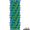
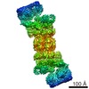
| Title | Structure of the Campylobacter jejuni G508A Flagellar Filament | |||||||||
 Map data Map data | ||||||||||
 Sample Sample |
| |||||||||
 Keywords Keywords | Helical Symmetry / Bacterial Flagellar Filament / STRUCTURAL PROTEIN | |||||||||
| Function / homology |  Function and homology information Function and homology informationbacterial-type flagellum / structural molecule activity / extracellular region Similarity search - Function | |||||||||
| Biological species |   Campylobacter jejuni (Campylobacter) Campylobacter jejuni (Campylobacter) | |||||||||
| Method | helical reconstruction / cryo EM / Resolution: 3.5 Å | |||||||||
 Authors Authors | Kreutzberger MAB / Wang F / Egelman EH | |||||||||
| Funding support |  United States, 2 items United States, 2 items
| |||||||||
 Citation Citation | Journal: Proc Natl Acad Sci U S A / Year: 2020 Title: Atomic structure of the flagellar filament reveals how ε Proteobacteria escaped Toll-like receptor 5 surveillance. Authors: Mark A B Kreutzberger / Cheryl Ewing / Frederic Poly / Fengbin Wang / Edward H Egelman / Abstract: Vertebrates, from zebra fish to humans, have an innate immune recognition of many bacterial flagellins. This involves a conserved eight-amino acid epitope in flagellin recognized by the Toll-like ...Vertebrates, from zebra fish to humans, have an innate immune recognition of many bacterial flagellins. This involves a conserved eight-amino acid epitope in flagellin recognized by the Toll-like receptor 5 (TLR5). Several important human pathogens, such as and , have escaped TLR5 activation by mutations in this epitope. When such mutations were introduced into flagellin, motility was abolished. It was previously argued, using very low-resolution cryoelectron microscopy (cryo-EM), that accommodated these mutations by forming filaments with 7 protofilaments, rather than the 11 found in other bacteria. We have now determined the atomic structure of the G508A flagellar filament from a 3.5-Å-resolution cryo-EM reconstruction, and show that it has 11 protofilaments. The residues in the TLR5 epitope have reduced contacts with the adjacent subunit compared to other bacterial flagellar filament structures. The weakening of the subunit-subunit interface introduced by the mutations in the TLR5 epitope is compensated for by extensive interactions between the outer domains of the flagellin subunits. In other bacteria, these outer domains can be nearly absent or removed without affecting motility. Furthermore, we provide evidence for the stabilization of these outer domain interactions through glycosylation of key residues. These results explain the essential role of glycosylation in motility, and show how the outer domains have evolved to play a role not previously found in other bacteria. | |||||||||
| History |
|
- Structure visualization
| Movie |
Movie viewer |
|---|---|
| Structure viewer | EM map: SurfViewMolmilJmol/JSmol |
| Supplemental images |

- Downloads & links
-EMDB archive
| Map data | emd_22088.map.gz | 46.8 MB | EMDB map data format | |
|---|---|---|---|---|
| Header (meta data) | emd-22088-v30.xmlemd-22088.xml | 15.9 KB 15.9 KB | Display Display | EMDB header |
| Images | 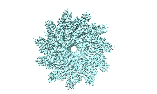 emd_22088.png emd_22088.png | 205.1 KB | ||
| Filedesc metadata | emd-22088.cif.gz | 6.2 KB | ||
| Archive directory |  http://ftp.pdbj.org/pub/emdb/structures/EMD-22088ftp://ftp.pdbj.org/pub/emdb/structures/EMD-22088 http://ftp.pdbj.org/pub/emdb/structures/EMD-22088ftp://ftp.pdbj.org/pub/emdb/structures/EMD-22088 | HTTPS FTP |
-Related structure data
| Related structure data | 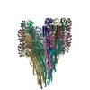 6x80MC M: atomic model generated by this map C: citing same article ( |
|---|---|
| Similar structure data |


-Links
| EMDB pages | EMDB (EBI/PDBe) / EMDataResource |
|---|
-Map
| File | Download / File: emd_22088.map.gz / Format: CCP4 / Size: 236.3 MB / Type: IMAGE STORED AS FLOATING POINT NUMBER (4 BYTES) | ||||||||||||||||||||||||||||||||||||||||||||||||||||||||||||||||||||
|---|---|---|---|---|---|---|---|---|---|---|---|---|---|---|---|---|---|---|---|---|---|---|---|---|---|---|---|---|---|---|---|---|---|---|---|---|---|---|---|---|---|---|---|---|---|---|---|---|---|---|---|---|---|---|---|---|---|---|---|---|---|---|---|---|---|---|---|---|---|
| Projections & slices | Image control
Images are generated by Spider. generated in cubic-lattice coordinate | ||||||||||||||||||||||||||||||||||||||||||||||||||||||||||||||||||||
| Voxel size | X=Y=Z: 1.08 Å | ||||||||||||||||||||||||||||||||||||||||||||||||||||||||||||||||||||
| Density |
| ||||||||||||||||||||||||||||||||||||||||||||||||||||||||||||||||||||
| Symmetry | Space group: 1 | ||||||||||||||||||||||||||||||||||||||||||||||||||||||||||||||||||||
| Details | EMDB XML:
CCP4 map header:
| ||||||||||||||||||||||||||||||||||||||||||||||||||||||||||||||||||||
 Z (Sec.)
 Y (Row.)
 X (Col.)


-Supplemental data
- Sample components
-Entire : Bacterial Flagellar Filament
| Entire | Name: Bacterial Flagellar Filament |
|---|---|
| Components |
|
-Supramolecule #1: Bacterial Flagellar Filament
| Supramolecule | Name: Bacterial Flagellar Filament / type: organelle_or_cellular_component / ID: 1 / Parent: 0 / Macromolecule list: #1 |
|---|---|
| Source (natural) | Organism: Campylobacter jejuni (Campylobacter) |
-Macromolecule #1: Flagellin A
| Macromolecule | Name: Flagellin A / type: protein_or_peptide / ID: 1 / Number of copies: 22 / Enantiomer: LEVO |
|---|---|
| Source (natural) | Organism: Campylobacter jejuni (Campylobacter) |
| Molecular weight | Theoretical: 59.590762 KDa |
| Recombinant expression | Organism: Campylobacter jejuni (Campylobacter) |
| Sequence | String: MGFRINTNVA ALNAKANSDL NAKSLDASLS RLSSGLRINS AADDASGMAI ADSLRSQANT LGQAISNGND ALGILQTADK AMDEQLKIL DTIKTKATQA AQDGQSLKTR TMLQADINKL MEELDNIANT TSFNGKQLLS GNFTNQEFQI GASSNQTVKA T IGATQSSK ...String: MGFRINTNVA ALNAKANSDL NAKSLDASLS RLSSGLRINS AADDASGMAI ADSLRSQANT LGQAISNGND ALGILQTADK AMDEQLKIL DTIKTKATQA AQDGQSLKTR TMLQADINKL MEELDNIANT TSFNGKQLLS GNFTNQEFQI GASSNQTVKA T IGATQSSK IGVTRFETGA QSFTSGVVGL TIKNYNGIED FKFDNVVIST SVGTGLGALA EEINKSADKT GVRATYDVKT TG VYAIKEG TTSQDFAING VTIGKIEYKD GDGNGSLISA INAVKDTTGV QASKDENGKL VLTSADGRGI KITGDIGVGS GIL ANQKEN YGRLSLVKND GRDINISGTN LSAIGMGTTD MISQSSVSLR ESKGQISATN ADAMGFNSYK GGGKFVFTQN VSSI SAFMS AQGSGFSRGS GFSVGSGKNL SVGLSQGIQI ISSAASMSNT YVVSAGSGFS SGSGNSQFAA LKTTAANTTD ETAGV TTLK GAMAVMDIAE TAITNLDQIR ADIASIQNQV TSTINNITVT QVNVKAAESQ IRDVDFASES ANYSKANILA QSGSYA MAQ ANSSQQNVLR LLQ UniProtKB: Flagellin A |
-Macromolecule #2: 5,7-diamino-3,5,7,9-tetradeoxy-L-glycero-alpha-L-manno-non-2-ulop...
| Macromolecule | Name: 5,7-diamino-3,5,7,9-tetradeoxy-L-glycero-alpha-L-manno-non-2-ulopyranosonic acid type: ligand / ID: 2 / Number of copies: 374 / Formula: P8E |
|---|---|
| Molecular weight | Theoretical: 250.249 Da |
| Chemical component information |  ChemComp-P8E: |
-Experimental details
-Structure determination
| Method | cryo EM |
|---|---|
 Processing Processing | helical reconstruction |
| Aggregation state | filament |
-Sample preparation
| Buffer | pH: 7 |
|---|---|
| Vitrification | Cryogen name: ETHANE |
- Electron microscopy
| Microscope | FEI TITAN KRIOS |
|---|---|
| Image recording | Film or detector model: GATAN K3 (6k x 4k) / Average electron dose: 51.0 e/Å2 |
| Electron beam | Acceleration voltage: 300 kV / Electron source:  FIELD EMISSION GUN FIELD EMISSION GUN |
| Electron optics | Illumination mode: FLOOD BEAM / Imaging mode: BRIGHT FIELD |
| Experimental equipment |  Model: Titan Krios / Image courtesy: FEI Company |
-Image processing
| Final reconstruction | Applied symmetry - Helical parameters - Δz: 4.8 Å Applied symmetry - Helical parameters - Δ&Phi: 65.32 ° Applied symmetry - Helical parameters - Axial symmetry: C1 (asymmetric) Resolution.type: BY AUTHOR / Resolution: 3.5 Å / Resolution method: OTHER / Software: (Name: SPIDER, RELION) / Details: MODEL:MAP FSC using a threshold of 0.5 / Number images used: 116959 |
|---|---|
| CTF correction | Type: PHASE FLIPPING AND AMPLITUDE CORRECTION |
| Startup model | Type of model: OTHER Details: averaged cylinder using all segments, with random azimuthal angles |
| Final angle assignment | Type: NOT APPLICABLE |
