 Movie
Movie Controller

[English] 日本語
 Yorodumi
- EMDB-2035: 8.4 A single-particle reconstruction of the 26S Proteasome from S... -
+ Open data

- Basic information
| Entry | Database: EMDB / ID: EMD-2035 | |||||||||
|---|---|---|---|---|---|---|---|---|---|---|
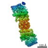
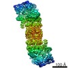
| Title | 8.4 A single-particle reconstruction of the 26S Proteasome from Schizosaccharomyces pombe | |||||||||
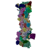
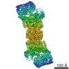
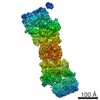
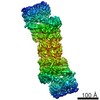
 Map data Map data | This is an image of a surface rendered S.pombe 26S proteasome | |||||||||
 Sample Sample |
| |||||||||
 Keywords Keywords | 26S proteasome / AAA-ATPase / PCI-domain / solenoid | |||||||||
| Biological species |  | |||||||||
| Method | single particle reconstruction / cryo EM / Resolution: 8.4 Å | |||||||||
 Authors Authors | Bohn S / Beck F / Lasker K / Forster F / Walzthoeni T / Villa E / Unverdorben P / Aebersold R / Sali A / Baumeister W | |||||||||
 Citation Citation | Journal: Proc Natl Acad Sci U S A / Year: 2012 Title: Molecular architecture of the 26S proteasome holocomplex determined by an integrative approach. Authors: Keren Lasker / Friedrich Förster / Stefan Bohn / Thomas Walzthoeni / Elizabeth Villa / Pia Unverdorben / Florian Beck / Ruedi Aebersold / Andrej Sali / Wolfgang Baumeister /  Abstract: The 26S proteasome is at the executive end of the ubiquitin-proteasome pathway for the controlled degradation of intracellular proteins. While the structure of its 20S core particle (CP) has been ...The 26S proteasome is at the executive end of the ubiquitin-proteasome pathway for the controlled degradation of intracellular proteins. While the structure of its 20S core particle (CP) has been determined by X-ray crystallography, the structure of the 19S regulatory particle (RP), which recruits substrates, unfolds them, and translocates them to the CP for degradation, has remained elusive. Here, we describe the molecular architecture of the 26S holocomplex determined by an integrative approach based on data from cryoelectron microscopy, X-ray crystallography, residue-specific chemical cross-linking, and several proteomics techniques. The "lid" of the RP (consisting of Rpn3/5/6/7/8/9/11/12) is organized in a modular fashion. Rpn3/5/6/7/9/12 form a horseshoe-shaped heterohexamer, which connects to the CP and roofs the AAA-ATPase module, positioning the Rpn8/Rpn11 heterodimer close to its mouth. Rpn2 is rigid, supporting the lid, while Rpn1 is conformationally variable, positioned at the periphery of the ATPase ring. The ubiquitin receptors Rpn10 and Rpn13 are located in the distal part of the RP, indicating that they were recruited to the complex late in its evolution. The modular structure of the 26S proteasome provides insights into the sequence of events prior to the degradation of ubiquitylated substrates. | |||||||||
| History |
|
- Structure visualization
| Movie |
 Movie viewer Movie viewer |
|---|---|
| Structure viewer | EM map: SurfViewMolmilJmol/JSmol |
| Supplemental images |

- Downloads & links
-EMDB archive
| Map data | emd_2035.map.gz | 4.7 MB | EMDB map data format | |
|---|---|---|---|---|
| Header (meta data) | emd-2035-v30.xmlemd-2035.xml | 9.3 KB 9.3 KB | Display Display | EMDB header |
| Images |  emdb2035_26S_proteasome.png emdb2035_26S_proteasome.png | 1 MB | ||
| Masks | emd_2035_msk_1.map | 64 MB | Mask map | |
| Archive directory |  http://ftp.pdbj.org/pub/emdb/structures/EMD-2035ftp://ftp.pdbj.org/pub/emdb/structures/EMD-2035 http://ftp.pdbj.org/pub/emdb/structures/EMD-2035ftp://ftp.pdbj.org/pub/emdb/structures/EMD-2035 | HTTPS FTP |
-Related structure data
| Similar structure data |
|---|

-Links
| EMDB pages | EMDB (EBI/PDBe) / EMDataResource |
|---|
-Map
| File | Download / File: emd_2035.map.gz / Format: CCP4 / Size: 62.5 MB / Type: IMAGE STORED AS FLOATING POINT NUMBER (4 BYTES) | ||||||||||||||||||||||||||||||||||||||||||||||||||||||||||||||||||||
|---|---|---|---|---|---|---|---|---|---|---|---|---|---|---|---|---|---|---|---|---|---|---|---|---|---|---|---|---|---|---|---|---|---|---|---|---|---|---|---|---|---|---|---|---|---|---|---|---|---|---|---|---|---|---|---|---|---|---|---|---|---|---|---|---|---|---|---|---|---|
| Annotation | This is an image of a surface rendered S.pombe 26S proteasome | ||||||||||||||||||||||||||||||||||||||||||||||||||||||||||||||||||||
| Projections & slices | Image control
Images are generated by Spider. | ||||||||||||||||||||||||||||||||||||||||||||||||||||||||||||||||||||
| Voxel size | X=Y=Z: 2.22 Å | ||||||||||||||||||||||||||||||||||||||||||||||||||||||||||||||||||||
| Density |
| ||||||||||||||||||||||||||||||||||||||||||||||||||||||||||||||||||||
| Symmetry | Space group: 1 | ||||||||||||||||||||||||||||||||||||||||||||||||||||||||||||||||||||
| Details | EMDB XML:
CCP4 map header:
| ||||||||||||||||||||||||||||||||||||||||||||||||||||||||||||||||||||
 Z (Sec.)
 Y (Row.)
 X (Col.)


-Supplemental data
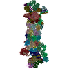
-Segmentation: This is a binary mask of the 26S proteasome
| Annotation | This is a binary mask of the 26S proteasome | ||||||||||||
|---|---|---|---|---|---|---|---|---|---|---|---|---|---|
| File | emd_2035_msk_1.map | ||||||||||||
| Projections & Slices |
| ||||||||||||
| Density Histograms |


- Sample components
-Entire : 26S proteasomes purified from S.pombe cells
| Entire | Name: 26S proteasomes purified from S.pombe cells |
|---|---|
| Components |
|
-Supramolecule #1000: 26S proteasomes purified from S.pombe cells
| Supramolecule | Name: 26S proteasomes purified from S.pombe cells / type: sample / ID: 1000 / Details: The sample was monodisperse / Number unique components: 1 |
|---|---|
| Molecular weight | Experimental: 2.5 MDa / Theoretical: 2.5 MDa |
-Supramolecule #1: 26S proteasome
| Supramolecule | Name: 26S proteasome / type: organelle_or_cellular_component / ID: 1 / Name.synonym: 26S proteasome / Recombinant expression: No |
|---|---|
| Source (natural) | Organism: |
-Experimental details
-Structure determination
| Method | cryo EM |
|---|---|
 Processing Processing | single particle reconstruction |
| Aggregation state | particle |
-Sample preparation
| Concentration | 0.3 mg/mL |
|---|---|
| Buffer | pH: 7.3 |
| Vitrification | Cryogen name: ETHANE / Instrument: OTHER |
- Electron microscopy
| Microscope | FEI TECNAI F20 |
|---|---|
| Electron beam | Acceleration voltage: 200 kV / Electron source:  FIELD EMISSION GUN FIELD EMISSION GUN |
| Electron optics | Illumination mode: FLOOD BEAM / Imaging mode: BRIGHT FIELD / Cs: 2 mm |
| Sample stage | Specimen holder: Eucentric / Specimen holder model: GATAN LIQUID NITROGEN |
| Experimental equipment |  Model: Tecnai F20 / Image courtesy: FEI Company |
-Image processing
| Final reconstruction | Applied symmetry - Point group: C2 (2 fold cyclic) / Resolution.type: BY AUTHOR / Resolution: 8.4 Å / Resolution method: FSC 0.5 CUT-OFF / Software - Name: XMIPP / Number images used: 380000 |
|---|
