 Movie
Movie Controller


+ Open data

- Basic information
| Entry | Database: EMDB / ID: EMD-5669 | |||||||||
|---|---|---|---|---|---|---|---|---|---|---|
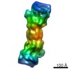
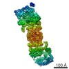
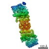
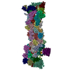
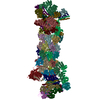
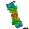
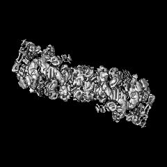
| Title | Substrate-bound 26S proteasome Rpn11AXA Rpn13-delta mutant | |||||||||
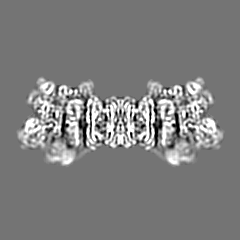
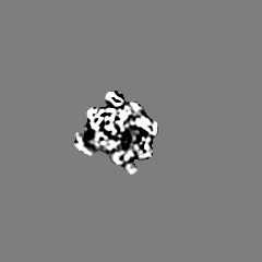
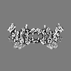
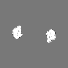
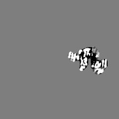
 Map data Map data | Reconstruction of substrate-bound Rpn11AXA Rpn13-delta yeast 26S proteasome | |||||||||
 Sample Sample |
| |||||||||
 Keywords Keywords | 26S proteasome / 19S regulatory particle / Rpn11 / deubiquitination / AAA+ ATPase / protein translocation / cryoEM / UPS | |||||||||
| Function / homology | proteasome regulatory particle / Proteasome, subunit alpha/beta Function and homology information Function and homology information | |||||||||
| Biological species |  | |||||||||
| Method | single particle reconstruction / cryo EM / Resolution: 9.0 Å | |||||||||
 Authors Authors | Matyskiela ME / Lander GC / Martin A | |||||||||
 Citation Citation | Journal: Nat Struct Mol Biol / Year: 2013 Title: Conformational switching of the 26S proteasome enables substrate degradation. Authors: Mary E Matyskiela / Gabriel C Lander / Andreas Martin /  Abstract: The 26S proteasome is the major eukaryotic ATP-dependent protease, responsible for regulating the proteome through degradation of ubiquitin-tagged substrates. Its regulatory particle, containing the ...The 26S proteasome is the major eukaryotic ATP-dependent protease, responsible for regulating the proteome through degradation of ubiquitin-tagged substrates. Its regulatory particle, containing the heterohexameric AAA+ ATPase motor and the essential deubiquitinase Rpn11, recognizes substrates, removes their ubiquitin chains and translocates them into the associated peptidase after unfolding, but detailed mechanisms remain unknown. Here we present the 26S proteasome structure from Saccharomyces cerevisiae during substrate degradation, showing that the regulatory particle switches from a preengaged to a translocation-competent conformation. This conformation is characterized by a rearranged ATPase ring with uniform subunit interfaces, a widened central channel coaxially aligned with the peptidase and a spiral orientation of pore loops that suggests a rapid progression of ATP-hydrolysis events around the ring. Notably, Rpn11 moves from an occluded position to directly above the central pore, thus facilitating substrate deubiquitination concomitant with translocation. | |||||||||
| History |
|
- Structure visualization
| Movie |
Movie viewer |
|---|---|
| Structure viewer | EM map: SurfViewMolmilJmol/JSmol |
| Supplemental images |

- Downloads & links
-EMDB archive
| Map data | emd_5669.map.gz | 2.2 MB | EMDB map data format | |
|---|---|---|---|---|
| Header (meta data) | emd-5669-v30.xmlemd-5669.xml | 10.5 KB 10.5 KB | Display Display | EMDB header |
| Images | 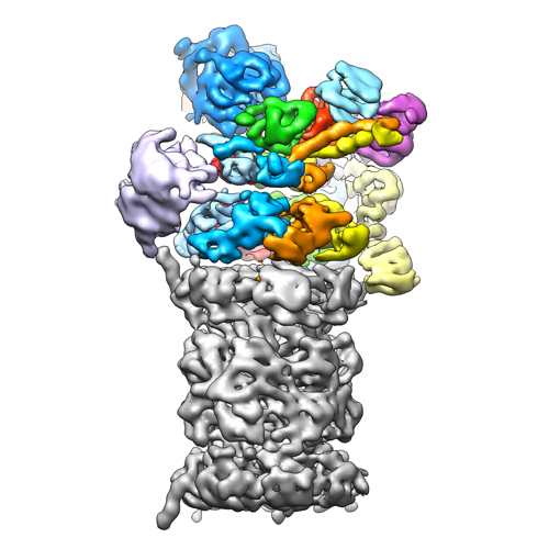 emd_5669_1.png emd_5669_1.png | 164 KB | ||
| Archive directory |  http://ftp.pdbj.org/pub/emdb/structures/EMD-5669ftp://ftp.pdbj.org/pub/emdb/structures/EMD-5669 http://ftp.pdbj.org/pub/emdb/structures/EMD-5669ftp://ftp.pdbj.org/pub/emdb/structures/EMD-5669 | HTTPS FTP |
-Related structure data

-Links
| EMDB pages | EMDB (EBI/PDBe) / EMDataResource |
|---|
-Map
| File | Download / File: emd_5669.map.gz / Format: CCP4 / Size: 51.5 MB / Type: IMAGE STORED AS FLOATING POINT NUMBER (4 BYTES) | ||||||||||||||||||||||||||||||||||||||||||||||||||||||||||||||||||||
|---|---|---|---|---|---|---|---|---|---|---|---|---|---|---|---|---|---|---|---|---|---|---|---|---|---|---|---|---|---|---|---|---|---|---|---|---|---|---|---|---|---|---|---|---|---|---|---|---|---|---|---|---|---|---|---|---|---|---|---|---|---|---|---|---|---|---|---|---|---|
| Annotation | Reconstruction of substrate-bound Rpn11AXA Rpn13-delta yeast 26S proteasome | ||||||||||||||||||||||||||||||||||||||||||||||||||||||||||||||||||||
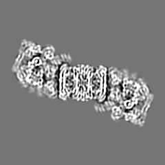
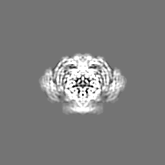
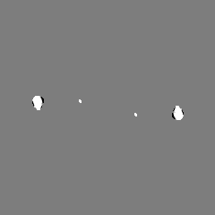
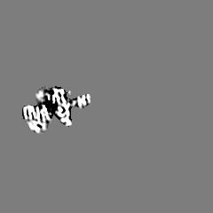
| Projections & slices | Image control
Images are generated by Spider. | ||||||||||||||||||||||||||||||||||||||||||||||||||||||||||||||||||||
| Voxel size | X=Y=Z: 2.17 Å | ||||||||||||||||||||||||||||||||||||||||||||||||||||||||||||||||||||
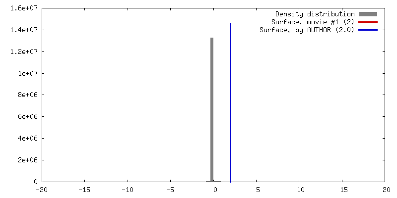
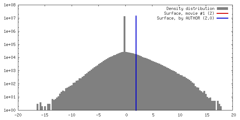
| Density |
| ||||||||||||||||||||||||||||||||||||||||||||||||||||||||||||||||||||
| Symmetry | Space group: 1 | ||||||||||||||||||||||||||||||||||||||||||||||||||||||||||||||||||||
| Details | EMDB XML:
CCP4 map header:
| ||||||||||||||||||||||||||||||||||||||||||||||||||||||||||||||||||||
 Z (Sec.)
 Y (Row.)
 X (Col.)


-Supplemental data
- Sample components
-Entire : Yeast Rpn11AXA Rpn13-delta mutant 26S proteasome
| Entire | Name: Yeast Rpn11AXA Rpn13-delta mutant 26S proteasome |
|---|---|
| Components |
|
-Supramolecule #1000: Yeast Rpn11AXA Rpn13-delta mutant 26S proteasome
| Supramolecule | Name: Yeast Rpn11AXA Rpn13-delta mutant 26S proteasome / type: sample / ID: 1000 / Details: mutant proteasome was purified from yeast / Oligomeric state: holoenzyme / Number unique components: 1 |
|---|---|
| Molecular weight | Experimental: 1.5 MDa / Theoretical: 1.5 MDa / Method: Sedimentation |
-Macromolecule #1: 26S proteasome
| Macromolecule | Name: 26S proteasome / type: protein_or_peptide / ID: 1 / Name.synonym: proteasome / Details: Mutant proteasome was purified from yeast / Number of copies: 1 / Oligomeric state: monomer / Recombinant expression: No / Database: NCBI |
|---|---|
| Source (natural) | Organism: |
| Molecular weight | Experimental: 1.5 MDa / Theoretical: 1.5 MDa |
| Sequence | GO: proteasome regulatory particle / InterPro: Proteasome, subunit alpha/beta |
-Experimental details
-Structure determination
| Method | cryo EM |
|---|---|
 Processing Processing | single particle reconstruction |
| Aggregation state | particle |
-Sample preparation
| Concentration | 2 mg/mL |
|---|---|
| Buffer | pH: 7.6 Details: 60mM HEPES, 50mM NaCl, 50mM KCl, 5mM MgCl2, 0.5mM EDTA, 1mM DTT, 2mM ATP, 0.05% NP40 |
| Grid | Details: 400-mesh C-flats, 2 um holes with 2 um spacing (Protochips Inc.) |
| Vitrification | Cryogen name: ETHANE / Chamber humidity: 50 % / Chamber temperature: 86 K / Instrument: FEI VITROBOT MARK II / Method: Blot 3 seconds with -1 offset |
- Electron microscopy
| Microscope | FEI TECNAI F20 |
|---|---|
| Temperature | Min: 78 K / Max: 80 K / Average: 79 K |
| Alignment procedure | Legacy - Astigmatism: objective lens astigmatism was corrected at 210,000 times magnification |
| Details | Data acquired with Leginon |
| Date | Sep 10, 2012 |
| Image recording | Category: CCD / Film or detector model: GATAN ULTRASCAN 4000 (4k x 4k) / Number real images: 5328 / Average electron dose: 20 e/Å2 |
| Electron beam | Acceleration voltage: 120 kV / Electron source:  FIELD EMISSION GUN FIELD EMISSION GUN |
| Electron optics | Calibrated magnification: 100000 / Illumination mode: FLOOD BEAM / Imaging mode: BRIGHT FIELD / Cs: 2.2 mm / Nominal defocus max: 2.5 µm / Nominal defocus min: 1.2 µm / Nominal magnification: 100000 |
| Sample stage | Specimen holder: Gatan 626, 70 degree cryoholder / Specimen holder model: GATAN LIQUID NITROGEN |
| Experimental equipment |  Model: Tecnai F20 / Image courtesy: FEI Company |
-Image processing
| Details | 3D reconstruction with EMAN2/SPARX libraries; final alignment of particles with FREALIGN. Sharpened with SPIDER; low-pass filtered to local resolution with BSOFT. |
|---|---|
| CTF correction | Details: each particle |
| Final reconstruction | Algorithm: OTHER / Resolution.type: BY AUTHOR / Resolution: 9.0 Å / Resolution method: FSC 0.5 CUT-OFF / Software - Name: EMAN2/SPARX, FREALIGN Details: Final map filtered to local resolution using the blocfilt function in Bsoft. Image processing performed in the Appion processing environment. Number images used: 188400 |
