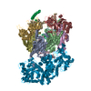
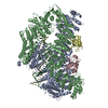
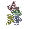
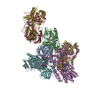
ジャーナル: Nature / 年: 2015 タイトル: Structural basis for retroviral integration into nucleosomes. 著者: Daniel P Maskell / Ludovic Renault / Erik Serrao / Paul Lesbats / Rishi Matadeen / Stephen Hare / Dirk Lindemann / Alan N Engelman / Alessandro Costa / Peter Cherepanov / 要旨: Retroviral integration is catalysed by a tetramer of integrase (IN) assembled on viral DNA ends in a stable complex, known as the intasome. How the intasome interfaces with chromosomal DNA, which ...Retroviral integration is catalysed by a tetramer of integrase (IN) assembled on viral DNA ends in a stable complex, known as the intasome. How the intasome interfaces with chromosomal DNA, which exists in the form of nucleosomal arrays, is currently unknown. Here we show that the prototype foamy virus (PFV) intasome is proficient at stable capture of nucleosomes as targets for integration. Single-particle cryo-electron microscopy reveals a multivalent intasome-nucleosome interface involving both gyres of nucleosomal DNA and one H2A-H2B heterodimer. While the histone octamer remains intact, the DNA is lifted from the surface of the H2A-H2B heterodimer to allow integration at strongly preferred superhelix location ±3.5 positions. Amino acid substitutions disrupting these contacts impinge on the ability of the intasome to engage nucleosomes in vitro and redistribute viral integration sites on the genomic scale. Our findings elucidate the molecular basis for nucleosome capture by the viral DNA recombination machinery and the underlying nucleosome plasticity that allows integration.
生物種: Escherichia coli BL21(DE3) (大腸菌) / 組換プラスミド: pet
-
分子 #3: DNA
分子
名称: DNA / タイプ: dna / ID: 3 / 分類: DNA / Structure: SINGLE STRANDED / Synthetic?: Yes
由来(天然)
生物種: synthetic construct (人工物)
-
実験情報
-
構造解析
手法
クライオ電子顕微鏡法
解析
単粒子再構成法
試料の集合状態
particle
-
試料調製
濃度
0.1 mg/mL
緩衝液
pH: 7.45 / 詳細: 320 mM NaCl, 25 mM BisTris propane-HCl
グリッド
詳細: 400 mesh C-flat copper grids CF-1/1
凍結
凍結剤: ETHANE / チャンバー内湿度: 80 % / チャンバー内温度: 101 K / 装置: GATAN CRYOPLUNGE 3 手法: The sample was incubated for 1 minute on the grid and blotted for 3.8 seconds before plunging.
Particles were picked in Xmipp 3.0; Contrast Transfer Function was estimated using CTFFIND3. All further processing was performed within the RELION 1.2 environment.
CTF補正
詳細: each particle
最終 再構成
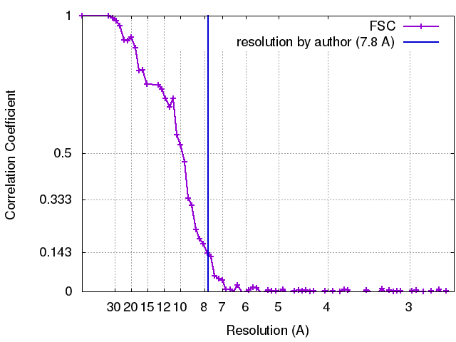
想定した対称性 - 点群: C1 (非対称) / 解像度のタイプ: BY AUTHOR / 解像度: 7.8 Å / 解像度の算出法: OTHER / ソフトウェア - 名称: RELION / 使用した粒子像数: 53887
 ムービー
ムービー コントローラー


 データを開く
 基本情報
基本情報 マップデータ
 試料
試料 キーワード
キーワード 機能・相同性情報
 Homo sapiens (ヒト) / synthetic construct (人工物)
 データ登録者
 引用
引用


 構造の表示
構造の表示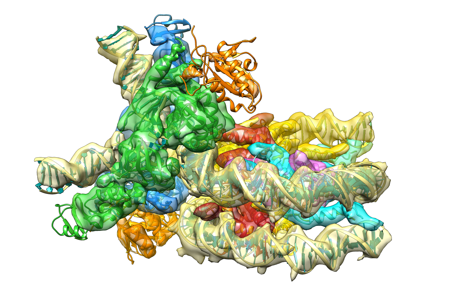
 ダウンロードとリンク
 emd_2992.png
 http://ftp.pdbj.org/pub/emdb/structures/EMD-2992


 Z (Sec.)
 Y (Row.)
 X (Col.)


 試料の構成要素
 解析
解析 電子顕微鏡法
 FIELD EMISSION GUN